. 2020 Nov 6;15(9):950-964.
doi: 10.1093/scan/nsaa122.

Fear conditioning prompts sparser representations of conditioned threat in primary visual cortex

Affiliations

Fear conditioning prompts sparser representations of conditioned threat in primary visual cortex

Siyang Yin et al. Soc Cogn Affect Neurosci. .

Abstract

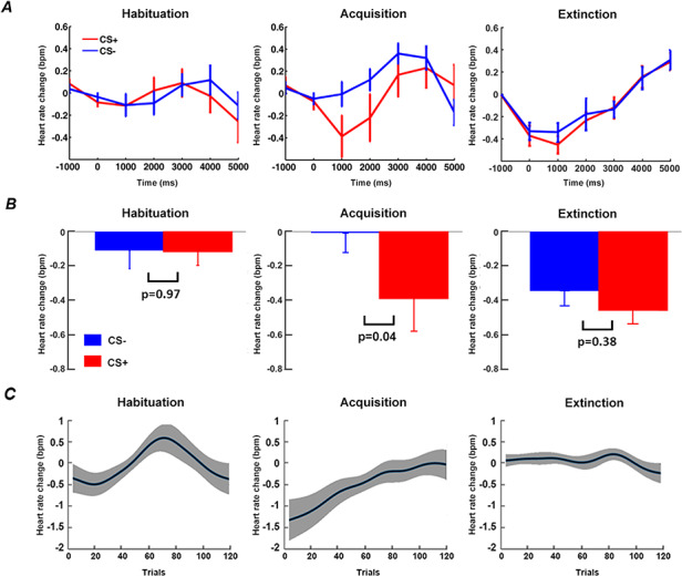
Repeated exposure to threatening stimuli alters sensory responses. We investigated the underlying neural mechanism by re-analyzing previously published simultaneous electroencephalogram-functional magnetic resonance imaging (EEG-fMRI) data from humans viewing oriented gratings during Pavlovian fear conditioning. In acquisition, one grating (CS+) was paired with a noxious noise, the unconditioned stimulus (US). The other grating (CS-) was never paired with the US. In habituation, which preceded acquisition, and in extinction, the same two gratings were presented without US. Using fMRI multivoxel patterns in primary visual cortex during habituation as reference, we found that during acquisition, aversive learning selectively prompted systematic changes in multivoxel patterns evoked by CS+. Specifically, CS+ evoked voxel patterns in V1 became sparser as aversive learning progressed, and the sparsified pattern appeared to be preserved in extinction. Concomitant with the voxel pattern changes, occipital alpha oscillations were increasingly more desynchronized during CS+ (but not CS-) trials. Across acquisition trials, the rate of change in CS+-related alpha desynchronization was correlated with the rate of change in multivoxel pattern representations of CS+. Furthermore, alpha oscillations co-varied with blood-oxygen-level-dependent (BOLD) data in the ventral attention network, but not with BOLD in the amygdala. Thus, fear conditioning prompts persistent sparsification of voxel patterns evoked by threat, likely mediated by attention-related mechanisms.

Keywords: alpha oscillations; attention; fear conditioning; sparsification; visual representation.


Figures

Fig. 1.
Fig. 1.
Experimental paradigm. Top: Temporal order of the three blocks. Bottom: timeline and stimuli used during the acquisition block. For the habituation block and the extinction block, the two Gabor patches and inter-trial interval (ITI) were the same, except that no US was presented.
Fig. 2.
Fig. 2.
ROI definition. (A) V1 ROI defined according to a recently published retinotopic atlas of the visual cortex by Wang et al. (2015). (B) rTPJ and rVLPFC ROIs defined according to previously published coordinates (Geng and Vossel, 2013; Yin et al., 2018). (C) Right amygdala ROI defined according to previously published coordinates based on US activation (Yin et al., 2018).
Fig. 3.
Fig. 3.
Heart rate (HR) analysis. (A) Event-related HR changes during habituation, acquisition and extinction. (B) Statistical comparison of HR between CS+ and CS- at time = 1 s (0.5 s to 1.5 s). (C) Time course of relative event-related HR changes (CS+ minus CS-) over trials in habituation, acquisition, and extinction. Note: Figure 3A (left and middle), 3B (left and middle) and 3C (left and middle) are adapted from Yin et al. (2018) under CC BY 4.0 and included here for comparison with Figure 3A (right), 3B (right) and 3C (right).
Fig. 4.
Fig. 4.
Pattern similarity changes during acquisition in V1. (A) Time course of pattern similarity change in V1 for CS- trials (Subject 8 in (C)). (B) Time course of pattern similarity change in V1 for CS+ trials from the same subject. (C) Slopes of linear fits to pattern similarity curves such as the ones in (A) and (B) for each participant. (D) Slopes of similarity curves between CS+ and CS- in V1 were significantly different.
Fig. 5.
Fig. 5.
Pattern sparsity analysis for CS+ trials. (A) No significant difference in number of representational voxels for CS+ in V1 between early and late habituation. (B) No significant difference in average BOLD activation between early and late habituation. (C) Number of representational voxels for CS+ in V1 was significantly lower in late acquisition than early acquisition. (D) No significant difference in average BOLD activation between early and late acquisition. (E) Schematic illustration of increasing sparsity observed during CS+ trials over time: CS+ evoked multivoxel patterns of beta values in habituation, early acquisition and late acquisition.
Fig. 6.
Fig. 6.
Event-related alpha desynchronization during habituation and acquisition. (A) Alpha-band (8–12 Hz) power averaged across CS+ trials and across CS- trials during habituation, the early period of acquisition and the late period of acquisition. (B) CS +-evoked alpha ERD and the difference in CS+ and CS- alpha-band power for early and late acquisition periods. (C) The slope of linear fit to the time course of alpha-band power across acquisition trials. (D) Relation between the rate of event-related alpha-band power decrease and the rate of pattern similarity change in V1 (each point in the plot represents one participant).
Fig. 7.
Fig. 7.
EEG-BOLD coupling in acquisition. (A) Across-participant correlations between alpha ERD and BOLD in the rTPJ and rVLPFC, both of the ventral attention network, and the right amygdala. A negative correlation was observed between alpha ERD difference (CS+ minus CS-) and the difference in rTPJ beta values (CS+ minus CS-). No correlation was observed between alpha ERD difference and the estimated beta difference in rVLPFC and right amygdala. Each point in the plots represents a participant. (B) Across-trial correlations between alpha ERD and BOLD in rTPJ, rVLPFC and right amygdala. There was a significant negative correlation between trial-wise alpha power and trial-wise beta value from rTPJ and rVLPFC, but no correlation between trial-wise alpha power and trial-wise beta from right amygdala. Each point in the plots represents a trial.
Fig. 8.
Fig. 8.
Neural dynamics in V1 during extinction. (A) Slopes of linear fits to pattern similarity curves for each participant. (B) Slopes of similarity curves were not significantly different between CS+ and CS-. (C) No significant difference in number of representational voxels for CS+ between early and late extinction. (D) No significant difference in average BOLD activation for CS+ between early and late extinction. (E) and (F) Alpha-band ERD for CS+ and CS- trials during early extinction and late extinction. (G) and (H) No significant difference in alpha ERD between CS+ and CS- in either early or late extinction.

Similar articles

Cited by

References

    1. Allen P.J., Polizzi G., Krakow K., Fish D.R., Lemieux L. (1998). Identification of EEG events in the MR scanner: the problem of pulse artifact and a method for its subtraction. NeuroImage, 8(3), 229–39. Headley and Weinberger, 2011; Moran and Katz, 2014. - PubMed
    1. Amaral D.G., Behniea H., Kelly J.L. (2003). Topographic organization of projections from the amygdala to the visual cortex in the macaque monkey. Neuroscience, 118(4), 1099–120. - PubMed
    1. Antov M.I., Plog E., Bierwirth P., Keil A., Stockhorst U. (2020). Visuocortical tuning to a threat-related feature persists after extinction and consolidation of conditioned fear. Scientific Reports, 10, 1–15. - PMC - PubMed
    1. Armony J.L., Dolan R.J. (2002). Modulation of spatial attention by fear-conditioned stimuli: an event-related fMRI study. Neuropsychologia, 40(7), 817–26. - PubMed
    1. Auksztulewicz R., Friston K.J., Nobre A.C. (2017). Task relevance modulates the behavioural and neural effects of sensory predictions. PLoS Biology, 15(12), e2003143. doi: 10.1371/journal.pbio.2003143 - DOI - PMC - PubMed

Publication types