. 2020 Nov;46(5):1733-1742.
doi: 10.3892/ijmm.2020.4718. Epub 2020 Sep 2.

Heterogeneous nuclear ribonucleoprotein K is overexpressed and contributes to radioresistance irrespective of HPV status in head and neck squamous cell carcinoma

Affiliations

Heterogeneous nuclear ribonucleoprotein K is overexpressed and contributes to radioresistance irrespective of HPV status in head and neck squamous cell carcinoma

Justus Kähler et al. Int J Mol Med. 2020 Nov.

Abstract

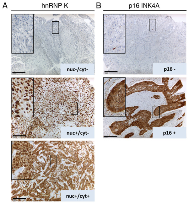
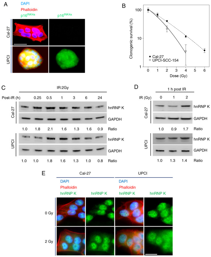
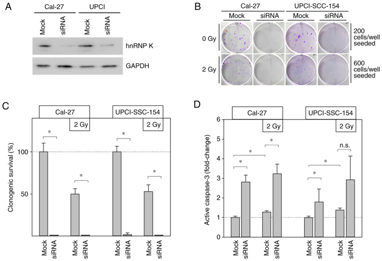
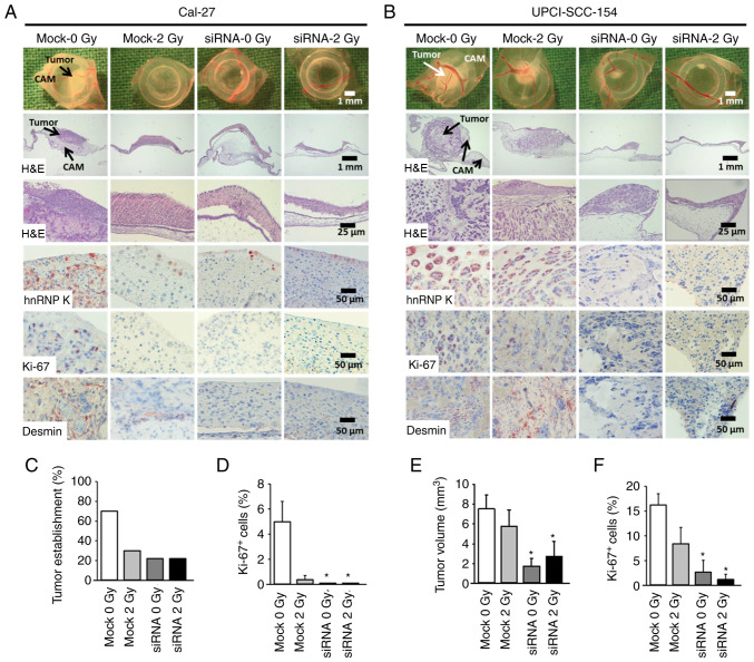
Radiotherapy is a major treatment option for head and neck squamous cell carcinoma (HNSCC). However, the success of radiotherapy is limited by tumor cell resistance to ionizing radiation (IR). Clinical studies have demonstrated an overall improved prognosis and higher susceptibility to radiotherapy of high‑risk human papillomavirus (HPV)‑associated HNSCC compared with classic HNSCC, as well as worse overall survival for male HNSCC patients. Overexpression of heterogeneous nuclear ribonucleoprotein (hnRNP) K has been associated with resistance to radiotherapy in melanoma and colorectal carcinoma. The aim of the present study was to analyze the impact of hnRNP K expression on the aggressiveness and radioresistance of HNSCC with respect to patient sex and HPV status. Immunohistochemical staining of HNSCC tissue specimens revealed elevated hnRNP K levels compared with those in the non‑neoplastic epithelium. Cytoplasmic hnRNP K accumulation was associated with advanced tumor stage and male sex. Exposure of HNSCC cells to IR was followed by rapid upregulation of hnRNP K at the protein level, along with re‑localization from the tumor cell nucleus to the cytoplasm. siRNA‑based knockdown of hnRNP K induced apoptosis and abolished tumor formation after xenotransplantation of HNSCC cells onto the chick egg chorioallantoic membrane (CAM). The observed effects were independent of the respective HPV status of the cell lines. These results indicated a tumorigenic and anti‑apoptotic role of hnRNP K in HNSCC, which appeared to be enhanced in male patients and contributed to the radioresistance of these tumors. However, the radioprotective effects of hnRNP K were found to be independent of the tumor's HPV status.


Figures

Figure 1
Figure 1
(A) Representative images of HNSCC tissue samples for hnRNP K IHC staining focusing on nuclear (nuc) and cytoplasmic (cyt) hnRNP K expression (scale bar, 200 μm): hnRNP K-negative (upper panel), nuclear hnRNP K expression (middle panel) and hnRNP K nuclear and cytoplasmic expression (lower panel). (B) Representative images of p16 INK4A-negative (upper panel) and -positive (lower panel) HNSCC samples. Staining for p16INK4A served as a surrogate marker for HPV-positive HNSCC. HNSCC, head and neck squamous cell carcinoma; hnRNP K, heterogenous nuclear ribonucleoprotein K; IHC, immunohistochemistry; HPV, human papillomavirus.
Figure 2
Figure 2
(A) Representative images of HNSCC cell clusters stained for p16INK4a using immunofluorescence illustrate the HPV status of Cal-27 (HPV-negative) and UPCI-SCC 154 (HPV-positive) cells (scale bar, 40 μm). (B) Clonogenic survival assay of the HNSCC cell lines Cal-27 and UPCI-SCC 154 after 9 days. Data are presented as mean ± SD of 4 independent experiments. (C) Representative immunoblots demonstrate a rapid increase in cellular hnRNP K levels induced by IR (2 Gy), reaching maximum levels after 30-60 min before normalization of cellular hnRNP K levels within 24 h. Ratios represent hnRNP K/GAPDH referenced to non-irradiated control. (D) Dose-dependent accumulation of cellular hnRNP K 1 h after IR. (E) Immunofluorescence microscopy indicated cytoplasmic hnRNP K accumulation 1 h after irradiating cells with 2 Gy (scale bar, 20 μm). HNSCC, head and neck squamous cell carcinoma; hnRNP K, heterogenous nuclear ribonucleoprotein K; HPV, human papillomavirus; IR, ionizing radiation.
Figure 3
Figure 3
(A) Effective hnRNP K knockdown by transient transfection was verified by immunoblotting. Mock transfection served as control. (B) Representative images of colonies after 9 days of incubation. (C) Statistical analysis of clonogenic survival assays during hnRNP K knockdown. All experiments were carried out in quadruplicate. Data are presented as mean ± SD. *P<0.05 (Kruskal Wallis test, Tukey's post hoc test). (D) ELISA showed significantly increased levels of cellular active caspase-3 in Cal-27 and UPCI-SCC-154 cells parallel to hnRNP K knockdown. Data are presented as mean ± SD. *P<0.05; n.s., not significant; n=6 (Kruskal Wallis test, Holm-Sidak post hoc test). hnRNP K, heterogenous nuclear ribonucleoprotein K.
Figure 4
Figure 4
Knockdown of hnRNP K inhibits growth of HNSSC xenografts on the chick egg CAM in vivo. Cells (mock or siRNA, ± irradiation) were seeded on the CAM of fertilized chick eggs 7 days after the start of incubation (1.5×10⁶ cells/egg in medium/Matrigel 1:1). After an incubation period of 4 days at 37°C, the tumors were collected, imaged, fixed and embedded in paraffin for immunohistochemical analysis. Sections (5 μm) were stained for hnRNP K, proliferation marker Ki-67 and the angiogenesis marker desmin. Each group contained 9-10 tumor-bearing eggs. (A and B) Representative images of tumor xenografts immediately after extraction (1st row), overview of tumor and underlying CAM tissue (H&E staining, 2nd and 3rd rows), immunohistochemical staining of hnRNP K-expressing cells (4th row), Ki-67+ proliferative cells (5th row) and desmin+ pericytes indicating angiogenesis (6th row). (C) Percentage of solid tumor formation of Cal-27 cells 4 days after xenotransplantation (9-10 tumors/group). (D) Percentage of proliferating Ki-67+ cells in Cal-27 xenografts. A total of 261-358 cells from each tumor were evaluated. Data are presented as the mean ± SEM of 4 tumors/group. (E) Mean tumor volume of UPCI-SCC-154 cancer xenografts 4 days after xenotransplantation as assessed immediately after extraction. Tumor volume was calculated according to the formula: π/6 × length × width2. Mean of 9-10 tumors/group. (F) Percentage of proliferating Ki-67+ cells. A total of 298-632 cells from each tumor were evaluated. Data are presented as the mean ± SEM of 5 tumors/group. *P<0.05 vs. control (mock 0 Gy) (Kruskal Wallis test, Dunnett's post hoc test). hnRNP K, heterogenous nuclear ribonucleoprotein K; HNSCC, head and neck squamous cell carcinoma; CAM, chorioallantoic membrane; H&E, hematoxylin and eosin.

Similar articles

Cited by

References

    1. Torre LA, Bray F, Siegel RL, Ferlay J, Lortet-Tieulent J, Jemal A. Global cancer statistics, 2012. CA Cancer J Clin. 2015;65:87–108. doi: 10.3322/caac.21262. - DOI - PubMed
    1. Bray F, Ferlay J, Soerjomataram I, Siegel RL, Torre LA, Jemal A. Global cancer statistics 2018: GLOBOCAN estimates of incidence and mortality worldwide for 36 cancers in 185 countries. CA Cancer J Clin. 2018;68:394–424. doi: 10.3322/caac.21492. - DOI - PubMed
    1. Franceschi S, Talamini R, Barra S, Baron AE, Negri E, Bidoli E, Serraino D, La Vecchia C. Smoking and drinking in relation to cancers of the oral cavity, pharynx, larynx, and esophagus in northern Italy. Cancer Res. 1990;50:6502–6507. - PubMed
    1. Ang KK, Sturgis EM. Human papillomavirus as a marker of the natural history and response to therapy of head and neck squamous cell carcinoma. Semin Radiat Oncol. 2012;22:128–142. doi: 10.1016/j.semradonc.2011.12.004. - DOI - PubMed
    1. Chaturvedi AK. Epidemiology and clinical aspects of HPV in head and neck cancers. Head Neck Pathol. 2012;6(Suppl 1):S16–S24. doi: 10.1007/s12105-012-0377-0. - DOI - PMC - PubMed

MeSH terms

Substances