

lncRNA small nucleolar RNA host gene 12 promotes renal cell carcinoma progression by modulating the miR‑200c‑5p/collagen type XI α1 chain pathway
- PMID: 32901847
- PMCID: PMC7533520
- DOI: 10.3892/mmr.2020.11490
lncRNA small nucleolar RNA host gene 12 promotes renal cell carcinoma progression by modulating the miR‑200c‑5p/collagen type XI α1 chain pathway
Abstract
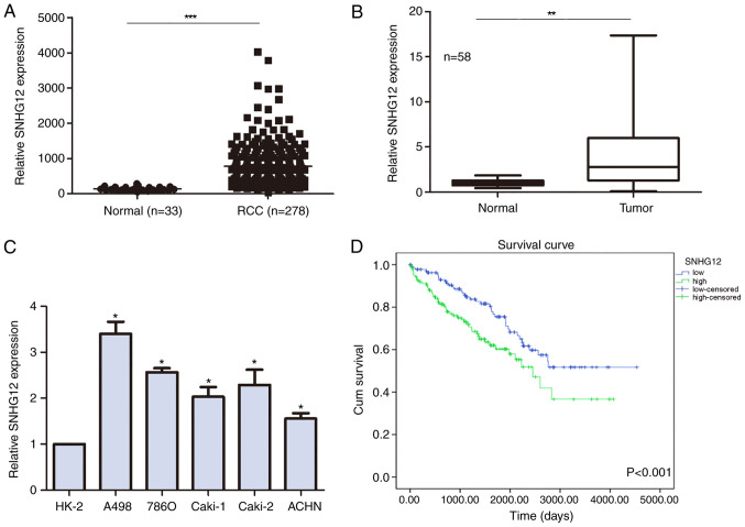
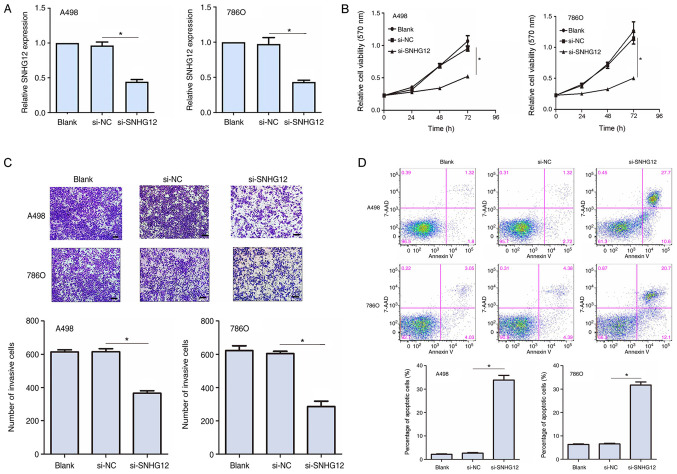
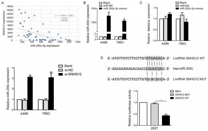
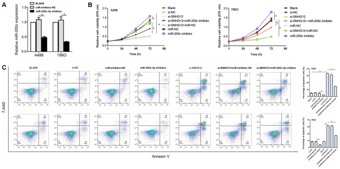
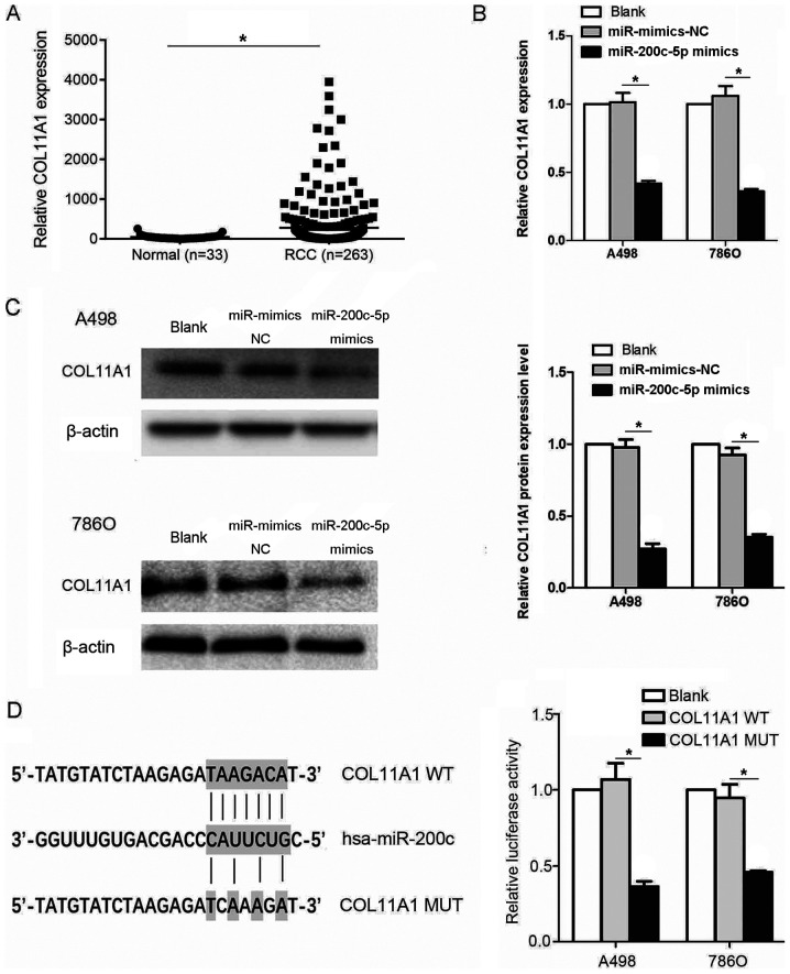
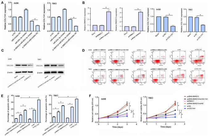
Renal cell carcinoma (RCC) is a primary malignant kidney cancer subtype. It has been suggested that long non‑coding RNAs (lncRNAs) serve important roles in the progression of kidney cancer. In fact, the lncRNA small nucleolar RNA host gene 12 (SNHG12) was discovered to be overexpressed in various types of cancer. However, to the best of our knowledge, the role of SNHG12 in RCC remains unclear. The present study aimed to investigate the function of SNHG12 and its underlying molecular mechanism of action in RCC. In patient samples and datasets from The Cancer Genome Atlas. Reverse transcription‑quantitative PCR, demonstrated that SNHG12 expression levels were upregulated in RCC tumor tissues, but not in normal kidney tissues. SNHG12 upregulation was also observed in RCC cell lines. Kaplan‑Meier survival analysis indicated a poor prognosis for those patients with RCC who had upregulated SNHG12 expression levels. Following lentivirus transduction, SNHG12 was successfully knocked down (validated by western blot analysis) and cell migration and invasion assays were performed. SNHG12 knockdown markedly inhibited cell viability and invasion, while increasing apoptosis in both A498 and 786O cell lines. The results of the luciferase reporter assay suggested that SNHG12 exerted its role by sponging microRNA (miR)‑200c‑5p, which led to the upregulation of its target gene, collagen type XI α1 chain (COL11A1). This was further validated, as miR‑200c‑5p inhibition reduced the effects of SNHG12 downregulation on cell viability and apoptosis, without affecting SNHG12 expression levels. Furthermore, the findings indicated that SNHG12 may partially exert its role through COL11A1, which was also upregulated in RCC. In conclusion, the results of the present study suggested that the SNHG12/miR‑200c‑5p/COL11A1 axis may be crucial for RCC progression, which provided an insight into potential therapeutic strategies for RCC treatment.
Figures
Similar articles
-
Long noncoding RNA SNHG12 promotes tumour progression and sunitinib resistance by upregulating CDCA3 in renal cell carcinoma.Cell Death Dis. 2020 Jul 8;11(7):515. doi: 10.1038/s41419-020-2713-8. Cell Death Dis. 2020. PMID: 32641718 Free PMC article.
-
SNHG12 promotes carcinogenesis of human renal cell cancer via functioning as a competing endogenous RNA and sponging miR-30a-3p.J Cell Mol Med. 2021 May;25(10):4696-4708. doi: 10.1111/jcmm.16417. Epub 2021 Mar 30. J Cell Mol Med. 2021. PMID: 33787057 Free PMC article.
-
Overexpressed lncRNA FTX promotes the cell viability, proliferation, migration and invasion of renal cell carcinoma via FTX/miR‑4429/UBE2C axis.Oncol Rep. 2022 Sep;48(3):163. doi: 10.3892/or.2022.8378. Epub 2022 Jul 22. Oncol Rep. 2022. PMID: 35866591 Free PMC article.
-
The relationship between Long Noncoding RNA (lncRNA) Small Nucleolar RNA Host Gene 12 (SNHG12) expression in solid malignant tumors and prognosis of tumor patients: A systematic review and meta-analysis.Medicine (Baltimore). 2020 Oct 9;99(41):e22247. doi: 10.1097/MD.0000000000022247. Medicine (Baltimore). 2020. PMID: 33031264 Free PMC article.
-
lncRNA PVT1 in the Pathogenesis and Clinical Management of Renal Cell Carcinoma.Biomolecules. 2021 Apr 29;11(5):664. doi: 10.3390/biom11050664. Biomolecules. 2021. PMID: 33947142 Free PMC article. Review.
Cited by
-
miR-335 Restrains the Aggressive Phenotypes of Ovarian Cancer Cells by Inhibiting COL11A1.Cancers (Basel). 2021 Dec 13;13(24):6257. doi: 10.3390/cancers13246257. Cancers (Basel). 2021. PMID: 34944877 Free PMC article.
-
Collagen XI Alpha 1 Chain, a Novel Therapeutic Target for Cancer Treatment.Front Oncol. 2022 Jun 29;12:925165. doi: 10.3389/fonc.2022.925165. eCollection 2022. Front Oncol. 2022. PMID: 35847935 Free PMC article. Review.
-
Identification of novel snoRNA-based biomarkers for clear cell renal cell carcinoma from urine-derived extracellular vesicles.Biol Direct. 2024 May 13;19(1):38. doi: 10.1186/s13062-024-00467-0. Biol Direct. 2024. PMID: 38741178 Free PMC article.
-
Non-coding transcriptome profiles in clear-cell renal cell carcinoma.Nat Rev Urol. 2025 Mar;22(3):151-174. doi: 10.1038/s41585-024-00926-3. Epub 2024 Sep 6. Nat Rev Urol. 2025. PMID: 39242964 Review.
-
Construction of a prognostic risk model for clear cell renal cell carcinomas based on centrosome amplification-related genes.Mol Genet Genomics. 2025 Mar 13;300(1):30. doi: 10.1007/s00438-025-02237-7. Mol Genet Genomics. 2025. PMID: 40075035 Free PMC article.
References
MeSH terms
Substances
LinkOut - more resources
Full Text Sources
Medical
Miscellaneous
