. 2020 Nov;44(5):1851-1862.
doi: 10.3892/or.2020.7750. Epub 2020 Sep 3.

SRSF6 regulates alternative splicing of genes involved in DNA damage response and DNA repair in HeLa cells

Affiliations

SRSF6 regulates alternative splicing of genes involved in DNA damage response and DNA repair in HeLa cells

Xiao Yang et al. Oncol Rep. 2020 Nov.

Abstract

Alternative splicing (AS) occurs in nearly all human genes and abnormal AS has a close association with cancer. Serine and arginine‑rich splicing factor 6 (SRSF6), a canonical member of the serine/arginine‑rich protein family, has been characterized as an important regulator of AS. However, the role of SRSF6 in regulating AS in cancers has remained to be fully elucidated. In the present study, the median expression of SRSF6 in tumors was determined to be higher compared with that in matched normal tissues in 13 out of 16 cancer types from The Cancer Genome Atlas. To investigate the biological effects of SRSF6 overexpression, an SRSF6‑overexpression model of HeLa cells was constructed and it was revealed that SRSF6 overexpression resulted in significantly higher apoptosis and lower proliferation compared to control cells. Transcriptome analysis indicated that overexpression of SRSF6 in cancer cells induced large‑scale changes in transcriptional expression levels and AS. Two groups of cervical cancer tumor samples in which SRSF6 was differentially expressed were then selected to analyze potential SRSF6‑regulated AS. It was determined that the pattern of SRSF6‑regulated AS in clinical samples was similar to that in cancer cells and AS genes were enriched in DNA damage response (DDR) pathways, including DNA repair and double‑strand break repair via homologous recombination. Furthermore, AS events regulated by SRSF6 were validated using reverse transcription‑quantitative PCR. The present results highlighted that SRSF6 is able to trigger the activation of DDR pathways via regulation of AS to influence cancer progression. These results markedly expand the current understanding of the mechanisms underlying SRSF6‑mediated gene regulation and suggest the potential use of SRSF6 as a therapeutic target in cancer.

Keywords: SRSF6/SRP55; alternative splicing; cancer; DNA damage response; DNA repair.


Figures

Figure 1.
Figure 1.
Differential expression of SRSF6 in tumor samples from The Cancer Genome Atlas. (A) Relative expression (TPM) of SRSF6 in tumor samples (red) compared with normal samples (green) from 16 cancer types. *P<0.05. (B) Association of SRSF6 expression with the survival rates in various types of cancer. SRSF6, serine and arginine-rich splicing factor 6; HR, hazard ratio; TPM, transcripts per million; BLCA, bladder urothelial carcinoma; BRCA, breast invasive carcinoma; COAD, colon adenocarcinoma; ESCA, esophageal carcinoma; HNSC, head and neck squamous cell carcinoma; KICH, kidney chromophobe; KIRC, kidney renal clear-cell carcinoma; KIRP, kidney renal papillary cell carcinoma; LIHC, liver hepatocellular carcinoma; LUAD, lung adenocarcinoma; LUSC, lung squamous-cell carcinoma; PRAD, prostate adenocarcinoma; READ, rectum adenocarcinoma; STAD, stomach adenocarcinoma; THCA, thyroid carcinoma; UCEC, uterine corpus endometrial carcinoma.
Figure 2.
Figure 2.
SRSF6 promotes HeLa cell apoptosis and inhibits cell proliferation. (A) Relative mRNA expression of SRSF6 was validated by RT-qPCR in HeLa cells after it was overexpressed. (B) The level of FLAG-tagged SRSF6 protein expression was determined by western blot analysis using β-actin as a housekeeping protein. (C and D) Apoptosis of SRSF6-overexpressing cells and controls was measured by flow cytometry following 7-AAD and annexin V staining. (C) Quantitative results and (D) representative flow cytometry dot plots. (E) Cell proliferation of SRSF6-overexpressing cells and controls was measured by MTT assay. ***P<0.001. SRSF6, serine and arginine-rich splicing factor 6; RT-qPCR, reverse transcription-quantitative PCR; OE, overexpression; Ctrl, control; PE, phycoerythrin; 7-AAD, 7-aminoactinomycin; OD, optical density.
Figure 3.
Figure 3.
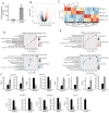
RNA-seq analysis of gene expression regulated by SRSF6 overexpression. (A) Following SRSF6 overexpression the mRNA expression level of SRSF6 was measured by RNA-seq and FPKM values were calculated. (B) Volcano plot of the genes regulated by SRSF6; upregulated genes (FC≥2; FDR<0.05) are labeled in red and downregulated genes (FC≤-2; FDR<0.05) are labeled in blue. (C) Heatmap of 837 DEGs between SRSF6 overexpression and control samples. Expression levels (FPKM) were log2-transformed and then median-centered for each gene. (D and E) The top 10 representative (D) GO biological process terms and (E) KEGG pathways of upregulated and downregulated genes following SRSF6 overexpression. (F) Reverse transcription-qPCR validation of DEGs regulated by SRSF6 in cancer cells; black bars are for the control group and grey bars for SRSF6 overexpression. ***P<0.001. Ctrl_1st and Ctrl_2nd, SRSF6_1st and SRSF6_2nd are two biological replicates. RNA-seq, RNA sequencing; SRSF6, serine and arginine-rich splicing factor 6; FPKM, fragments per kilobase of transcript per million mapped reads; qPCR, quantitative PCR; FC, fold change; FDR, false discovery rate; Ctrl, control; Up/Down, up-/downregulated genes; KEGG, Kyoto Encyclopedia of Genes and Genomes; ECM, extracellular matrix; GO, gene ontology; DEG, differentially expressed gene; POLR1G, RNA polymerase I subunit G; DLEU2L, deleted in lymphocytic leukemia 2 like; IFIT2, interferon induced protein with tetratricopeptide repeats 2; KRT13, keratin 13; PCDHB14, protocadherin beta 14; RUNDC3B, RUN domain containing 3B; YTHDF1, YTH N6-methyladenosine RNA binding protein 1.
Figure 4.
Figure 4.
AS analysis of cancer cells. (A) Classification of differential AS types regulated by SRSF6 overexpression. (B) Overlap of DEGs and RASGs following SRSF6 overexpression. (C and D) The top 10 representative (C) GO biological process terms and (D) KEGG pathways of RASGs. SRSF6, serine and arginine-rich splicing factor 6; AS, alternative splicing; DEG, differentially expressed gene; RASG, regulated AS gene; GO, gene ontology; KEGG, Kyoto Encyclopedia of Genes and Genomes; MXE, mutual exclusive exon skipping; 3pMXE, alternative last exon; 5pMXE, alternative first exon; A3SS, alternative 3′ splice site; A5SS, alternative 5′ splice site; cassetteExon, exon included; ES, exon skipping; IntronR, intron retention.
Figure 5.
Figure 5.
Validation of ASEs in cancer cells. (A and B) Genome visualization (left panel) indicates SRSF6-regulated ASEs in SRSF6-overexpression and control cells. (A) BRCA2 and (B) CHEK1. The number of junction reads was marked on the line representing splice junctions composing ASEs. The structures of the ASEs are depicted in the top-right panel. The altered ratio of ASEs according to RNA-seq and reverse transcription-qPCR was calculated and plotted (right panel, bottom). Ctrl_1st and Ctrl_2nd, SRSF6_1st and SRSF6_2nd are two biological replicates. (C) Validation results of the other four ASEs as in the right panels in A and B. Black bars are for the control group and grey bars for SRSF6 OE. *P<0.05, **P<0.01, ***P<0.001 vs. Ctrl. SRSF6, serine and arginine-rich splicing factor 6; OE, overexpression; Ctrl, control; chr, chromosome; qPCR, quantitative PCR; RNA-seq, RNA sequencing; CHEK1, checkpoint kinase 1; ASE, alternative splicing event; PARP3, poly(ADP-ribose) polymerase family member 3; PALB2, partner and localizer of BRCA2; CHEK1, checkpoint kinase 1; PARPBP, PARP1 binding protein; BRCA2, BRCA2 DNA repair associated; ATF2, activating transcription factor 2; ES, exon skipping; cassetteExon, exon included; IR, intron retention; A5SS, alternative 5′ splice site.
Figure 6.
Figure 6.
Analysis of potential SRSF6-regulated AS in cervical tumor samples. (A) A total of 16 tumor samples were divided into two groups based on the expression level of SRSF6. Box plots indicate the expression (RPKM) of SRSF6 in the two groups with high or low expression of SRSF6. The horizontal lines indicate the median, the boxes indicate the interquartile range and the vertical lines indicate the standard deviation. (B) Classification of regulated AS types in cervical tumors. (C) The top 10 representative GO biological process terms and (D) Reactome pathways in which RASGs were enriched following SRSF6 overexpression. SRSF6, serine and arginine-rich splicing factor 6; AS, alternative splicing; RASG, regulated AS gene; GO, gene ontology; RUNX1, RUNX family transcription factor 1; PD-1, programmed cell death 1; RPKM, reads per kilobase of transcript per million reads mapped; 3pMXE, alternative last exon; 5pMXE, alternative first exon; A3SS, alternative 3′ splice site; A5SS, alternative 5′ splice site; cassetteExon, exon included; ES, exon skipping; IntronR, intron retention.
Figure 7.
Figure 7.
Comparison of regulated ASEs in cancer cells (left) and in tumor samples (right). Splicing ratio of ASEs involved in DNA damage response-associated terms, which were detected in cancer cell pathways, were compared. *P<0.05, **P<0.01 vs. control. RNA-seq, RNA sequencing; Ctrl, control; OE, overexpression; ASE, alternative splicing event; MCM8, minichromosome maintenance 8 homologous recombination repair factor; MEN1, menin 1; MDC1, mediator of DNA damage checkpoint 1; NEIL1, nei-like DNA glycosylase 1; SLX1A, SLX1 homolog A, structure-specific endonuclease subunit; IR, intron retention; A5SS, alternative 5′ splice site; MXE, mutual exclusive exon skipping.

Similar articles

Cited by

References

    1. Bray F, Ferlay J, Soerjomataram I, Siegel RL, Torre LA, Jemal A. Global cancer statistics 2018: GLOBOCAN estimates of incidence and mortality worldwide for 36 cancers in 185 countries. CA Cancer J Clin. 2018;68:394–424. doi: 10.3322/caac.21492. - DOI - PubMed
    1. DePinho RA. The age of cancer. Nature. 2000;408:248–254. doi: 10.1038/35041694. - DOI - PubMed
    1. Preston DL, Mattsson A, Holmberg E, Shore R, Hildreth NG, Boice JD., Jr Radiation effects on breast cancer risk: A pooled analysis of eight cohorts. Radiat Res. 2002;158:220–235. doi: 10.1667/0033-7587(2002)158[0220:REOBCR]2.0.CO;2. - DOI - PubMed
    1. Barbieri CE, Baca SC, Lawrence MS, Demichelis JF, Blattner M, Theurillat JP, White TA, Stojanov P, Van Allen E, Stransky N, et al. Exome sequencing identifies recurrent SPOP, FOXA1 and MED12 mutations in prostate cancer. Nat Genet. 2012;44:685–689. doi: 10.1038/ng.2279. - DOI - PMC - PubMed
    1. Kaczkowski B, Tanaka Y, Kawaji H, Sandelin A, Andersson R, Itoh M, Lassmann T, Hayashizaki Y, Carninci P, Forrest AR, FANTOM5 Consortium Transcriptome analysis of recurrently deregulated genes across multiple cancers identifies new pan-cancer biomarkers. Cancer Res. 2016;76:216–226. doi: 10.1158/0008-5472.CAN-15-0484. - DOI - PubMed