. 2020 Nov;44(5):1971-1984.
doi: 10.3892/or.2020.7751. Epub 2020 Sep 4.

MicroRNA‑216a‑5p suppresses esophageal squamous cell carcinoma progression by targeting KIAA0101

Affiliations

MicroRNA‑216a‑5p suppresses esophageal squamous cell carcinoma progression by targeting KIAA0101

Tuanhe Sun et al. Oncol Rep. 2020 Nov.

Abstract

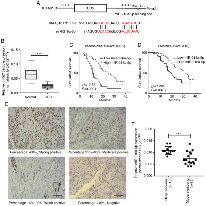
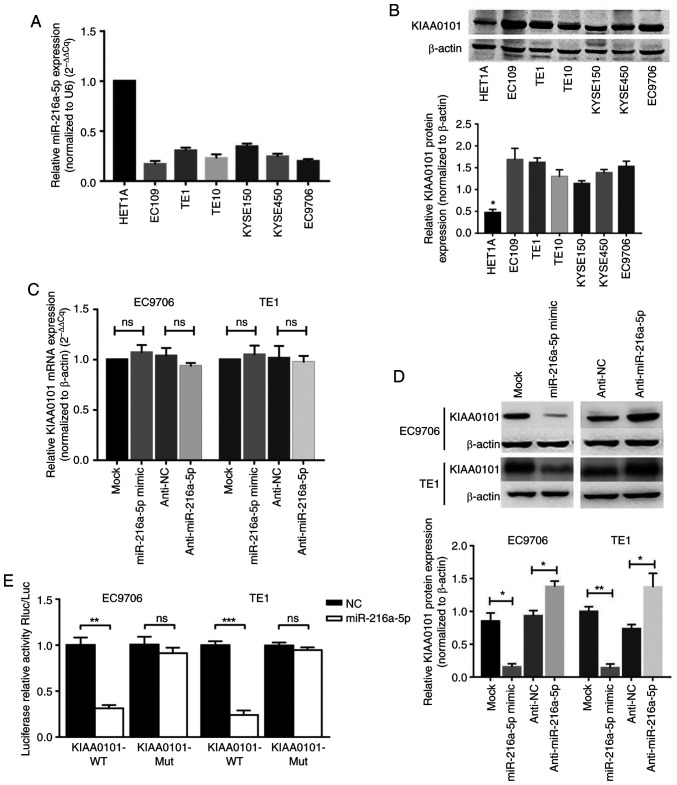
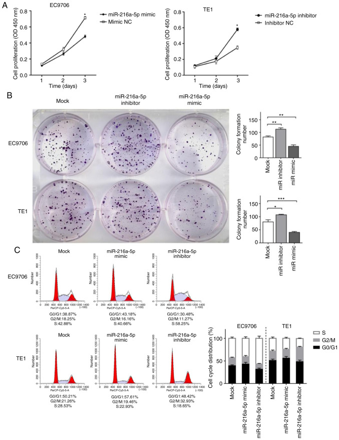
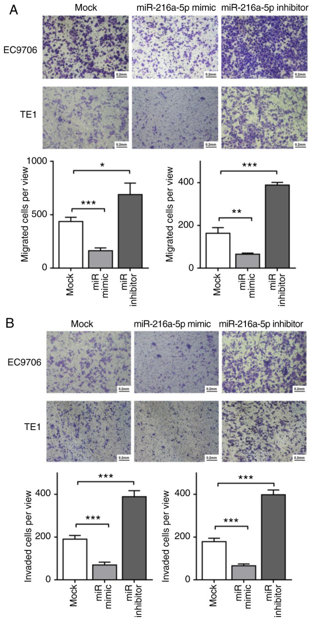
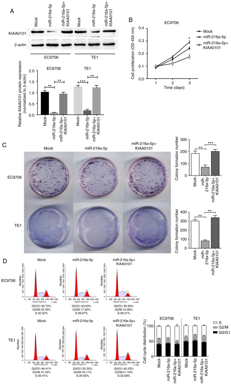
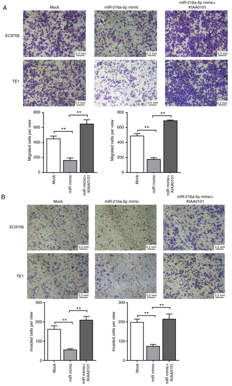
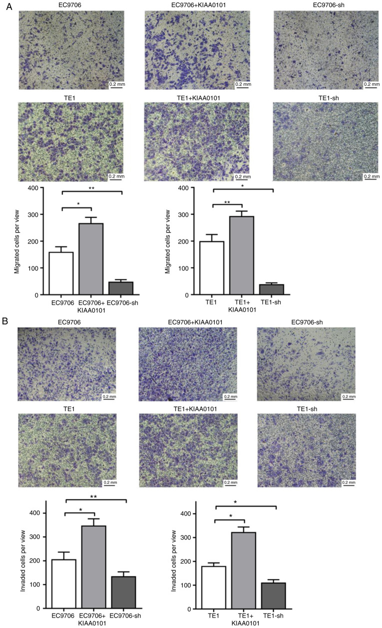
The KIAA0101 protein (also referred to as NS5ATP9 or Paf15) is overexpressed in esophageal squamous cell carcinoma (ESCC) and is associated with disease progression and poor patient survival, but how KIAA0101 expression is regulated remains unknown. The relationship between tumor miR‑216a‑5p expression and prognosis in patients with ESCC was revealed by survival analyses. Quantitative reverse‑transcriptase PCR and western blot analysis were used to evaluate miR‑216a‑5p and KIAA0101 expression in human ESCC tissues and cell lines. The targeting of KIAA0101 by miR‑216a‑5p was verified by dual‑luciferase reporter assays. The EC9706 and TE1 cell lines were transfected with miR‑216a‑5p mimics and inhibitor, or KIAA0101‑specific shRNA and KIAA0101‑expressing plasmids, in order to evaluate the effect of manipulating miR‑216a‑5p and KIAA0101 expression on ESCC cell proliferation, cell cycle progression, migration, and invasion. miR‑216a‑5p was lowly expressed and inversely correlated with KIAA0101 protein expression in ESCC tissues and cell lines. Lower miR‑216a‑5p expression was associated with worse prognosis in patients with ESCC. miR‑216a‑5p negatively regulated KIAA0101 expression by directly targeting the 3'‑untranslated region of the KIAA0101 mRNA. Overexpression of miR‑216a‑5p suppressed the proliferation, migration, and invasion of the ESCC cell lines, whereas inhibition of miR‑216a‑5p had the opposite effects. Meanwhile, KIAA0101 promoted ESCC migration and invasion, and its overexpression abolished the antitumor effects of miR‑216a‑5p mimics. As a tumor suppressor, miR‑216a‑5p targets KIAA0101 to inhibit the proliferation, migration, and invasion of ESCC. Therefore, the miR‑216a‑5p/KIAA0101 axis may be a potential target for ESCC treatment.

Keywords: microRNA; miR-216a-5p; esophageal squamous cell carcinoma; KIAA0101; disease progression.


Figures

Figure 1.
Figure 1.
miR-216a-5p is expressed at a lower level in ESCC specimens, and its low expression predicts a worse prognosis of ESCC. (A) The potential binding site of miR-216a-5p in the KIAA0101 3′UTR. (B) The relative expression levels of miR-216a-5p in ESCC specimens and adjacent normal tissues were quantified by qPCR; n=29 for the control group; n=29 for the ESCC group. (C and D) The Kaplan-Meier cumulative survival curves of 83 patients with ESCC who were grouped by their median expression levels of miR-216a-5p. The ESCC patients with lower miR-216a-5p expression displayed shorter disease-free survival (DFS; C) and overall survival (OS; D) (both P<0.0001 for DFS, P=0.0073 for OS); n=41 for the miR-216-5p low group; n=42 for the miR-216-5p high group. (E) Representative IHC images showing the different staining patterns of KIAA0101 expression with negative/weak/moderate/strong intensities. Scale bar, 50 µm. (F) miR-216a-5p expression in the ESCC specimens with moderate/strong KIAA0101 staining (n=16) and negative/weak KIAA0101 staining (n=13) was quantified by qPCR. ***P<0.001. ESCC, esophageal squamous cell carcinoma; IHC, immunohistochemistry.
Figure 2.
Figure 2.
KIAA0101 is a downstream target of miR-216a-5p, and its expression is inversely correlated with miR-216a-5p expression level in ESCC. (A) miR-216a-5p expression level in the human normal esophageal epithelial HET1A cells and six human ESCC cell lines (EC109, TE1, TE10, KYSE150, KYSE450, and EC9706) were quantified by qPCR. Values were calculated using the 2−ΔΔCq method, normalized to U6. (B) Expression of KIAA0101 protein (15 kDa) and β-actin (42 kDa) in the indicated cell lines was quantified by western blot analysis. Representative western blot images are shown (upper panel), and the band intensities were summarized (lower panel; n=3 for each group). *P<0.05, HET1A cells compared with the ESCC cell lines. (C) The mRNA level of KIAA0101 in EC9706 and TE1 cells transfected with miR-216a-5p mimic, or inhibitor (Anti-miR-216a-5p), or the corresponding controls (Mock and Anti-NC) was determined by qPCR. n=3 for each group. Values were calculated using the 2−ΔΔCq method, normalized to β-actin. ns not significant. (D) The protein levels of KIAA0101 in EC9706 and TE1 cells with the indicated transfection were determined by western blot analysis. Representative western blot images are shown (upper panel), and the band intensities were summarized (lower panel; n=3 for each group). *P<0.05 and **P<0.01. (E) Compared with the controls, miR-216a-5p inhibited the reporter activities of the vector with the wild-type (WT), but not the mutant-type (MUT) of 3′ UTR of KIAA0101 in EC9706 and TE1 cells in the dual-luciferase assays. **P<0.01 and ***P<0.001; ns, not significant. ESCC, esophageal squamous cell carcinoma.
Figure 3.
Figure 3.
miR-216a-5p suppresses the proliferation of ESCC cells in vitro. (A) The proliferation ability of EC9706 and TE1 cells transfected with the miR-216a-5p mimic, or the miR-216a-5p inhibitor, or the corresponding controls (mimic NC or inhibitor NC) were determined by CCK-8 assays. *P<0.05, compared with the mimic NC or inhibitor NC. (B) The proliferation abilities of EC9706 and TE1 cells after the indicated transfections were evaluated by colony formation assays. The representative images of cells in a 6-well plate after crystal violet staining are shown. *P<0.05, **P<0.01 and ***P<0.001. (C) The cell cycle progression of EC9706 and TE1 cells after the indicated transfections were determined by flow cytometry. The representative flow profiles of DNA content after PI staining are shown. ESCC, esophageal squamous cell carcinoma.
Figure 4.
Figure 4.
miR-216a-5p suppresses the migration and invasion of ESCC cells. (A and B) The migration (A) and invasion (B) of EC9706 and TE1 cells with mock transfection, or transfection of the miR-216a-5p mimic, or transfection of the miR-216a-5p inhibitor were evaluated by Transwell assays without Matrigel and Transwell assays with Matrigel, respectively. Representative images after crystal violet staining in each group are shown, and the numbers of migrated cells (A) or invaded cells (B) are summarized from three independent experiments with three replicates in each group. *P<0.05, **P<0.01 and ***P<0.001. ESCC, esophageal squamous cell carcinoma.
Figure 5.
Figure 5.
Ectopic KIAA0101 expression attenuates the anti-proliferative effects of miR-216a-5p in ESCC cells. (A) Western blot analysis was used to determine the expression level of KIAA0101 protein after mock transfection, or transfection of miR-216a-5p mimic alone (miR-216-5p), or combined transfection of miR-216a-5p mimic and KIAA0101-expressing plasmid (miR-216-5p+KIAA0101). Representative western blot images are shown, and the relative expression level of KIAA0101 from three independent experiments was summarized. **P<0.01 and ***P<0.001. (B) The proliferation of EC9706 cells after the indicated transfections was determined by CCK-8 assay. n=3 for each group at each time point. *P<0.05, compared with the Mock group. (C) The proliferation of EC9706 cells after the indicated transfections was determined by colony formation assays. The representative images of crystal violet staining are shown, and the numbers of colonies per well were summarized. n=3 for each group. **P<0.01 and ***P<0.001. (D) The cell cycle progression of EC9706 and TE1 cells after the indicated transfections was determined by flow cytometry. The representative flow profiles of DNA content after PI staining are shown. ESCC, esophageal squamous cell carcinoma.
Figure 6.
Figure 6.
Ectopic KIAA0101 expression attenuates the effects of miR-216a-5p on the migration and invasion of ESCC cells. (A and B) The migration (A) and invasion (B) of EC9706 and TE1 cells with mock transfection, or transfection of the miR-216a-5p mimic alone, or combined transfection of the miR-216a-5p mimic and KIAA0101-expressing plasmid were evaluated by Transwell assays without Matrigel and Transwell assays with Matrigel, respectively. Representative images after crystal violet staining in each group are shown, and the numbers of migrated cells (A) or invaded cells (B) are summarized from three independent experiments with three replicates in each group. **P<0.01. ESCC, esophageal squamous cell carcinoma.
Figure 7.
Figure 7.
KIAA0101 promotes the migration and invasion of ESCC cells. (A and B) The migration (A) and invasion (B) of EC9706 and TE1 cells without transfection, or with transfection of the KIAA0101-expressing plasmid (+KIAA0101), or with transfection of the KIAA0101-specific shRNA-expressing plasmid (-sh), were evaluated by Transwell assays without Matrigel and Transwell assays with Matrigel, respectively. Representative images after crystal violet staining in each group are shown, and the numbers of migrated cells (A) or invaded cells (B) per well are summarized from three independent experiments with three replicates in each group. *P<0.05, **P<0.01. ESCC, esophageal squamous cell carcinoma.

References

    1. Pennathur A, Gibson MK, Jobe BA, Luketich JD. Oesophageal carcinoma. Lancet. 2013;381:400–412. doi: 10.1016/S0140-6736(12)60643-6. - DOI - PubMed
    1. Bray F, Ferlay J, Soerjomataram I, Siegel RL, Torre LA, Jemal A. Global cancer statistics 2018: GLOBOCAN estimates of incidence and mortality worldwide for 36 cancers in 185 countries. CA Cancer J Clin. 2018;68:394–424. doi: 10.3322/caac.21492. - DOI - PubMed
    1. Lin Y, Totsuka Y, He Y, Kikuchi S, Qiao Y, Ueda J, Wei W, Inoue M, Tanaka H. Epidemiology of esophageal cancer in Japan and China. J Epidemiol. 2013;23:233–242. doi: 10.2188/jea.JE20120162. - DOI - PMC - PubMed
    1. Talukdar F, di Pietro M, Secrier M, Moehler M, Goepfert K, Lima SSC, Pinto LFR, Hendricks D, Parker MI, Herceg Z. Molecular landscape of esophageal cancer: Implications for early detection and personalized therapy. Ann N Y Acad Sci. 2018;1434:342–359. doi: 10.1111/nyas.13876. - DOI - PubMed
    1. Yu P, Huang B, Shen M, Lau C, Chan E, Michel J, Xiong Y, Payan DG, Luo Y. p15(PAF), a novel PCNA associated factor with increased expression in tumor tissues. Oncogene. 2001;20:484–489. doi: 10.1038/sj.onc.1204113. - DOI - PubMed

MeSH terms