

Perfluorobutane sulfonate exposure disrupted human placental cytotrophoblast cell proliferation and invasion involving in dysregulating preeclampsia related genes
- PMID: 32901980
- PMCID: PMC9012146
- DOI: 10.1096/fj.202000716RR
Perfluorobutane sulfonate exposure disrupted human placental cytotrophoblast cell proliferation and invasion involving in dysregulating preeclampsia related genes
Abstract
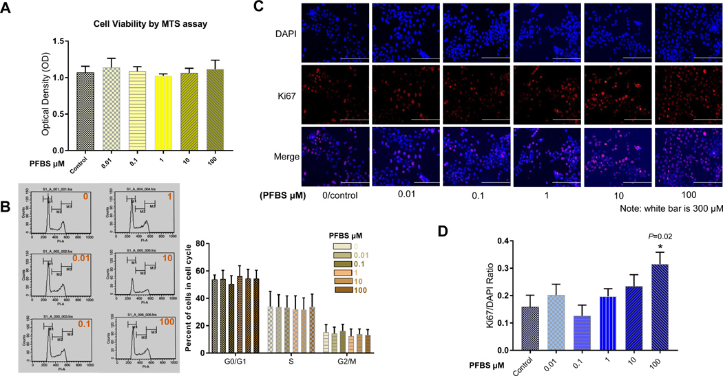
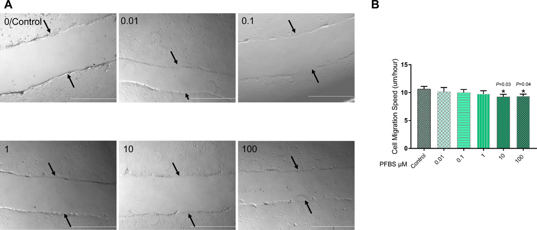
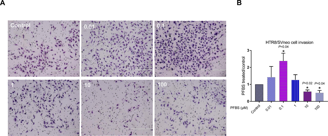
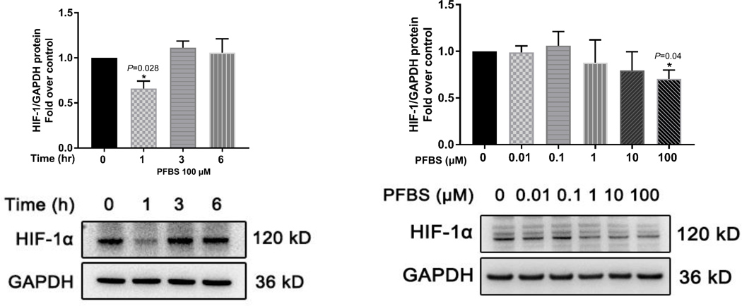
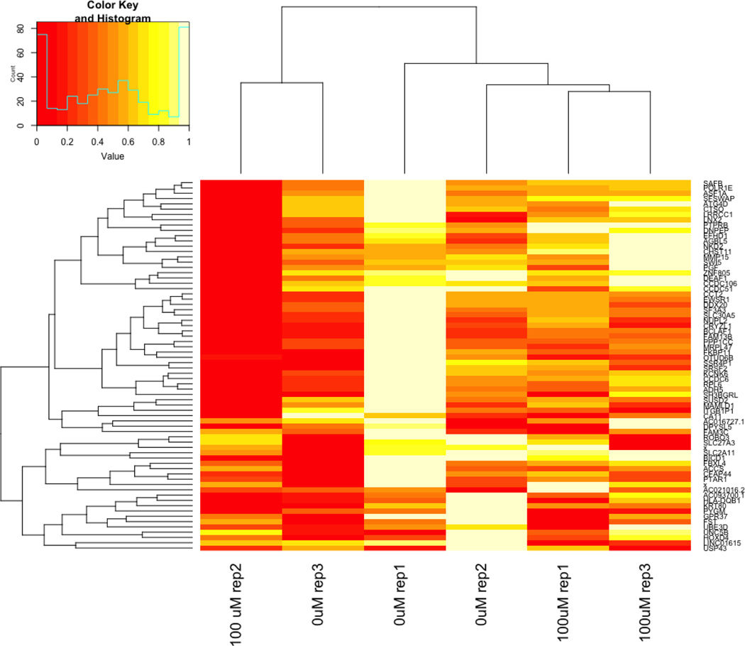
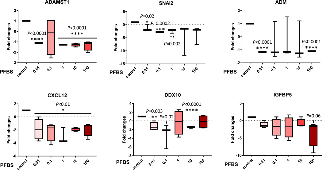
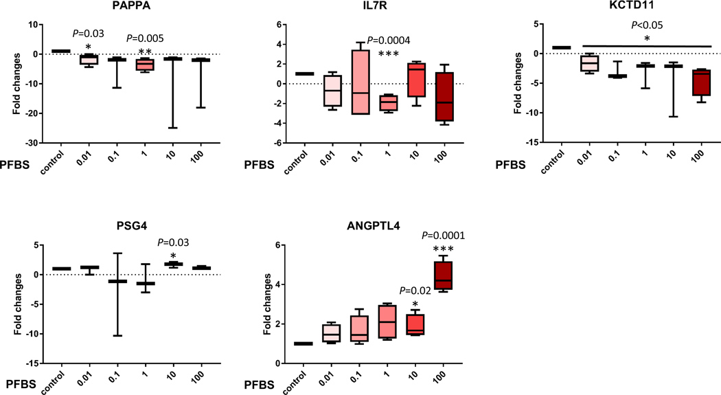
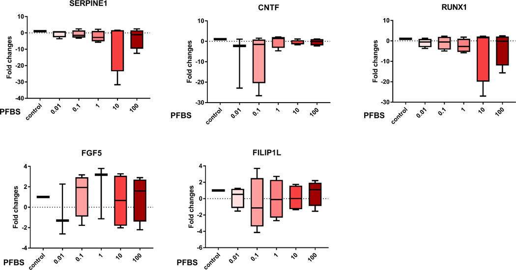
We reported that maternal PFBS, an emerging pollutant, exposure is positively associated with preeclampsia which can result from aberrant trophoblasts invasion and subsequent placental ischemia. In this study, we investigated the effects of PFBS on trophoblasts proliferation/invasion and signaling pathways. We exposed a human trophoblast line, HTR8/SVneo, to PFBS. Cell viability, proliferation, and cell cycle were evaluated by the MTS assay, Ki-67 staining, and flow cytometry, respectively. We assessed cell migration and invasion with live-cell imaging-based migration assay and matrigel invasion assay, respectively. Signaling pathways were examined by Western blot, RNA-seq, and qPCR. PFBS exposure interrupted cell proliferation and invasion in a dose-dependent manner. PFBS (100 μM) did not cause cell death but instead significant cell proliferation without cell cycle disruption. PFBS (10 and 100 μM) decreased cell migration and invasion, while PFBS (0.1 μM) significantly increased cell invasion but not migration. Further, RNA-seq analysis identified dysregulated HIF-1α target genes that are relevant to cell proliferation/invasion and preeclampsia, while Western Blot data showed the activation of HIF-1α, but not Notch, ERK1/2, (PI3K)AKT, and P38 pathways. PBFS exposure altered trophoblast cell proliferation/invasion which might be mediated by preeclampsia-related genes, suggesting a possible association between prenatal PFBS exposure and adverse placentation.
Keywords: PFBS; invasion; placenta cytotrophoblast; preeclampsia; proliferation.
© 2020 Federation of American Societies for Experimental Biology.
Figures
Similar articles
-
Downregulation of receptor tyrosine kinase-like orphan receptor 1 in preeclampsia placenta inhibits human trophoblast cell proliferation, migration, and invasion by PI3K/AKT/mTOR pathway accommodation.Placenta. 2019 Jul;82:17-24. doi: 10.1016/j.placenta.2019.05.002. Epub 2019 May 12. Placenta. 2019. PMID: 31174622
-
KLF15 alleviates oxidative stress and apoptosis of H/R-induced trophoblast cells to improve invasion and migration capacity via the activation of IGF1R.Tissue Cell. 2024 Oct;90:102485. doi: 10.1016/j.tice.2024.102485. Epub 2024 Jul 20. Tissue Cell. 2024. PMID: 39067323
-
Oleic acid stimulation of motility of human extravillous trophoblast cells is mediated by stearoyl-CoA desaturase-1 activity.Mol Hum Reprod. 2017 Nov 1;23(11):755-770. doi: 10.1093/molehr/gax051. Mol Hum Reprod. 2017. PMID: 29117333
-
Research Progress on Extracellular Matrix Involved in the Development of Preeclampsia.Curr Protein Pept Sci. 2024;25(7):527-538. doi: 10.2174/0113892037284176240302052521. Curr Protein Pept Sci. 2024. PMID: 38561606 Review.
-
Trophoblast Fusion in Hypertensive Disorders of Pregnancy and Preeclampsia.Int J Mol Sci. 2025 Mar 21;26(7):2859. doi: 10.3390/ijms26072859. Int J Mol Sci. 2025. PMID: 40243430 Free PMC article. Review.
Cited by
-
Maternal exposure to perfluorobutane sulfonate (PFBS) during pregnancy: evidence of adverse maternal and fetoplacental effects in New Zealand White (NZW) rabbits.Toxicol Sci. 2023 Feb 17;191(2):239-252. doi: 10.1093/toxsci/kfac126. Toxicol Sci. 2023. PMID: 36453863 Free PMC article.
-
Extravillous trophoblast migration and invasion: Impact of environmental chemicals and pharmaceuticals.Reprod Toxicol. 2022 Jan;107:60-68. doi: 10.1016/j.reprotox.2021.11.008. Epub 2021 Nov 25. Reprod Toxicol. 2022. PMID: 34838982 Free PMC article. Review.
-
Per- and polyfluoroalkyl substances (PFAS) and hypertensive disorders of Pregnancy- integration of epidemiological and mechanistic evidence.Reprod Toxicol. 2024 Dec;130:108702. doi: 10.1016/j.reprotox.2024.108702. Epub 2024 Aug 31. Reprod Toxicol. 2024. PMID: 39222887 Review.
-
Early life exposure to per- and polyfluoroalkyl substances (PFAS) and latent health outcomes: A review including the placenta as a target tissue and possible driver of peri- and postnatal effects.Toxicology. 2020 Oct;443:152565. doi: 10.1016/j.tox.2020.152565. Epub 2020 Aug 27. Toxicology. 2020. PMID: 32861749 Free PMC article. Review.
-
Exposure to perfluorobutane sulfonate and perfluorooctanesulfonic acid disrupts the production of angiogenesis factors and stress responses in human placental syncytiotrophoblast.Reprod Toxicol. 2020 Dec;98:269-277. doi: 10.1016/j.reprotox.2020.10.013. Epub 2020 Nov 2. Reprod Toxicol. 2020. PMID: 33144174 Free PMC article.
References
-
- Pelch KE, Reade A, Wolffe TAM, and Kwiatkowski CF (2019) PFAS health effects database: Protocol for a systematic evidence map. Environment international 130, 104851. - PubMed
-
- Zushi Y, Hogarh JN, and Masunaga S (2012) Progress and perspective of perfluorinated compound risk assessment and management in various countries and institutes. Clean Technologies and Environmental Policy 14, 9–20
-
- Gorrochategui E, Perez-Albaladejo E, Casas J, Lacorte S, and Porte C (2014) Perfluorinated chemicals: differential toxicity, inhibition of aromatase activity and alteration of cellular lipids in human placental cells. Toxicol Appl Pharmacol 277, 124–130 - PubMed
-
- Fromme H, Tittlemier SA, Volkel W, Wilhelm M, and Twardella D (2009) Perfluorinated compounds--exposure assessment for the general population in Western countries. International journal of hygiene and environmental health 212, 239–270 - PubMed
-
- Parsons JR, Sáez M, Dolfing J, and de Voogt P (2008) Biodegradation of Perfluorinated Compounds. In Reviews of Environmental Contamination and Toxicology Vol 196 (Whitacre DM, ed) pp. 53–71, Springer US, New York, NY: - PubMed
Publication types
MeSH terms
Substances
Grants and funding
LinkOut - more resources
Full Text Sources
Miscellaneous
