Review
. 2020 Jul;80(7-8):277-301.
doi: 10.1002/dneu.22780. Epub 2020 Sep 28.

Axon growth and synaptic function: A balancing act for axonal regeneration and neuronal circuit formation in CNS trauma and disease

Affiliations
Review

Axon growth and synaptic function: A balancing act for axonal regeneration and neuronal circuit formation in CNS trauma and disease

Conrad Kiyoshi et al. Dev Neurobiol. 2020 Jul.

Abstract

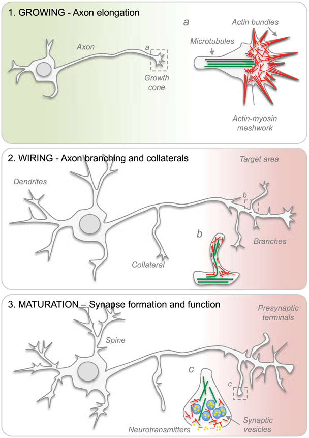
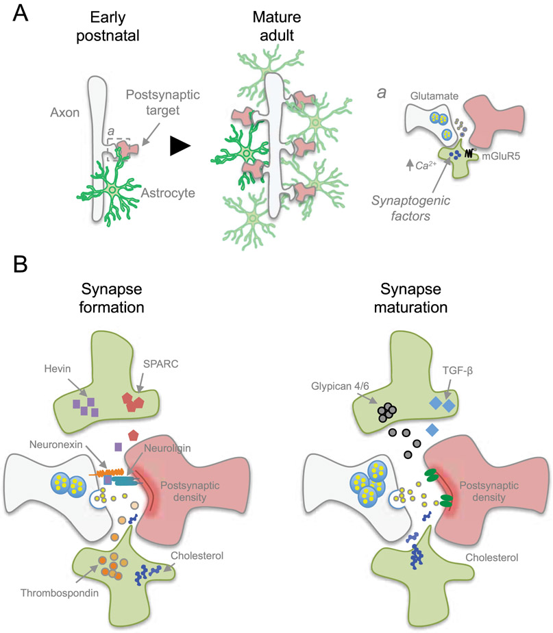
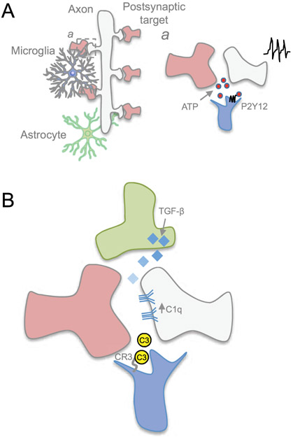
Axons in the adult mammalian central nervous system (CNS) fail to regenerate inside out due to intrinsic and extrinsic neuronal determinants. During CNS development, axon growth, synapse formation, and function are tightly regulated processes allowing immature neurons to effectively grow an axon, navigate toward target areas, form synaptic contacts and become part of information processing networks that control behavior in adulthood. Not only immature neurons are able to precisely control the expression of a plethora of genes necessary for axon extension and pathfinding, synapse formation and function, but also non-neuronal cells such as astrocytes and microglia actively participate in sculpting the nervous system through refinement, consolidation, and elimination of synaptic contacts. Recent evidence indicates that a balancing act between axon regeneration and synaptic function may be crucial for rebuilding functional neuronal circuits after CNS trauma and disease in adulthood. Here, we review the role of classical and new intrinsic and extrinsic neuronal determinants in the context of CNS development, injury, and disease. Moreover, we discuss strategies targeting neuronal and non-neuronal cell behaviors, either alone or in combination, to promote axon regeneration and neuronal circuit formation in adulthood.

Keywords: astrocyte; axon growth and regeneration; branching and synapse formation; microglia.


Conflict of interest statement

CONFLICT OF INTEREST

The authors declare that the research was conducted in the absence of any commercial or financial relationships that could be construed as a potential conflict of interest.

Figures

Figure 1.
Figure 1.. The developmental transition from a growing to a transmitting phase.
Schematic representation of axon elongation, branching, synaptic transmission and synaptogenesis during neuronal maturation. Cytoskeletal arrangements present in (a) growth cones and (b) axon branches. (c) Detailed view of a few structural elements found within presynaptic terminals.
Figure 2.
Figure 2.. Lipid raft microdomains of the plasma membrane.
Lipid rafts are highly organized subdomains of the plasma membrane enriched in cholesterol and glycosphingolipids. Rafts serve as a platform for the recruitment, insertion and interaction of membrane receptors and biomolecules involved in a variety of cellular processes including signal transduction, membrane trafficking and cytoskeletal organization. Membrane proteins are either free to move or immobilized to the actin cytoskeleton.
Figure 3.
Figure 3.. Astrocytes regulate formation and function of synapses.
A) During early postnatal development, astrocytes begin participating in synaptogenesis while proliferating to expand the astrocyte network. This rapid expansion of astrocytes coincides with the increase in the number of synapses as the nervous system matures. a) Astrocytes control synaptogenesis by releasing synaptogenic factors in a calcium-dependent response after sensing neurotransmitters, such as through Gq-metabotropic glutamate receptor mGluR5. B) Astrocytes release factors to facilitate structural formation of synapses. Cholesterol provides building material for new membrane production, while other factors such as Hevin and Thrombospondins promote synapse formation, or opposing synapse formation via SPARC. Additional factors are involved in synapse maturation and function, such as Glypican 4/6 and TGF-β that induces functional synapses.
Figure 4.
Figure 4.. Microglia refines the circuit by eliminating synapses.
A) Microglia constantly surveils the neural environment, and a) its motile processes respond to synaptic activity through P2Y12 receptor by sensing ATP released during neurotransmission. B) Microglia, through its C3 receptor, identifies synapses to be pruned through complement molecules C1q and C3. C1q expression on neurites may be regulated through astrocyte-secreted TGF-β.

References

    1. Agarwal A, Wu PH, Hughes EG, Fukaya M, Tischfield MA, Langseth AJ, … Bergles DE (2017). Transient Opening of the Mitochondrial Permeability Transition Pore Induces Microdomain Calcium Transients in Astrocyte Processes. Neuron, 93(3), 587–605 e587. doi: 10.1016/j.neuron.2016.12.034 - DOI - PMC - PubMed
    1. Allahyari RV, Clark KL, Shepard KA, & Garcia ADR (2019). Sonic hedgehog signaling is negatively regulated in reactive astrocytes after forebrain stab injury. Sci Rep, 9(1), 565. doi: 10.1038/s41598-018-37555-x - DOI - PMC - PubMed
    1. Allen NJ, Bennett ML, Foo LC, Wang GX, Chakraborty C, Smith SJ, & Barres BA (2012). Astrocyte glypicans 4 and 6 promote formation of excitatory synapses via GluA1 AMPA receptors. Nature, 486(7403), 410–414. doi: 10.1038/nature11059 - DOI - PMC - PubMed
    1. Allen NJ, & Eroglu C (2017). Cell Biology of Astrocyte-Synapse Interactions. Neuron, 96(3), 697–708. doi: 10.1016/j.neuron.2017.09.056 - DOI - PMC - PubMed
    1. Allen NJ, & Lyons DA (2018). Glia as architects of central nervous system formation and function. Science, 362(6411), 181–185. doi: 10.1126/science.aat0473 - DOI - PMC - PubMed

Publication types

MeSH terms