. 2020 Dec 1;103(6):1186-1198.
doi: 10.1093/biolre/ioaa155.

Transforming growth factor beta signaling and decidual integrity in mice†

Affiliations

Transforming growth factor beta signaling and decidual integrity in mice†

Xin Fang et al. Biol Reprod. .

Abstract

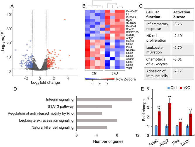
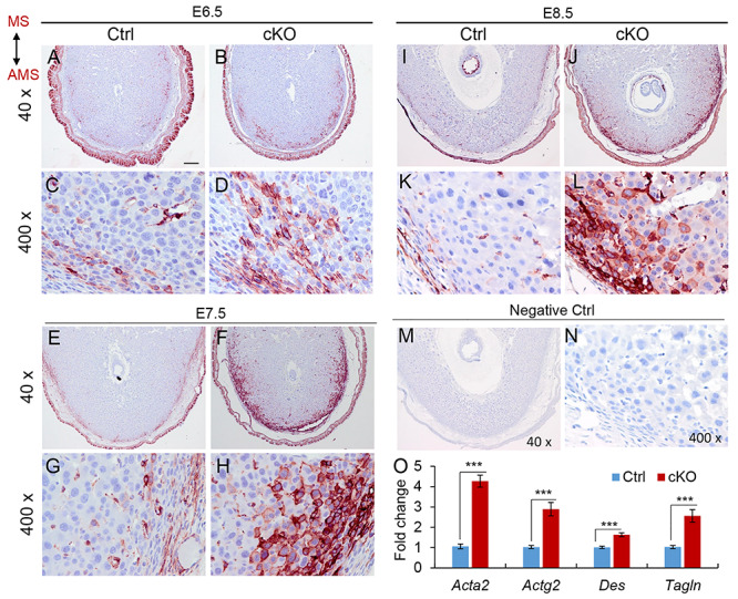
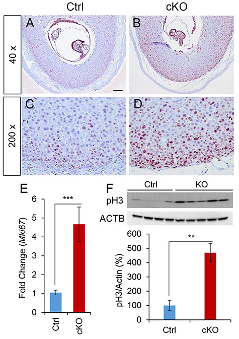
Transforming growth factor beta (TGFβ) signaling regulates multifaceted reproductive processes. It has been shown that the type 1 receptor of TGFβ (TGFBR1) is indispensable for female reproductive tract development, implantation, placental development, and fertility. However, the role of TGFβ signaling in decidual development and function remains poorly defined. Our objective is to determine the impact of uterine-specific deletion of Tgfbr1 on decidual integrity, with a focus on the cellular and molecular properties of the decidua during development. Our results show that the developmental dynamics of the decidua is altered in TGFBR1 conditionally depleted uteri from embryonic day (E) 5.5 to E8.5, substantiated by downregulation of genes associated with inflammatory responses and uterine natural killer cell abundance, reduced presence of nondecidualized fibroblasts in the antimesometrial region, and altered decidual cell development. Notably, conditional ablation of TGFBR1 results in the formation of decidua containing more abundant alpha smooth muscle actin (ACTA2)-positive cells at the peripheral region of the antimesometrial side versus controls at E6.5-E8.5. This finding is corroborated by upregulation of a subset of smooth muscle marker genes in Tgfbr1 conditionally deleted decidua at E6.5 and E8.5. Moreover, increased cell proliferation and enhanced decidual ERK1/2 signaling were found in Tgfbr1 conditional knockout mice upon decidual regression. In summary, conditional ablation of TGFBR1 in the uterus profoundly impacts the cellular and molecular properties of the decidua. Our results suggest that TGFBR1 in uterine epithelial and stromal compartments is important for the integrity of the decidua, a transient but crucial structure that supports embryo development.

Keywords: TGFBR1; TGFβ signaling; decidualization; endometrium; uterus.


Figures

Figure 1
Figure 1
Altered decidual differentiation in mice with conditional deletion of Tgfbr1. (A–L) Immunostaining of DES in the decidua of control and Tgfbr1 Pgr-Cre cKO mice at E6.5 (A–D), E7.5 (E–H), and E8.5 (I–L). Panels (C, D, G, H, K, and L) represent high power images for panels (A, B, E, F, I, and J), respectively. Cellular localization of DES in the mesometrial side was comparable between control and Tgfbr1 Pgr-Cre cKO mice (not shown). AMS, antimesometrial side; MS, mesometrial side. At least three independent samples were analyzed. Scale bar is representatively shown in (A) and equals 10 μm (C, D, G, H, K, and L) and 100 μm (A, B, E, F, I, and J). (M) QRT-PCR analysis of Alpl, Bmp2, Wnt4, Ptgs2, and Tgfbr1 expression in the decidua of control and Tgfbr1 Pgr-Cre cKO mice at E6.5. Data are mean ± SEM. n = 4 per group. ***P < 0.001.
Figure 2
Figure 2
Differential gene expression analysis using RNA-seq. (A) Volcano plot of genes identified by RNA-seq using E6.5 decidual tissues of control and Tgfbr1 Pgr-Cre cKO mice. n = 4 per group. Blue and red data points represent downregulated and upregulated genes (FDR adjusted P value <0.05 and fold change >2), respectively. Adj.P, FDR-adjusted P value. (B) Heatmap of the top 10 upregulated and downregulated genes. (C) Selected cellular functions associated with immune cell dysregulation identified by IPA. (D) Selected pathways associated with immune cell recruitment and activation and decidual development identified by IPA. (E) Increased expression of smooth muscle-related genes in the decidua of Tgfbr1 Pgr-Cre cKO mice at E6.5. Data are mean ± SEM. n = 4 per group. **P < 0.01.
Figure 3
Figure 3
Accumulation of ACTA2-positive cells in the periphery of implantation sites from mice with TGFBR1 depletion. (A–L) Increased number of ACTA2-positive cells in the peripheral region of the antimesometrial decidua of Tgfbr1 Pgr-Cre cKO mice at E6.5 (A–D), E7.5 (E–H), and E8.5 (I–L). Panels (C, D, G, H, K, and L) represent high power images for panels (A, B, E, F, I, and J), respectively. (M and N) Representative negative controls where anti-ACTA2 antibody was replaced by isotype-matched IgG. Three independent samples per genotype were examined. Scale bar is representatively shown in (A) and equals 20 μm (C, D, G, H, K, L, and N) and 200 μm (A, B, E, F, I, J, and M). (O) QRT-PCR analysis of Acta2, Actg2, Des, and Tagln in the decidua of control and Tgfbr1 Pgr-Cre cKO mice at E8.5. Data are mean ± SEM. n = 10 per group. ***P < 0.001.
Figure 4
Figure 4
Double immunofluorescence of ACTA2 and DES in the decidua of Tgfbr1 conditionally deleted mice. (A–H) Immunofluorescence staining of DES (green) and ACTA2 (red) in the decidua of control and Tgfbr1 Pgr-Cre cKO mice at E7.5. Negative controls using isotype-matched IgG to replace the primary antibodies are shown in Supplementary Figure S7. DAPI (blue) counterstained nuclei (C, D, G, and H). Three independent samples per genotype were examined. Scale bar is representatively shown in (A) and equals 20 μm (A–H).
Figure 5
Figure 5
Enhanced BMP signaling in TGFBR1 conditionally ablated decidua at E8.5. (A–F) QRT-PCR analysis of Bmp2 and BMP target genes in the decidua of control and Tgfbr1 Pgr-Cre cKO mice at E8.5. Data are mean ± SEM. n = 10 per group. ***P < 0.001. (G) Increased phospho-SMAD1/5/8 levels in the decidua of Tgfbr1 Pgr-Cre cKO mice at E8.5 by western blot. The upper panel shows the western blot image and the lower one shows the quantification result by Image J. Data are normalized to the control group, which is set to 100%. n = 4–5 per group. *P < 0.05.
Figure 6
Figure 6
Increased cell proliferation in the decidua of mice with conditional depletion of TGFBR1. (A–D) Immunohistochemical staining of MKI67 in implantation sites of control and Tgfbr1 Pgr-Cre cKO mice at E8.5. At least three independent samples per genotype were examined. Scale bar is representatively shown in (A) and equals 40 μm (C and D) and 200 μm (A and B). (E) QRT-PCR analysis of Mki67 in the decidua of control and Tgfbr1 Pgr-Cre cKO mice at E8.5. Data are mean ± SEM. n = 10 per group. ***P < 0.001. (F) Increased phospho-histone H3 (pH 3) expression in the decidua of Tgfbr1 Pgr-Cre cKO mice compared with controls at E8.5. The upper panel shows a representative western blot image, whereas the lower one depicts the quantification result. Data are normalized to the control group that is set to 100%. n = 4–5 per group. **P < 0.01.
Figure 7
Figure 7
Altered ERK1/2 signaling and CASP3 in the decidua of mice with conditional depletion of TGFBR1. (A) Western blot analysis of cleaved CASP3, phospho-ERK1/2, and total ERK1/2 in the decidua of control and Tgfbr1 Pgr-Cre cKO mice at E8.5. ACTB was included as an internal control. (B and C) Quantification results of western blots of cleaved CASP3 and phospho-ERK1/2. Data are normalized to the control group that is set to 100%. n = 4–5 per group. *P < 0.05 and ***P < 0.001. (D) A model of TGFBR1-mediated signaling in endometrial cells during pregnancy. TGFβ ligands signal through TGFBR1 and ERK1/2 pathway to regulate endometrial cell properties and decidual integrity. Conditional deletion of Tgfbr1 potentiates ERK1/2 signaling and BMP signaling, suggesting an antagonism between TGFBR1-mediated signaling and the BMP pathway. The detailed interactions between TGFBR1, ERK1/2, and BMP signaling require further experimental testing.

Similar articles

Cited by

References

    1. Salamonsen LA, Dimitriadis E, Jones RL, Nie G. Complex regulation of decidualization: a role for cytokines and proteases--a review. Placenta 2003; 24:S76–S85. - PubMed
    1. Sroga JM, Ma X, Das SK. Developmental regulation of decidual cell polyploidy at the site of implantation. Front Biosci (Schol Ed) 2012; 4:1475–1486. - PMC - PubMed
    1. Wang H, Dey SK. Roadmap to embryo implantation: clues from mouse models. Nat Rev Genet 2006; 7:185–199. - PubMed
    1. Cha J, Sun X, Dey SK. Mechanisms of implantation: strategies for successful pregnancy. Nat Med 2012; 18:1754–1767. - PMC - PubMed
    1. Guzeloglu-Kayisli O, Kayisli UA, Taylor HS. The role of growth factors and cytokines during implantation: endocrine and paracrine interactions. Semin Reprod Med 2009; 27:62–79. - PMC - PubMed

Publication types

MeSH terms

Substances