. 2021 Feb;383(2):835-852.
doi: 10.1007/s00441-020-03270-1. Epub 2020 Sep 9.

Camel regulates development of the brain ventricular system

Affiliations

Camel regulates development of the brain ventricular system

Shulan Yang et al. Cell Tissue Res. 2021 Feb.

Abstract

Development of the brain ventricular system of vertebrates and the molecular mechanisms involved are not fully understood. The developmental genes expressed in the elements of the brain ventricular system such as the ependyma and circumventricular organs act as molecular determinants of cell adhesion critical for the formation of brain ventricular system. They control brain development and function, including the flow of cerebrospinal fluid. Here, we describe the novel distantly related member of the zebrafish L1-CAM family of genes-camel. Whereas its maternal transcripts distributed uniformly, the zygotic transcripts demonstrate clearly defined expression patterns, in particular in the axial structures: floor plate, hypochord, and roof plate. camel expresses in several other cell lineages with access to the brain ventricular system, including the midbrain roof plate, subcommissural organ, organum vasculosum lamina terminalis, median eminence, paraventricular organ, flexural organ, and inter-rhombomeric boundaries. This expression pattern suggests a role of Camel in neural development. Several isoforms of Camel generated by differential splicing of exons encoding the sixth fibronectin type III domain enhance cell adhesion differentially. The antisense oligomer morpholino-mediated loss-of-function of Camel affects cell adhesion and causes hydrocephalus and scoliosis manifested via the tail curled down phenotype. The subcommissural organ's derivative-the Reissner fiber-participates in the flow of cerebrospinal fluid. The Reissner fiber fails to form upon morpholino-mediated Camel loss-of-function. The Camel mRNA-mediated gain-of-function causes the Reissner fiber misdirection. This study revealed a link between Chl1a/Camel and Reissner fiber formation, and this supports the idea that CHL1 is one of the scoliosis factors.

Keywords: Ependyma; Flexural organ; Floor plate; Hypochord; Roof plate; Subcommissural organ.


Conflict of interest statement

The authors declare that they have no conflict of interest.

Figures

Fig. 1
Fig. 1
The comparison of Camel to other proteins of L1-CAM family of proteins and camel expression during development. Camel is the more evolutionarily distant of the two duplicated Chl1-related proteins of zebrafish. a The dendrogram comparison of similarity within a group of vertebrate CAM, where the Camel protein forms a distinct group different from the “Chl1b” one. b The temporal expression of camel during development as detected by RT-PCR. Abbreviations: d, Drosophila melanogaster; g, spotted gar (Lepisosteus oculatus); h, human (Homo sapiens); su, sea urchin (Strongylocentrotus purpuratus); tf, Takifugu rubripes; zf, zebrafish (Danio rerio)
Fig. 2
Fig. 2
The expression pattern of camel during early zebrafish development detected by whole-mount in situ hybridization. a, b camel is inherited as a maternal transcript distributed uniformly. c During gastrula, the embryonic shield (ES, arrow) is stained more intensely compared to other regions. d During somitogenesis, the expression is largely in the ventral midline (arrowhead). Scale bar = 100 μm
Fig. 3
Fig. 3
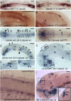
The expression pattern of camel in wild-type embryos and mutants detected by whole-mount in situ hybridization during neurogenesis. camel is expressed in the axial structures (fp, rp, hp), along segmental boundaries in the brain and in circumventricular organs. a, c 17 hpf, wild-type embryos. b, d 17 hpf, mibta52b mutant. e, g 24 hpf, wild-type embryos. f 24 hpf, mibta52b mutant. h 36 hpf, brain of a wild-type embryo. i Wild-type trunk, 48 hpf. h, j Wild-type brain, 48 hpf. Numbers define rhombomeres of the hindbrain, asterisk indicates gaps in the floor plate, and arrow indicates remains of hypochord. a, b, e, f Dorsal view. c, d, gj Lateral view. Abbreviations: ahp, adenohypophysis; b, brain; c, cerebellum; d, diencephalon; e, epiphysis; emp, eminentia thalami; epIII, ependyma of the third ventricle; ey, eye; hb, hindbrain; ht, hypothalamus; fo, flexural organ; fp, floor plate; hb, hindbrain; hc, hypochord; ht, hypothalamus; me, median eminence; mrp, midbrain roof plate; not, notochord; ot, optic tectum; ov, otic vesicle; ovlt, organum vasculosum lamina terminalis; pvo, paraventricular organ; rp, roof plate; sc, spinal cord; sco, subcommissural organ; t, telencephalon; tg, tegmentum. Scale bar = 100 μm
Fig. 4
Fig. 4
The expression pattern of camel during late development. camel expresses in the brain ventricular system, circumventricular organs, ventro-rostral patch (vrp), and the eye-differentiating retina. All images are in the lateral view. a 72 hpf, wild-type larvae. b 48 hpf, the left-hand side eye of the wild-type embryo. c 72 hpf, the left-hand side eye of the wild-type larvae. Abbreviations: c, cerebellum; chf, choroid fissure; cf, cephalic flexure; d, diencephalon; e, epiphysis; emt, eminentia thalami; fo, flexural organ; hb, hindbrain; ht, hypothalamus; mhb, midbrain-hindbrain boundary; mrp, midbrain roof plate; op, olfactory placode, ot, optic tectum; ovlt, organum vasculosum lamina terminalis; pvo, paraventricular organ; sco, subcommissural organ; t, telencephalon; tg, tegmentum. Scale bar = 100 μm except a’, where it is 25 μm
Fig. 5
Fig. 5
The schematics of organization of the camel genomic DNA and four differentially spliced isoforms. The putative proteins encoded by these mRNA isoforms vary at the level of the fourth fibronectin type III domain encoded by exons 24 and 25. a Organization of camel genomic DNA showing the target sites for morpholino and color-coded exons 24 (yellow) and 25 (blue). b Organization of the putative camel isoforms. The two halves of the sixth fibronectin type III domain are color-coded according to a. The Camel domain structure is generated using this software (http://expasy.org/prosite)
Fig. 6
Fig. 6
af Camel regulates cell adhesion in hanging drops. The control cells of different-color formed clusters mixed uniformly, unlike those in experimental conditions when Camel expression was affected in red cells by the anti-Camel morpholino. c (red, green), control; MO1 and MO2, pan-Camel morpholino; MS1, isoform 1,3-specific morpholino; MS2, isoform 1,2-specific morpholino. The combination of MS1 + MS2 blocks isoforms 1–3. Scale bar = 20 μm
Fig. 7
Fig. 7
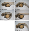
ae The effect of different anti-camel MOs varies depending on the target. MO1 targets all isoforms causing the most severe effect, including the hydrocephalus, curled trunk, and edema. MS1 and MS2 target specific mRNA isoforms (1 + 3 and 1 + 2, correspondingly). MS1 causes the hydrocephalus and mildly curled trunk. MS2 causes hydrocephalus. The effect of MS1 and MS2 is additive in enhancing the trunk curvature. 50 embryos were observed in each experiment repeated three times. The phenotypes representing 95% of experimental animals were selected. Scale bar = 50 μm
Fig. 8
Fig. 8
Rescue of hydrocephalus in Camel morphants by camel mRNA. a Control. bd Morphants with brain ventricles filled-in by Dextran-Texas Red. e Control wild-type embryo injected by camel mRNA. Notice that the size of the ventricular system is reduced compared to the intact control. fh Different morphants rescued by injection of different camel mRNA isoforms. All images are dorsal views of 54-hpf embryos with anterior to the left. Scale bar = 100 μm
Fig. 9
Fig. 9
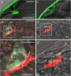
Camel required for the development of SCO and formation of Reissner fiber. a, b Control. c, d Pan-anti-Camel morphants (MO1). All images are of ET27 transgenics at 48 hpf. a, c In vivo images of GFP expression. b, d Double immunostaining for GFP and Reissner fiber. b’, d’ Blowup of boxed areas in b and d, respectively. b’ The AFRU staining in controls is mostly presented by the filamentous extracellular material with gaps between SCO and AFRU material (arrow) and in the AFRU material (arrowhead). Some faint AFRU staining was detected in the cytoplasm (asterisk). d’ In morphants, the AFRU material is disorganized; it covers the SCO more with some residual signal in the cytoplasm (asterisk). Scale bar = 100 μm (ad) and 50 μm (b’, d’)
Fig. 10
Fig. 10
Pan-Camel and isoform-specific anti-Camel morpholino–mediated LOF differentially affects the formation of RF. a Being generated by the SCO (green, GFP, Tg(ET33-mi2a) and the RF (detected by anti-AFRU antibody, red) and spanning the BVS. It may obtain additional contributions from FO and FP. It extends through the central canal to the posterior end of the spinal cord to be disassembled at the filum terminale. b MO1 blocks the formation of RF (b’) and causes the distortion in the distribution of RF+ material in FO (b”) and FP (b”’). c, d In comparison, the effect of isoform-specific morpholinos MS1 and MS2 is less obvious. All confocal images of Tg(ET33-mi2a) transgenic 48-hpf embryos are shown as lateral views with anterior to the left. a Stack of sections. a’ Selected sections to illustrate the primarily apical distribution of the RF material in the SCO. a”’ and a”” are the same as c and d shown for comparing the effect of MO. a’b Blowup of a’. a”b Blowup of a”. d’b Blowup of d’. d”b Blowup of d”. Asterisks indicate lack of RF, and arrows and arrowheads indicate RF. Abbreviations: fo, flexural organ; fp, floor plate; ft, filum terminale; rf, Reissner fiber; sco, subcommissural organ. Scale bar = 200 μm (upper left-hand side column, ad), 100 μm (four other upper columns marked ’ to ””), and 50 μm (blowups)
Fig. 11
Fig. 11
The overexpression of camel affects the development of the Reissner fiber. Upon camel overexpression, the RF+ material seems to increase in the BVS with its significant redistribution towards the choroid plexus of the fourth ventricle (CPIV) and ependyma (e, anterior and posterior of the SCO). The ectopic sprouts of RF (erf) in the Sylvius aqueduct (sa) formed. In the vIV, the RF changes the ventral trajectory (along FP) towards the CPIV in the dorsal position. In the posterior vIV, the trajectory of RF normalizes and leaves the vIV via the central canal. All images are lateral views of Tg(ET33-mi2a) transgenic 48-hpf embryos with anterior to the left. a, b Larvae after strong overexpression of Chl1a. Boxes in a represent SCO, FO, and CPIV and correspond to a’a”’. Scale bar = 100 μm
Fig. 12
Fig. 12
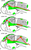
Schematics show organization of the Reissner fiber in respect of the ventricular system (based on Figs. 9, 10, and 11). ac 48 hpf. a Controls. b Anti-Camel morpholino–mediated loss-of-function. c Camel mRNA–mediated gain-of-function. Green color, midline structures and some CVOs; red, RF and AFRU+ material. Abbreviations: afp, anterior floor plate; ap, area postrema; cc, central canal; cpIII, choroid plexus of the third ventricle; cpIV, choroid plexus of the fourth ventricle; d, diencephalon; e, epiphysis; erf, ectopic Reissner fiber; h, hindbrain; fo, flexural organ; fp, floor plate; m, midbrain; mrp, midbrain roof plate; opc, optocoele (Sylvius aqueduct); po, pineal organ; rf, Reissner fiber; sco, subcommissural organ; t, telencephalon; vIII, third ventricle; vIV, fourth ventricle

Similar articles

Cited by

References

    1. Alsanie WF, Penna V, Schachner M, Thompson LH, Parish CL. Homophilic binding of the neural cell adhesion molecule CHL1 regulates development of ventral midbrain dopaminergic pathways. Sci Rep. 2017;7(1):9368. doi: 10.1038/s41598-017-09599-y. - DOI - PMC - PubMed
    1. Angeloni D, Lindor NM, Pack S, Latif F, Wei MH, Lerman MI. CALL gene is haploinsufficient in a 3p- syndrome patient. Am J Med Genet. 1999;86:482–485. doi: 10.1002/(sici)1096-8628(19991029)86:5<482::aid-ajmg15>3.0.co;2-l. - DOI - PubMed
    1. Becker CG, Lieberoth BC, Morellini F, Feldner J, Becker T, Schachner M. L1.1 is involved in spinal cord regeneration in adult zebrafish. J Neurosci. 2004;24:7837–7842. doi: 10.1523/JNEUROSCI.2420-04.2004. - DOI - PMC - PubMed
    1. Ben-Hur T, Rogister B, Murray K, Rougon G, Dubois-Dalcq M. Growth and fate of PSA-NCAM+ precursors of the postnatal brain. J Neurosci. 1998;18:5777–5788. doi: 10.1523/JNEUROSCI.18-15-05777.1998. - DOI - PMC - PubMed
    1. Boswell CW, Ciruna B. Understanding idiopathic scoliosis: a new Zebrafish School of Thought. Trends Genet. 2017;33:183–196. doi: 10.1016/j.tig.2017.01.001. - DOI - PubMed

MeSH terms

LinkOut - more resources