

Evaluation of perforating venous insufficiency with shear wave elastography: a preliminary study
- PMID: 32902811
- PMCID: PMC8572307
- DOI: 10.1007/s40477-020-00527-x
Evaluation of perforating venous insufficiency with shear wave elastography: a preliminary study
Abstract
Purpose: The aim of this study was to investigate the efficacy of shear wave elastography (SWE) in the diagnosis of perforating vein insufficiency, and to determine the applicability of these measurements.
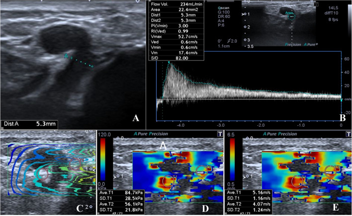
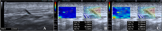
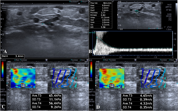
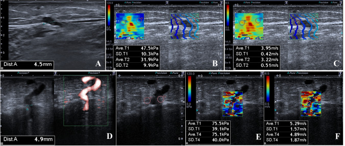
Methods: A total of 140 symptomatic patients with a total of 280 lower extremities were investigated. All patients presented with venous insufficiency (VI) symptoms, and received Doppler ultrasound assessment to determine VI and SWE measurements. The SWE values were measured in the adjacent perivenous tissue of the largest Cockett's perforating vein (PV) of both lower extremities, at the level where they pass the fascia. The Cockett's PV diameter and the presence of reflux in Cockett's PV and the great saphenous vein were compared with SWE values in perivenous tissue of PVs.
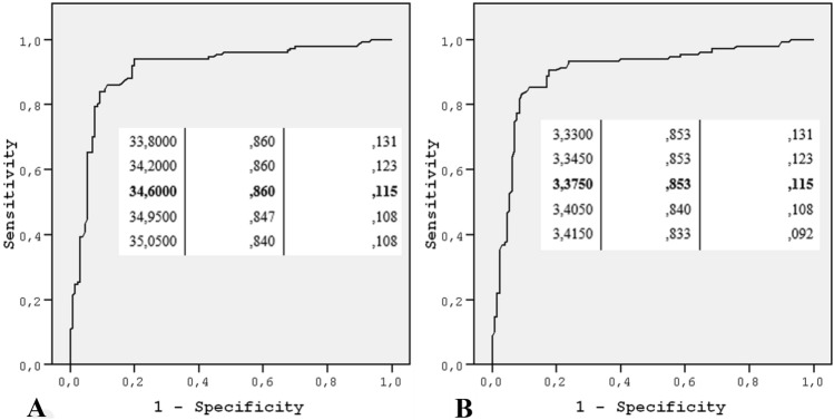
Results: The SWE values of the perforating vein insufficiency group were significantly higher than those of the normal PV without insufficiency group (P < 0.001). A significant and positive relation was seen between increased PV diameter and SWE values (P < 0.001) and there was a significant relationship between the presence of perforating vein insufficiency and increase in PV diameter. A statistically significant increase was detected in SWE values for the PV for those with reflux in the great saphenous vein (P < 0.001). The best cut-off values that can be used to detect perforating vein insufficiency were found 34.600 for kPa and 3.375 for m/s.
Conclusion: SWE may be used effectively in addition to conventional Doppler ultrasound examination in diagnosing and following perforating vein insufficiency.
Keywords: Doppler ultrasound; Perforating venous insufficiency; Shear wave elastography; Venous insufficiency.
© 2020. Società Italiana di Ultrasonologia in Medicina e Biologia (SIUMB).
Conflict of interest statement
The authors declare that there are no financial or other relations that could lead to a conflict of interest.
Figures
Similar articles
-
Diminished Sphenous Compartment Connective Tissue Elasticity has Little Impact on Low Grade Venous Insufficiency: An Ultrasound Shearwave Elastography Study.Curr Med Imaging. 2021;17(7):897-903. doi: 10.2174/1573405617666210507122819. Curr Med Imaging. 2021. PMID: 33966622 Free PMC article.
-
Experience of Using Shear Wave Elastography Imaging in Superficial Venous Insufficiency of the Lower Extremity.Ultrasound Q. 2018 Sep;34(3):176-182. doi: 10.1097/RUQ.0000000000000356. Ultrasound Q. 2018. PMID: 29634667
-
Color-Doppler sonography in chronic venous insufficiency: what the radiologist should know.Curr Probl Diagn Radiol. 2005 Mar-Apr;34(2):51-62. doi: 10.1067/j.cpradiol.2004.12.001. Curr Probl Diagn Radiol. 2005. PMID: 15753879
-
[A phlebologic dogma concerning the localization of the Cockett's perforating veins].Phlebologie. 1992 Apr-Jun;45(2):207-12. Phlebologie. 1992. PMID: 1528975 French.
-
Duplex mapping of 2036 primary varicose veins.J Vasc Surg. 2009 Mar;49(3):681-9. doi: 10.1016/j.jvs.2008.09.062. J Vasc Surg. 2009. PMID: 19268773
Cited by
-
Diminished Sphenous Compartment Connective Tissue Elasticity has Little Impact on Low Grade Venous Insufficiency: An Ultrasound Shearwave Elastography Study.Curr Med Imaging. 2021;17(7):897-903. doi: 10.2174/1573405617666210507122819. Curr Med Imaging. 2021. PMID: 33966622 Free PMC article.
References
-
- Van Neer PA, Veraart JC, Neumann HA. Venae perforantes: a clinical review. Dermatol Surg. 2003;29:931–942. - PubMed
MeSH terms
LinkOut - more resources
Full Text Sources
Research Materials
