. 2020 Oct;13(10):e008824.
doi: 10.1161/CIRCEP.120.008824. Epub 2020 Sep 9.

Prospective STAR-Guided Ablation in Persistent Atrial Fibrillation Using Sequential Mapping With Multipolar Catheters

Affiliations

Prospective STAR-Guided Ablation in Persistent Atrial Fibrillation Using Sequential Mapping With Multipolar Catheters

Shohreh Honarbakhsh et al. Circ Arrhythm Electrophysiol. 2020 Oct.

Abstract

Background: A novel stochastic trajectory analysis of ranked signals (STAR) mapping approach to guide atrial fibrillation (AF) ablation using basket catheters recently showed high rates of AF termination and subsequent freedom from AF.

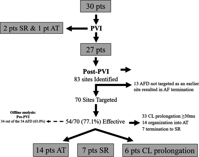
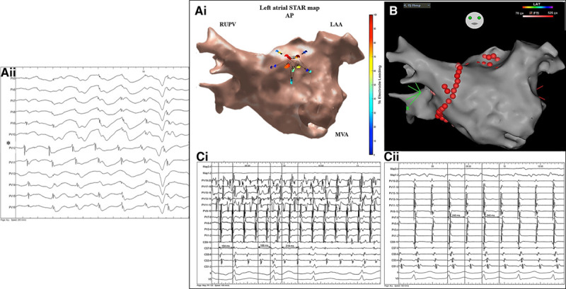
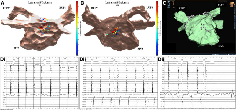
Methods: This study aimed to determine whether STAR mapping using sequential recordings from conventional pulmonary vein mapping catheters could achieve similar results. Patients with persistent AF<2 years were included. Following pulmonary vein isolation AF drivers (AFDs) were identified on sequential STAR maps created with PentaRay, IntellaMap Orion, or Advisor HD Grid catheters. Patients had a minimum of 10 multipolar recordings of 30 seconds each. These were processed in real-time and AFDs were targeted with ablation. An ablation response was defined as AF termination or cycle length slowing ≥30 ms.

Results: Thirty patients were included (62.4±7.8 years old, AF duration 14.1±4.3 months) of which 3 had AF terminated on pulmonary vein isolation, leaving 27 patients that underwent STAR-guided AFD ablation. Eighty-three potential AFDs were identified (3.1±1.1 per patient) of which 70 were targeted with ablation (2.6±1.2 per patient). An ablation response was seen at 54 AFDs (77.1% of AFDs; 21 AF termination and 33 cycle length slowing) and occurred in all 27 patients. No complications occurred. At 17.3±10.1 months, 22 out of 27 (81.5%) patients undergoing STAR-guided ablation were free from AF/atrial tachycardia off antiarrhythmic drugs.

Conclusions: STAR-guided AFD ablation through sequential mapping with a multipolar catheter effectively achieved an ablation response in all patients. AF terminated in a majority of patients, with a high freedom from AF/atrial tachycardia off antiarrhythmic drugs at long-term follow-up. Registration: URL: https://www.clinicaltrials.gov; Unique identifier: NCT02950844.

Keywords: atrial fibrillation; catheter ablation; coronary sinus; pulmonary vein; tachycardia.


Figures

Figure 1.
Figure 1.
Flow diagram summarizing the study findings. AF indicates atrial fibrillation; AFD, AF driver; AT, atrial tachycardia; CL, cycle length; pts, patients; PVI, pulmonary vein isolation; and SR, sinus rhythm.
Figure 2.
Figure 2.
Patient ID 16. A, i and ii, Stochastic trajectory analysis of ranked signals (STAR) map in (A, i) an anterior-posterior (AP) view and (A, ii) tilted roof view demonstrating an atrial fibrillation driver (AFD) mapped to the high anterior/roof. On this STAR map, the recording segment that demonstrated the AFD that was targeted with ablation is only displayed. B, i and ii, Rhythmia map in (B, i) an AP view and (B, ii) tilted roof view demonstrating ablation at the AFD. C, Bard signals including surface ECG, coronary sinus (CS), ablation (ABL), and selected IntellaMap Orion catheter electrograms demonstrating (i) AF termination to atrial tachycardia (AT) on ablation at the AFD and (ii) with further consolidating lesions at AFD resulting in AT termination to sinus rhythm. LUPV indicates left upper pulmonary vein; MVA, mitral valve annulus; and RUPV, right upper pulmonary vein.
Figure 3.
Figure 3.
Patient ID 18. A, i and ii, Stochastic trajectory analysis of ranked signals (STAR) map in a tilted roof view demonstrating (A, i) an atrial fibrillation driver (AFD) mapped to the midroof/right upper pulmonary vein (RUPV) and (A, ii) roof/left atrial appendage (LAA). A, iii, Demonstrates the electrograms obtained from the mapping catheter at the site of the AFD. The asterisk highlights the electrode that is leading the greatest proportion of time compared to its neighbors. On this STAR map the recording segments that demonstrated the AFD with ablation response are only displayed. B, Rhythmia map in a roof view demonstrating ablation at both AFDs (the blue tag highlights the site of intermittent organization and yellow tag highlights site of termination to sinus rhythm). Ablation at an AFD mapped to the high anterior wall did not result in a study-defined ablation response. C, i and ii, Bard signals including surface ECG, coronary sinus (CS), ablation (ABL), and selected IntellaMap Orion catheter electrograms demonstrating (C, i and ii) intermittent AF organization into atrial tachycardia during ablation at the AFD mapped to the midroof/RUPV and cycle length slowing and (C, ii) termination to sinus rhythm on ablation at the AFD mapped to the roof/LAA. LUPV indicates left upper pulmonary vein.
Figure 4.
Figure 4.
Patient ID 20. A, Stochastic trajectory analysis of ranked signals (STAR) map in a tilted lateral view demonstrating an atrial fibrillation driver (AFD) mapped to the midlateral wall. B, Rhythmia map in a titled lateral view demonstrating ablation at the AFD. C, Bard signals including surface ECG, coronary sinus (CS), ablation, and selected IntellaMap Orion catheter electrograms demonstrating AF termination to atrial tachycardia (AT) on ablation at the AFD. D, i and ii, Rhythmia LAT map in a (D, i) lateral valve view and (D, ii) posterior-anterior view demonstrating a mitral isthmus dependent tachycardia. E, Bard signals including surface ECG, CS, ablation (ABL), and selected IntellaMap Orion catheter electrograms (ME) demonstrating AT with a cycle length of 240 ms. F, i and ii, Bard electrograms demonstrating entrainment at (F, i) CS with a postpacing interval–tachycardia cycle length (PPI-TCL) of 14 ms and (F, ii) posterior wall with a PPI-TCL of 134 ms. LAA indicates left atrial appendage; LUPV, left upper pulmonary vein; and MVA, mitral valve annulus.
Figure 5.
Figure 5.
Patient ID 23. A, i, Demonstrates a stochastic trajectory analysis of ranked signals (STAR) left atrial (LA) map in an anterior-posterior view with an atrial fibrillation driver (AFD) identified midanterior wall of the LA. A, ii, Demonstrates the electrograms obtained from the mapping catheter at the site of the AFD. The asterisk highlights the electrode that is leading the greatest proportion of time compared to its neighbors. B, This AFD was targeted with ablation as shown on the CARTO LA map in an anterior-posterior view. On this STAR map, the recording segment that demonstrated this AFD is only displayed. C, i and ii, Ablation here resulted in AF termination to atrial tachycardia (AT) as shown on the Bard electrograms. The AT was mapped to a focal/micro-reentry AT in the close vicinity to the ablation lesions that organized the AF into AT. Further cluster ablation lesions at this site terminated the AT to sinus rhythm. The Bard electrograms include surface ECG, pulmonary vein (PV), Map, and coronary sinus (CS). LAA indicates left atrial appendage; MVA, mitral valve annulus; and RUPV, right upper pulmonary vein.
Figure 6.
Figure 6.
Patient ID 21. A, Demonstrates a stochastic trajectory analysis of ranked signals (STAR) left atrial (LA) map in a posterior-anterior view that highlights 2 atrial fibrillation drivers (AFDs) at the posterior wall in close vicinity to each other with the arrows demonstrating that the wavefront is spreading to away from each AFD in the opposite direction suggesting that a potential AF driver is present from which radial spread of activation occurs. This site was targeted with ablation that slowed the AF from ≥30 ms and intermittently organized it. On this STAR map, the two recording segments that demonstrated this wavefront activation pattern is only displayed. B, STAR LA map in an anterior-posterior view that highlights an AFD at the midanterior wall. On this STAR map, the recording segment that demonstrated this AFD is only displayed. C, Ablation was performed at this site as shown on the EnSite Precision system LA map in an anterior-posterior view. D, i and ii, Ablation here resulted in AF termination to an atrial tachycardia (AT) as shown on the Bard electrograms. D, iii, The AT was mapped to roof-dependent flutter and a roofline was ablated with a 10 ms slowing in AT cycle length. Remap was suggestive of mitral isthmus–dependent AT which was successfully ablated with a mitral line resulting in sinus rhythm as shown on the Bard electrograms. The Bard electrograms include surface ECG, pulmonary vein, ablation, and coronary sinus (CS). LUPV indicates left upper pulmonary vein; MVA, mitral valve annulus; and RUPV, right upper pulmonary vein.

Similar articles

Cited by

References

    1. Haissaguerre M, Hocini M, Denis A, Shah AJ, Komatsu Y, Yamashita S, Daly M, Amraoui S, Zellerhoff S, Picat MQ, et al. Driver domains in persistent atrial fibrillation. Circulation. 2014; 130:530–538. doi: 10.1161/CIRCULATIONAHA.113.005421 - PubMed
    1. Knecht S, Sohal M, Deisenhofer I, Albenque JP, Arentz T, Neumann T, Cauchemez B, Duytschaever M, Ramoul K, Verbeet T, et al. Multicentre evaluation of non-invasive biatrial mapping for persistent atrial fibrillation ablation: the AFACART study. Europace. 2017; 19:1302–1309. doi: 10.1093/europace/euw168 - PubMed
    1. Narayan SM, Baykaner T, Clopton P, Schricker A, Lalani GG, Krummen DE, Shivkumar K, Miller JM. Ablation of rotor and focal sources reduces late recurrence of atrial fibrillation compared with trigger ablation alone: extended follow-up of the CONFIRM trial (Conventional Ablation for Atrial Fibrillation With or Without Focal Impulse and Rotor Modulation). J Am Coll Cardiol. 2014; 63:1761–1768. doi: 10.1016/j.jacc.2014.02.543 - PMC - PubMed
    1. Buch E, Share M, Tung R, Benharash P, Sharma P, Koneru J, Mandapati R, Ellenbogen KA, Shivkumar K. Long-term clinical outcomes of focal impulse and rotor modulation for treatment of atrial fibrillation: a multicenter experience. Heart Rhythm. 2016; 13:636–641. doi: 10.1016/j.hrthm.2015.10.031 - PMC - PubMed
    1. Honarbakhsh S, Schilling RJ, Dhillon G, Ullah W, Keating E, Providencia R, Chow A, Earley MJ, Hunter RJ. A novel mapping system for panoramic mapping of the left atrium: application to detect and characterize localized sources maintaining atrial fibrillation. JACC Clin Electrophysiol. 2018; 4:124–134. doi: 10.1016/j.jacep.2017.09.177 - PMC - PubMed

Publication types

MeSH terms

Associated data