

Diffusion tensor imaging in renal artery stenosis: a preliminary report
- PMID: 32903036
- PMCID: PMC8519640
- DOI: 10.1259/bjr.20200101
Diffusion tensor imaging in renal artery stenosis: a preliminary report
Abstract
Objective: To investigate the diffusion properties in the kidneys affected by renal artery stenosis (RAS) using diffusion tensor imaging (DTI).
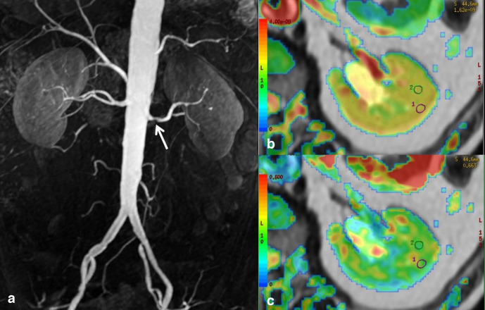
Methods: In this prospective study, 35 patients with RAS and 15 patients without renal abnormalities were enrolled and examined using DTI. Cortical and medullary regions of interest (ROIs) were located to obtain the corresponding values of the apparent diffusion coefficient (ADC) and fractional anisotropy (FA). The cortical and medullary ADC and FA were compared in the kidney affected by variable degrees of stenosis (RAS 50-75% and >75%) vs controls, using the one-way ANOVA and Student's t-test. The Spearman correlation test was used to correlate the mean ADC and FA values in the cortex and medulla with the estimate glomerular filtration rate (eGFR).
Results: For the controls, the ADC value was significantly (p = 0.03) higher in the cortex than in the medulla; the FA value was significantly (p = 0.001) higher in the medulla than in the cortex. Compared with the controls, a significant reduction in the cortical ADC was present with a RAS of 50-75% and >75% (p = 0.001 and 0.041, respectively); a significant reduction in the medullary FA was verified only for RAS >75% (p = 0.023). The Spearman correlation test did not show a statistically significant correlation between the cortical and medullary ADC and FA, and the eGFR.
Conclusion: The alterations of the diffusional parameters caused by RAS can be detected by DTI and could be useful in the diagnostic evaluation of these patients.
Advances in knowledge: 1. Magnetic resonance DTI could provide useful information about renal involvement in RAS.2. Magnetic resonance DTI allows non-invasive repeatable evaluation of the renal parenchyma, without contrast media.
Figures


Similar articles
-
Diffusion tensor imaging and tractography of the kidneys: assessment of chronic parenchymal diseases.Eur Radiol. 2013 Jun;23(6):1678-85. doi: 10.1007/s00330-012-2749-y. Epub 2013 Jan 9. Eur Radiol. 2013. PMID: 23300038
-
Assessment of renal allograft function early after transplantation with isotropic resolution diffusion tensor imaging.Eur Radiol. 2016 Feb;26(2):567-75. doi: 10.1007/s00330-015-3841-x. Epub 2015 May 28. Eur Radiol. 2016. PMID: 26017738
-
Diffusion tensor imaging of the kidney with parallel imaging: initial clinical experience.Invest Radiol. 2008 Oct;43(10):677-85. doi: 10.1097/RLI.0b013e31817d14e6. Invest Radiol. 2008. PMID: 18791409
-
Chronic kidney disease: pathological and functional assessment with diffusion tensor imaging at 3T MR.Eur Radiol. 2015 Mar;25(3):652-60. doi: 10.1007/s00330-014-3461-x. Epub 2014 Oct 11. Eur Radiol. 2015. PMID: 25304821
-
The role of diffusion tensor imaging and fractional anisotropy in the evaluation of patients with idiopathic normal pressure hydrocephalus: a literature review.Neurosurg Focus. 2016 Sep;41(3):E12. doi: 10.3171/2016.6.FOCUS16192. Neurosurg Focus. 2016. PMID: 27581308 Review.
References
MeSH terms
LinkOut - more resources
Full Text Sources
Research Materials
Miscellaneous
