Review
. 2021 Feb;216(2):275-294.
doi: 10.2214/AJR.20.24436. Epub 2020 Dec 23.

Screening Algorithms in Dense Breasts: AJR Expert Panel Narrative Review

Affiliations
Review

Screening Algorithms in Dense Breasts: AJR Expert Panel Narrative Review

Wendie A Berg et al. AJR Am J Roentgenol. 2021 Feb.

Abstract

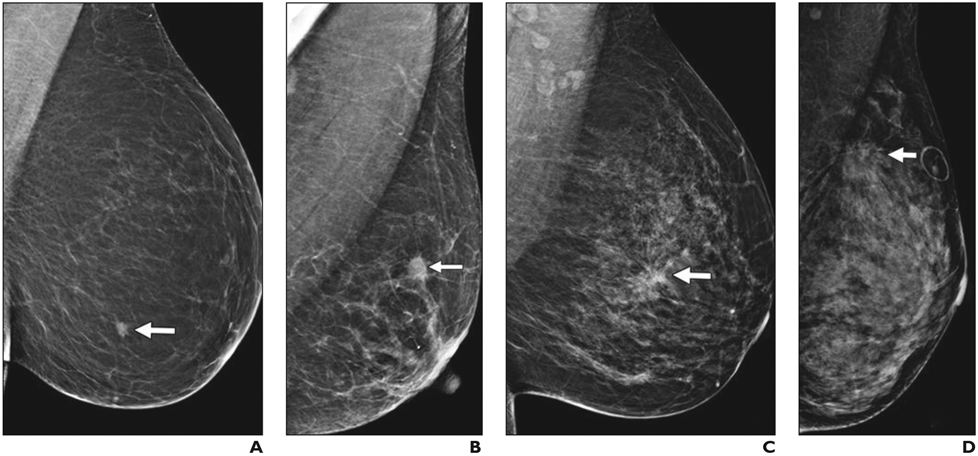
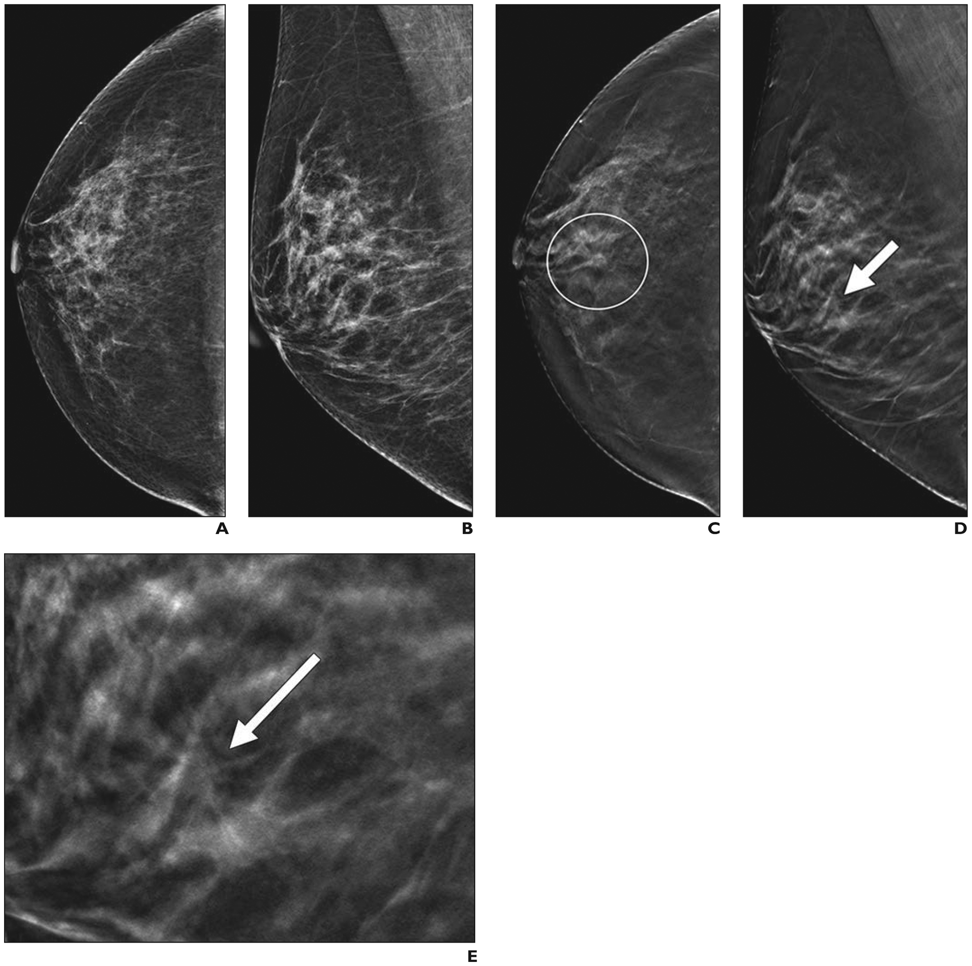
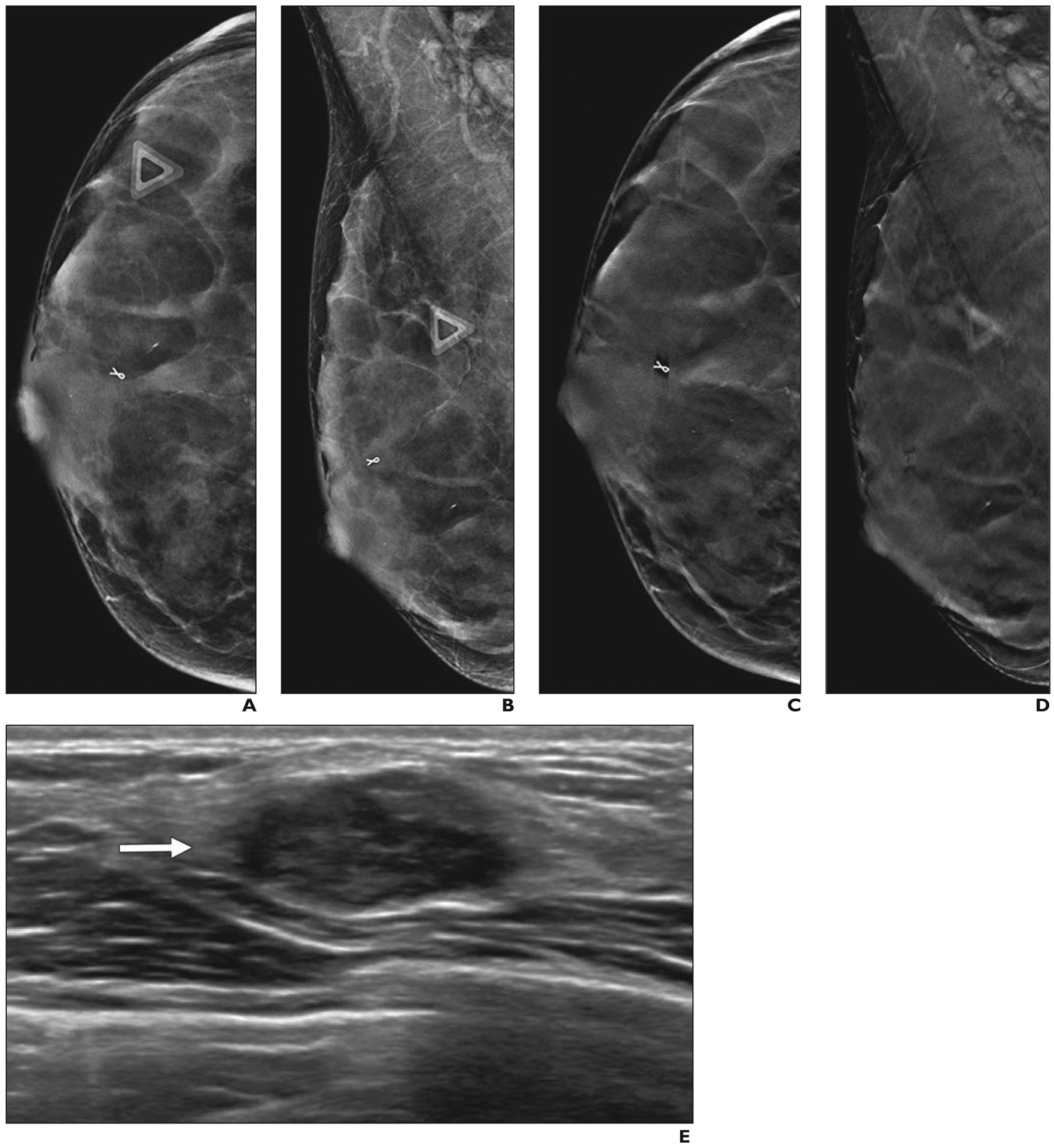
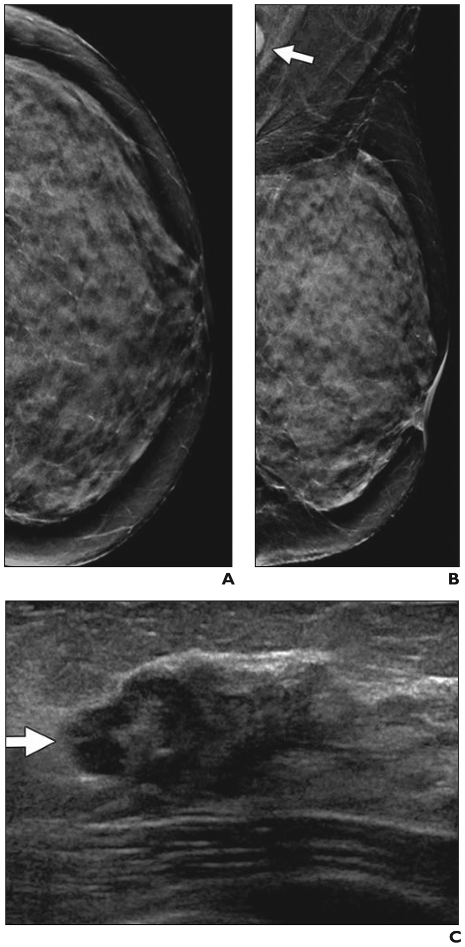
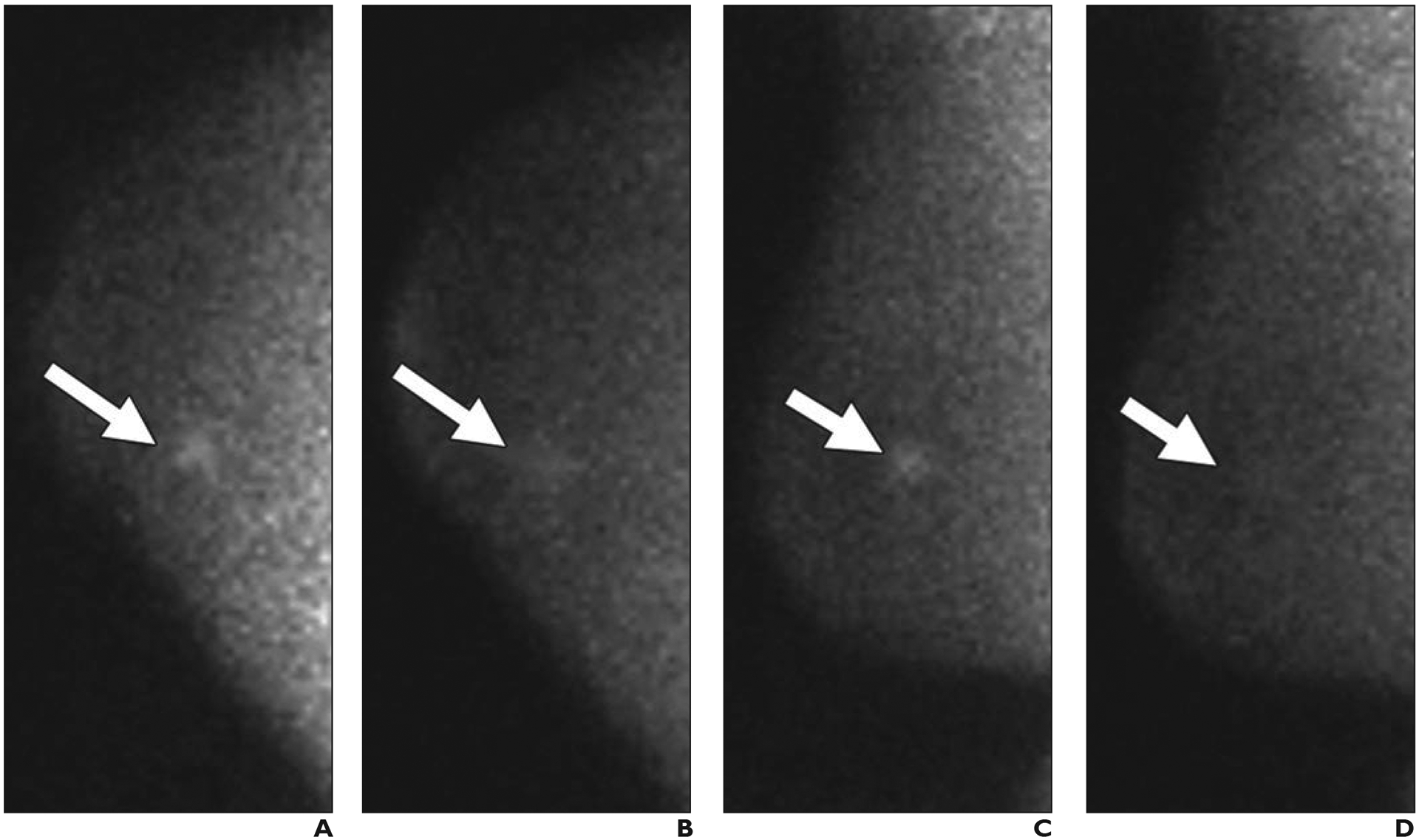
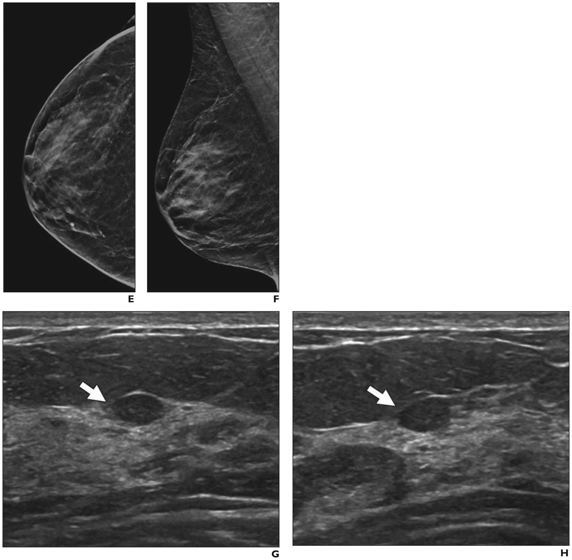
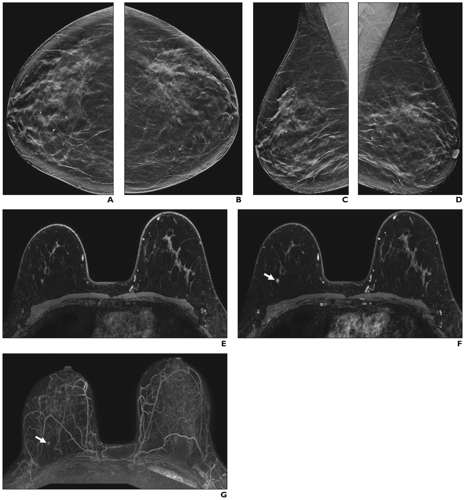
Screening mammography reduces breast cancer mortality; however, when used to examine women with dense breasts, its performance and resulting benefits are reduced. Increased breast density is an independent risk factor for breast cancer. Digital breast tomosynthesis (DBT), ultrasound (US), molecular breast imaging (MBI), MRI, and contrast-enhanced mammography (CEM) each have shown improved cancer detection in dense breasts when compared with 2D digital mammography (DM). DBT is the preferred mammographic technique for producing a simultaneous reduction in recalls (i.e., additional imaging). US further increases cancer detection after DM or DBT and reduces interval cancers (cancers detected in the interval between recommended screening examinations), but it also produces substantial additional false-positive findings. MBI improves cancer detection with an effective radiation dose that is approximately fourfold that of DM or DBT but is still within accepted limits. MRI provides the greatest increase in cancer detection and reduces interval cancers and late-stage disease; abbreviated techniques will reduce cost and improve availability. CEM appears to offer performance similar to that of MRI, but further validation is needed. Dense breast notification will soon be a national standard; therefore, understanding the performance of mammography and supplemental modalities is necessary to optimize screening for women with dense breasts.

Keywords: MRI; breast density; contrast-enhanced mammography; molecular breast imaging; tomosynthesis; ultrasound.


Figures

Fig. 1—
Fig. 1—
Left mediolateral oblique digital mammography (DM) images of cancer in breasts representing each of four BI-RADS breast density categories. A, 61-year-old woman with pathogenic BRCA1 mutation. Screening DM image shows fatty breast and spiculated mass (arrow) that proved to be a 0.9-cm grade 3 invasive ductal carcinoma (IDC) that was estrogen receptor (ER) negative, progesterone receptor (PR) negative, and human epidermal growth factor receptor 2 (HER2 [also known as ERBB2]) negative with associated ductal carcinoma in situ (DCIS). Ki-67 proliferation index was high (90%). Three sentinel nodes were negative for metastasis. B, 65-year-old woman with scattered fibroglandular breast tissue density. Screening DM image shows spiculated mass (arrow) that proved to be 1.1-cm grade 2 IDC with extensive DCIS that was ER positive, PR positive, and HER2 negative. Ki-67 proliferation index was high (30%), and metastatic sentinel node showed focal extracapsular extension. C, 40-year-old woman with heterogeneously dense parenchyma, which may obscure small masses. Baseline screening DM image shows irregular mass (arrow) with distortion in central left breast. Core biopsy specimen showed 1.2-cm IDC that was ER positive, PR positive, and HER2 negative. Ki-67 proliferation index was moderate (15%). Patient went elsewhere for treatment. D, 43-year-old woman with extremely dense parenchyma, which lowers sensitivity of mammography. Baseline screening DM image shows spiculated mass (arrow) with associated distortion in upper posterior left breast. This proved to be T2 (> 2 cm) grade 2 IDC with associated DCIS that was ER positive, PR positive, and HER2 negative. Ki-67 proliferation index was low (10%), and sentinel node biopsy was negative for metastasis.
Fig. 2—
Fig. 2—
46-year-old woman with cancer seen on annual screening tomosynthesis examination only. A and B, Two-dimensional craniocaudal (A) and mediolateral oblique (B) digital mammograms show heterogeneously dense breast tissue. C and D, Craniocaudal digital breast tomosynthesis image (C) shows spiculated mass (within circle), which has much more subtle appearance on mediolateral oblique image (arrow, D). E, Close-up of mediolateral oblique digital breast tomosynthesis image in D shows mass (arrow) in detail. Core biopsy and excision revealed 0.7-cm grade 2 invasive ductal carcinoma that was estrogen receptor positive, progesterone receptor positive, and human epidermal growth factor receptor 2 (HER2 [also known as ERBB2]) negative. Sentinel node was negative for metastasis.
Fig. 3—
Fig. 3—
51-year-old woman with right breast pain who had cancer that was masked by extremely dense breast tissue on tomosynthesis. A and B, Two-dimensional craniocaudal (A) and mediolateral oblique (B) digital mammograms show extremely dense parenchyma with ribbon clip from prior benign biopsy and no abnormality at site of pain (triangles). C and D, Craniocaudal (C) and mediolateral oblique (D) digital breast tomosynthesis images show no abnormality. E, Directed ultrasound image of area of focal pain shows 1.6-cm irregular, hypoechoic mass (arrow). Ultrasound-guided core needle biopsy revealed grade 3 invasive ductal carcinoma that was triple receptor negative. Excision after neoadjuvant chemotherapy showed no residual tumor. Sentinel node was negative for metastasis.
Fig. 4—
Fig. 4—
58-year-old woman with extremely dense breasts who had cancer seen on screening ultrasound (US) only. A and B, Craniocaudal (A) and mediolateral oblique (B) synthetic 2D mammograms show normal extremely dense parenchyma, which was also interpreted as negative on digital breast tomosynthesis. In retrospect, portion of dense node is seen in left axilla (arrow, B). Handheld screening US performed by technologist was conducted bilaterally with standard documentation. C, Radial US image of left breast shows irregular, hypoechoic 1.9-cm mass (arrow) located in 12-o’clock position 6 cm from nipple. US-guided core biopsy revealed grade 2 invasive ductal carcinoma that was estrogen receptor positive, progesterone receptor negative, and human epidermal growth factor receptor 2 (HER2 [also known as ERBB2]) amplified by fluorescence in situ hybridization. Ki-67 proliferation index was high (40%). US-guided core biopsy of left axillary node confirmed metastatic disease. Patient received primary chemotherapy and had no residual invasive carcinoma and few foci of ductal carcinoma in situ at lumpectomy. Targeted axillary dissection (with seed-localized excision of known metastatic node) showed one metastatic node with treatment effect and three normal nodes.
Fig. 5—
Fig. 5—
47-year-old woman with heterogeneously dense breasts who had cancer detected on screening molecular breast imaging (MBI) only. (Courtesy of Hunt KN, Mayo Clinic, Rochester, MN) AD, Craniocaudal views from upper (A) and lower (B) detector heads and mediolateral oblique views from medial (C) and lateral (D) detector heads obtained during MBI performed after IV injection of 7.5-mCi (277.5 MBq) 99mTc-sestamibi show 9-mm focal area of moderate-intensity uptake (arrows) in upper inner right breast at middle depth. E and F, Synthetic 2D craniocaudal (E) and mediolateral oblique (F) mammograms from digital breast tomosynthesis acquisition show heterogeneously dense parenchyma and no abnormality. Spot compression mediolateral oblique view (not shown) was also normal. G and H, Targeted transverse (G) and longitudinal (H) ultrasound images show oval, circumscribed 0.7-cm isoechoic mass (arrows) thought to correspond to MBI finding. Ultrasound-guided core biopsy and surgical excision revealed 0.4-cm grade 1 invasive ductal carcinoma that was estrogen receptor positive, progesterone receptor positive, and human epidermal growth factor receptor 2 (HER2 [also known as ERBB2]) negative. Ki-67 proliferation index was low at 8% and low-grade ductal carcinoma in situ was present. Sentinel node biopsy was negative for metastasis.
Fig. 5—
Fig. 5—
47-year-old woman with heterogeneously dense breasts who had cancer detected on screening molecular breast imaging (MBI) only. (Courtesy of Hunt KN, Mayo Clinic, Rochester, MN) AD, Craniocaudal views from upper (A) and lower (B) detector heads and mediolateral oblique views from medial (C) and lateral (D) detector heads obtained during MBI performed after IV injection of 7.5-mCi (277.5 MBq) 99mTc-sestamibi show 9-mm focal area of moderate-intensity uptake (arrows) in upper inner right breast at middle depth. E and F, Synthetic 2D craniocaudal (E) and mediolateral oblique (F) mammograms from digital breast tomosynthesis acquisition show heterogeneously dense parenchyma and no abnormality. Spot compression mediolateral oblique view (not shown) was also normal. G and H, Targeted transverse (G) and longitudinal (H) ultrasound images show oval, circumscribed 0.7-cm isoechoic mass (arrows) thought to correspond to MBI finding. Ultrasound-guided core biopsy and surgical excision revealed 0.4-cm grade 1 invasive ductal carcinoma that was estrogen receptor positive, progesterone receptor positive, and human epidermal growth factor receptor 2 (HER2 [also known as ERBB2]) negative. Ki-67 proliferation index was low at 8% and low-grade ductal carcinoma in situ was present. Sentinel node biopsy was negative for metastasis.
Fig. 6—
Fig. 6—
69-year-old woman with dense breasts and history of lobular carcinoma in situ that had been excised 2 years previously who had cancer seen on screening abbreviated MRI only. AD, Craniocaudal (A and B) and mediolateral oblique (C and D) synthetic 2D views from screening digital breast tomosynthesis show heterogeneously dense breasts with no abnormalities. EG, Images from baseline abbreviated MRI screening examination, which consisted of unenhanced 3D fat-suppressed T1-weighted acquisition (E) and single contrast-enhanced 3D T1-weighted acquisition (F) from which maximum-intensity-projection subtracted image (G) was created, show enhancing, round 0.6-cm mass (arrows, F and G) with indistinct margins and heterogeneous internal enhancement in upper outer right breast. MRI-guided biopsy and excision confirmed 0.6-cm grade 1 invasive ductal carcinoma that was estrogen receptor positive, progesterone receptor positive, and human epidermal growth factor receptor 2 (HER2 [also known as ERBB2]) negative. Sentinel node biopsy was negative for metastasis.
Fig. 7—
Fig. 7—
57-year-old woman with dense breasts who had invasive ductal cancer shown on contrast-enhanced mammography. A and B, Screening craniocaudal (A) and mediolateral oblique (B) 2D synthetic mammography images show heterogeneously dense parenchyma and few calcifications. C and D, Enlarged views of craniocaudal tomosynthesis image of inner right breast (C) and angled spot compression craniocaudal tomosynthesis image (D) show subtle distortion (arrows). E and F, Targeted radial (E) and antiradial (F) ultrasound images of right breast show irregular, hypoechoic 0.9-cm mass (arrows) in 1-o’clock position located 6 cm from nipple, although findings of screening handheld ultrasound examination were negative. Before biopsy, patient underwent research contrast-enhanced mammography performed 2.5 minutes after IV injection of 125 mL of iopamidol (370 mg I/mL; Isovue 370, Bracco). G and H, Craniocaudal (G) and mediolateral oblique (H) subtracted contrast-enhanced mammography images show strongly enhancing 1.2-cm irregular mass (arrows) and mild background parenchymal enhancement. Ultrasound-guided core biopsy revealed grade 2 invasive ductal carcinoma with ductal carcinoma in situ that was estrogen receptor positive, progesterone receptor positive, and human epidermal growth factor receptor 2 (HER2 [also known as ERBB2]) negative. Ki-67 proliferation index was low (10%). Invasive carcinoma measured 1.3 cm at lumpectomy, and four sentinel nodes were negative for metastasis.
Fig. 8—
Fig. 8—
51-year-old woman with extremely dense breasts who had invasive ductal carcinoma visualized using multiple modalities. A and B, Screening craniocaudal (A) and mediolateral oblique (B) mammography images of left breast show regional amorphous and pleomorphic calcifications (arrows) that are well seen despite extremely dense parenchyma. C and D, Subtracted craniocaudal (C) and mediolateral oblique (D) images from research contrast-enhanced mammography performed after IV injection of 125 mL of 300 mg I/mL iohexol (Omnipaque 300, GE Healthcare) show strong enhancement of irregular 2.7-cm mass (arrows) at site of calcifications. E, Targeted ultrasound image shows partially circumscribed, partially indistinctly marginated, slightly hypoechoic 3.1-cm mass (arrowhead) with echogenic calcifications (arrows). Stereotactic biopsy was performed to ensure optimal sampling of calcifications and showed grade 2 invasive ductal carcinoma with ductal carcinoma in situ that was estrogen receptor negative, progesterone receptor negative, and human epidermal growth factor receptor 2 (HER2 [also known as ERBB2]) negative. Ki-67 proliferation index was low (10%). F and G, Craniocaudal (F) and mediolateral oblique (G) views from research molecular breast imaging (10-minute acquisitions obtained after IV injection of 7.3 mCi [270.1 MBq] of 99mTc-sestamibi) performed after diagnosis shows intense uptake in irregular 3.4-cm mass (arrows). Note slight reduction in inclusion of extreme posterior tissues relative to mammography. H, Axial fat-suppressed T1-weighted MR image shows intense enhancement of irregular 3.0-cm mass (arrow) at site of known malignancy. Patient had partial response to primary chemotherapy with two sentinel nodes negative for metastases (or therapy-related changes).
Fig. 8—
Fig. 8—
51-year-old woman with extremely dense breasts who had invasive ductal carcinoma visualized using multiple modalities. A and B, Screening craniocaudal (A) and mediolateral oblique (B) mammography images of left breast show regional amorphous and pleomorphic calcifications (arrows) that are well seen despite extremely dense parenchyma. C and D, Subtracted craniocaudal (C) and mediolateral oblique (D) images from research contrast-enhanced mammography performed after IV injection of 125 mL of 300 mg I/mL iohexol (Omnipaque 300, GE Healthcare) show strong enhancement of irregular 2.7-cm mass (arrows) at site of calcifications. E, Targeted ultrasound image shows partially circumscribed, partially indistinctly marginated, slightly hypoechoic 3.1-cm mass (arrowhead) with echogenic calcifications (arrows). Stereotactic biopsy was performed to ensure optimal sampling of calcifications and showed grade 2 invasive ductal carcinoma with ductal carcinoma in situ that was estrogen receptor negative, progesterone receptor negative, and human epidermal growth factor receptor 2 (HER2 [also known as ERBB2]) negative. Ki-67 proliferation index was low (10%). F and G, Craniocaudal (F) and mediolateral oblique (G) views from research molecular breast imaging (10-minute acquisitions obtained after IV injection of 7.3 mCi [270.1 MBq] of 99mTc-sestamibi) performed after diagnosis shows intense uptake in irregular 3.4-cm mass (arrows). Note slight reduction in inclusion of extreme posterior tissues relative to mammography. H, Axial fat-suppressed T1-weighted MR image shows intense enhancement of irregular 3.0-cm mass (arrow) at site of known malignancy. Patient had partial response to primary chemotherapy with two sentinel nodes negative for metastases (or therapy-related changes).
Fig. 9—
Fig. 9—
Flowchart illustrates current approaches to supplemental screening in context of risk factors, including breast density. If not performed by age 30 years, genetic testing can be performed at time of diagnosis of breast or ovarian cancer, when appropriate family history is identified, or when family member is found to have pathogenic mutation. Women with high risk for breast cancer who are pregnant or lactating may consider screening ultrasound (US) during that time. Similar performance has been observed for abbreviated MRI and full-protocol diagnostic MRI. For women who cannot tolerate MRI, US is most widely available alternative but produces less gain in cancer detection than MRI. Molecular breast imaging (MBI) or contrast-enhanced mammography (CEM) appear to produce cancer detection similar to that of MRI but are not yet widely available alternatives when MRI is not possible; further validation is needed. If screening MRI is performed, additional supplemental screening with US, MBI, or CEM is not needed. Supplemental screening MRI should stop by age 75 even among high-risk women. PHBC = personal history of breast cancer, LCIS = lobular carcinoma in situ, ADH = atypical ductal hyperplasia, DBT = digital breast tomosynthesis.

Comment in

  • Breast MRI for "the Masses".
    Berg WA. Berg WA. Eur Radiol. 2022 Jun;32(6):4034-4035. doi: 10.1007/s00330-022-08782-8. Epub 2022 Apr 14. Eur Radiol. 2022. PMID: 35420302 No abstract available.

Similar articles

Cited by

References

    1. Coldman A, Phillips N, Wilson C, et al. Pan-Canadian study of mammography screening and mortality from breast cancer. J Natl Cancer Inst 2014; 106:dju261. - PubMed
    1. Tabár L, Vitak B, Chen TH, et al. Swedish two-county trial: impact of mammographic screening on breast cancer mortality during 3 decades. Radiology 2011; 260:658–663 - PubMed
    1. Monticciolo DL, Newell MS, Hendrick RE, et al. Breast cancer screening for average-risk women: recommendations from the ACR Commission on Breast Imaging. J Am Coll Radiol 2017; 14:1137–1143 - PubMed
    1. Oeffinger KC, Fontham ET, Etzioni R, et al.; American Cancer Society. Breast cancer screening for women at average risk: 2015 guideline update from the American Cancer Society. JAMA 2015; 314:1599–1614 - PMC - PubMed
    1. Siu AL US Preventive Services Task Force. Screening for breast cancer: U.S. Preventive Services Task Force recommendation statement. Ann Intern Med 2016; 164:279–296 - PubMed

Publication types

LinkOut - more resources