. 2019 May 7:1:11.
doi: 10.1186/s42490-019-0011-2. eCollection 2019.

A biomechanical test model for evaluating osseous and osteochondral tissue adhesives

Affiliations

A biomechanical test model for evaluating osseous and osteochondral tissue adhesives

Philip Procter et al. BMC Biomed Eng. .

Abstract

Background: Currently there are no standard models with which to evaluate the biomechanical performance of calcified tissue adhesives, in vivo. We present, herein, a pre-clinical murine distal femoral bone model for evaluating tissue adhesives intended for use in both osseous and osteochondral tissue reconstruction.

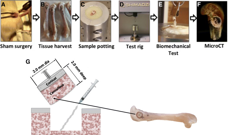
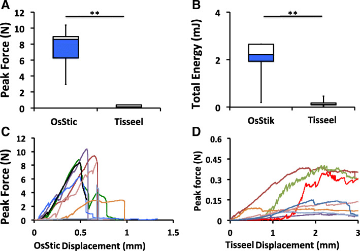
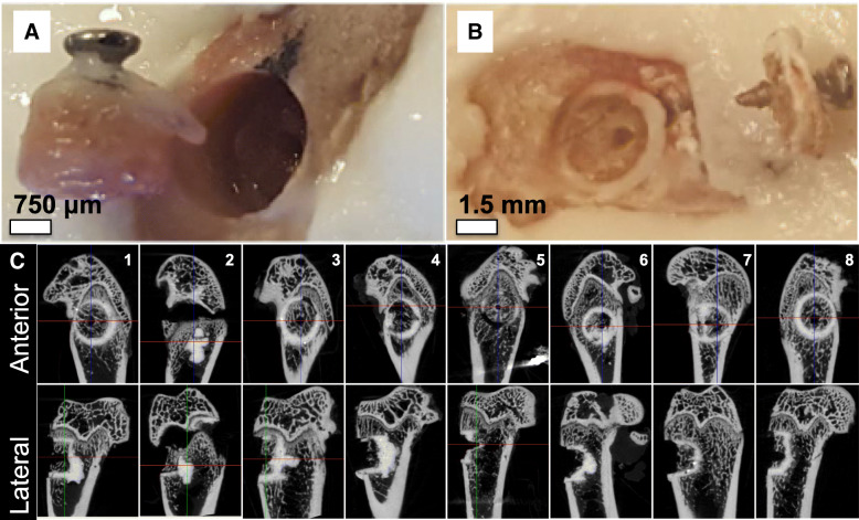
Results: Cylindrical cores (diameter (Ø) 2 mm (mm) × 2 mm depth), containing both cancellous and cortical bone, were fractured out from the distal femur and then reattached using one of two tissue adhesives. The adhesiveness of fibrin glue (Tisseeltm), and a novel, biocompatible, calcium phosphate-based tissue adhesive (OsStictm) were evaluated by pullout testing, in which glued cores were extracted and the peak force at failure recorded. The results show that Tisseel weakly bonded the metaphyseal bone cores, while OsStic produced > 30-fold higher mean peak forces at failure (7.64 Newtons (N) vs. 0.21 N). The failure modes were consistently disparate, with Tisseel failing gradually, while OsStic failed abruptly, as would be expected with a calcium-based material. Imaging of the bone/adhesive interface with microcomputed tomography revealed that, for OsStic, failure occurred more often within cancellous bone (75% of tested samples) rather than at the adhesive interface.

Conclusions: Despite the challenges associated with biomechanical testing in small rodent models the preclinical ex-vivo test model presented herein is both sensitive and accurate. It enabled differences in tissue adhesive strength to be quantified even for very small osseous fragments (<Ø4mm). Importantly, this model can easily be scaled to larger animals and adapted to fracture fragment fixation in human bone. The present model is also compatible with other long-term in vivo evaluation methods (i.e. in vivo imaging, histological analysis, etc.).

Keywords: Biomechanical model; Bone adhesive; Calcium phosphate cements; Fracture repair; Orthobiologics; Phosphoserine; Tissue adhesive.


Conflict of interest statement

Competing interestsThe following authors declare partial ownership in a company that owns all related intellectual property (GPBio LTD): Michael Pujari-Palmer (M.P.), Gerard Insley (G.I.), Philip Procter (P.P.), Håkan Engqvist (H.E.).

Figures

Fig. 1
Fig. 1
Surgical approach and experimental overview. The anatomical location of the defect and theoretical surgical approach is shown in a sham surgery (a, note that no actual surgeries were performed in the present study), whole femurs excised with defects clearly marked (b), after the fragment is reattached the femur is truncated to fit into the potting (c) and test rig (d), a tensile load is applied (e, Tisseel sample at failure point) and, after testing, a three dimensional reconstruction of each sample was used to analyze the bond thickness, adhesive failure mode and variability in tissue architecture (f, OsStic sample after failure). For clarity the dimensions of the excised/reattached fragment, and anatomical location are indicated (g)
Fig. 2
Fig. 2
Exotherms of potting compounds and epoxies. The total heat release of three potting compounds, Technovit 7100, Araldite, and Bostic Rapid were compared to a clinical grade polymethylmethacrylate (PMMA), V-Steady (a). Since each material produced excessive heat while curing, calcium phosphate was added to reduce the heat release (b). An equal mixture, by weight, was found to minimize heat without noticeably impairing the potting strength
Fig. 3
Fig. 3
Adhesive bond strength of OsStic or Tisseel to metaphyseal bone. Box plots of failure strength of OsStic or Tisseel, bonded to metaphyseal bone, following pull-out testing (a). The average total energy (force integrated over displacement), for each sample at failure, is shown in (b). The force/displacement curve of each individual sample are shown for OsStic (c) or Tisseel (d). Note that the true bond thickness, and true strain values, could only be estimated from micro Computed Tomography (microCT) images after testing. Therefore, absolute forces and displacement values were used to produce force/displacement curves, rather than stress/strain curves. In Fig. 3a and b white and blue regions represent failure strength, and total energy, of samples in the 75th and 25th percentile, while whiskers represent the highest and lowest values in each group)
Fig. 4
Fig. 4
Failure analysis of reconstructed metaphyseal bone. The different type of failure is shown for Tisseel (b) and OsStic (a). In all Tisseel samples failure clearly occurred at the Tisseel/tissue interface (adhesive failure) and the entire plug, including the Tisseel material, was pulled out completely. In 6/8 OsStic samples failure occurred in cancellous bone, rather than within OsStic (cohesive failure) or at the OsStic/tissue interface (adhesive failure) (c)

References

    1. Amin S, Achenbach SJ, Atkinson EJ, Khosla S, Melton LJ., 3rd Trends in fracture incidence: a population-based study over 20 years. J Bone Miner Res. 2014;29(3):581–589. doi: 10.1002/jbmr.2072. - DOI - PMC - PubMed
    1. Sozen T, Ozisik L, Basaran NC. An overview and management of osteoporosis. Eur J Rheumatol. 2017;4(1):46–56. doi: 10.5152/eurjrheum.2016.048. - DOI - PMC - PubMed
    1. van Staa TP, Dennison EM, Leufkens HG, Cooper C. Epidemiology of fractures in England and Wales. Bone. 2001;29(6):517–522. doi: 10.1016/S8756-3282(01)00614-7. - DOI - PubMed
    1. Kratzel C, Bergmann C, Duda G, Greiner S, Schmidmaier G, Wildemann B. Characterization of a rat osteotomy model with impaired healing. BMC Musculoskelet Disord. 2008;9:135. doi: 10.1186/1471-2474-9-135. - DOI - PMC - PubMed
    1. Kuhle J, Angele P, Balcarek P, Eichinger M, Feucht M, Haasper C, et al. Treatment of osteochondral fractures of the knee: a meta-analysis of available scientific evidence. Int Orthop. 2013;37(12):2385–2394. doi: 10.1007/s00264-013-2070-7. - DOI - PMC - PubMed

LinkOut - more resources