. 2020 Aug 12:11:992.
doi: 10.3389/fphys.2020.00992. eCollection 2020.

Heteromeric Channels Formed From Alternating Kv7.4 and Kv7.5 α-Subunits Display Biophysical, Regulatory, and Pharmacological Characteristics of Smooth Muscle M-Currents

Affiliations

Heteromeric Channels Formed From Alternating Kv7.4 and Kv7.5 α-Subunits Display Biophysical, Regulatory, and Pharmacological Characteristics of Smooth Muscle M-Currents

Lyubov I Brueggemann et al. Front Physiol. .

Abstract

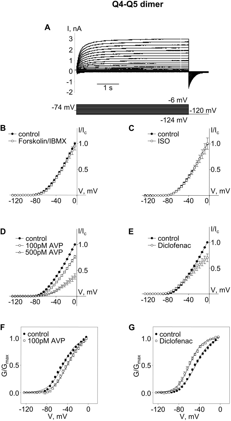
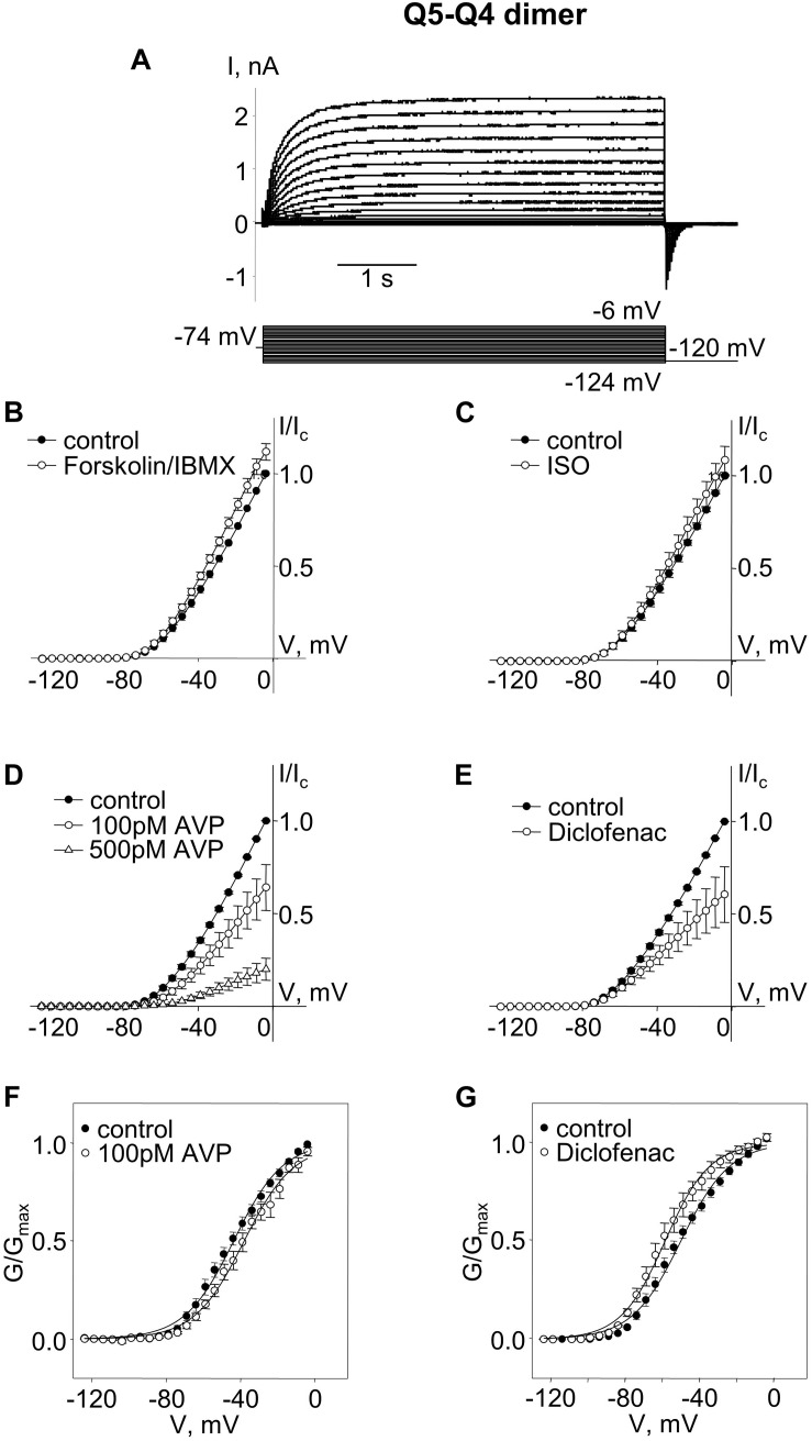
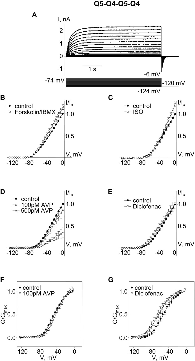
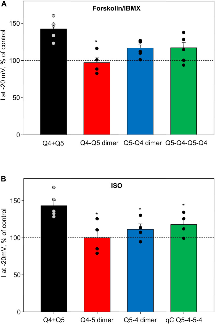
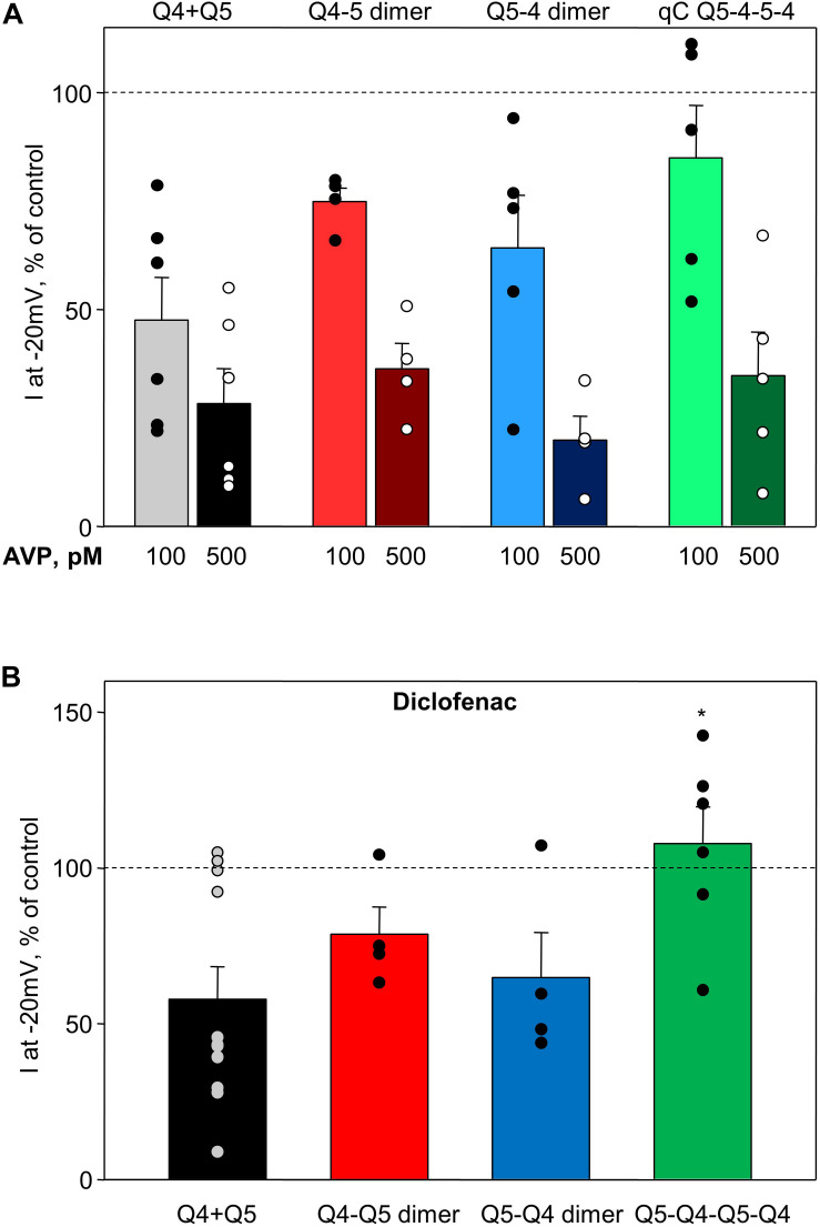
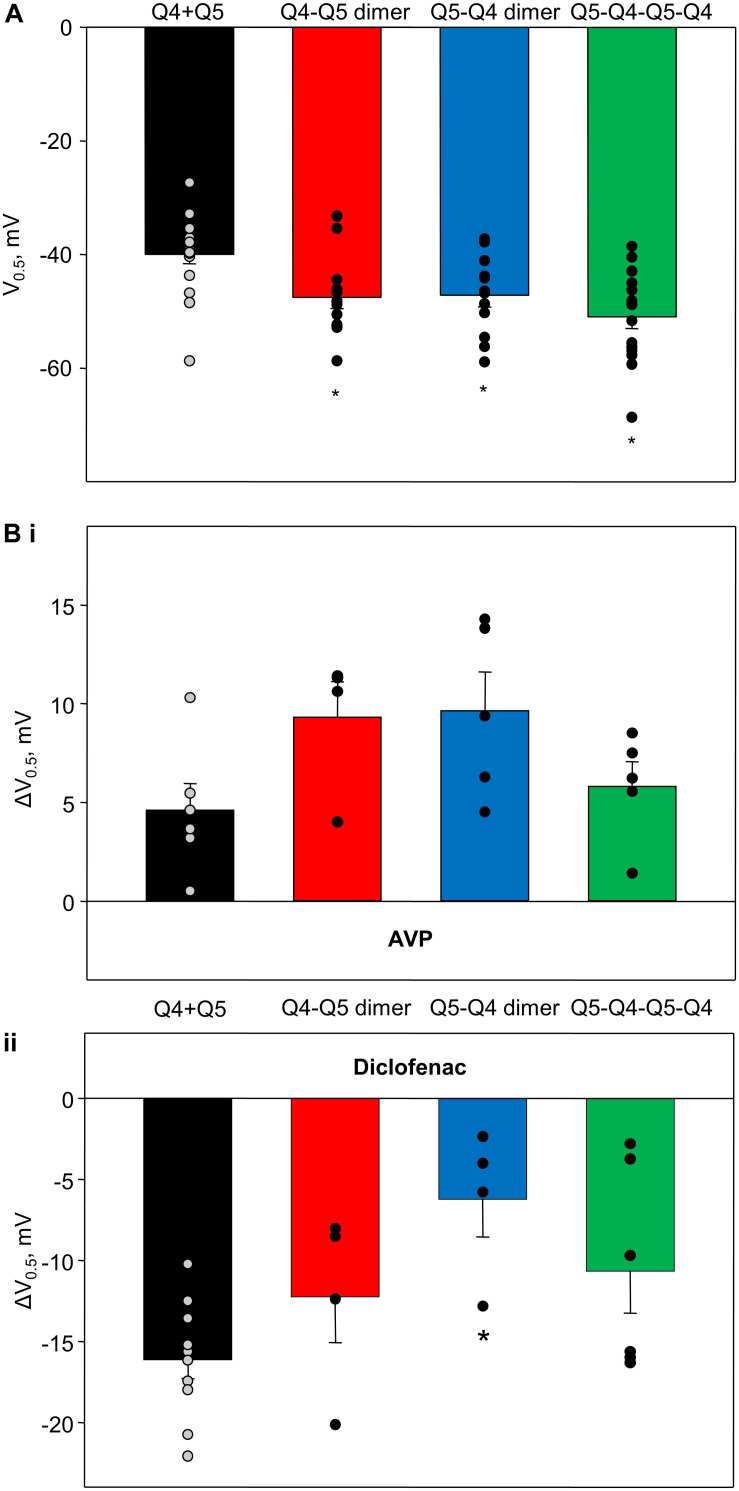
Smooth muscle cells of the vasculature, viscera, and lungs generally express multiple α-subunits of the Kv7 voltage-gated potassium channel family, with increasing evidence that both Kv7.4 and Kv7.5 can conduct "M-currents" that are functionally important for the regulation of smooth muscle contractility. Although expression systems demonstrate that functional channels can form as homomeric tetramers of either Kv7.4 or Kv7.5 α-subunits, there is evidence that heteromeric channel complexes, containing some combination of Kv7.4 and Kv7.5 α-subunits, may represent the predominant configuration natively expressed in some arterial myocytes, such as rat mesenteric artery smooth muscle cells (MASMCs). Our previous work has suggested that Kv7.4/Kv7.5 heteromers can be distinguished from Kv7.4 or Kv7.5 homomers based on their biophysical, regulatory, and pharmacological characteristics, but it remains to be determined how Kv7.4 and Kv7.5 α-subunits combine to produce these distinct characteristics. In the present study, we constructed concatenated dimers or tetramers of Kv7.4 and Kv7.5 α-subunits and expressed them in a smooth muscle cell line to determine if a particular α-subunit configuration can exhibit the features previously reported for natively expressed Kv7 currents in MASMCs. Several unique characteristics of native smooth muscle M-currents were reproduced under conditions that constrain channel formation to a Kv7.4:Kv7.5 stoichiometry of 2:2, with alternating Kv7.4 and Kv7.5 α-subunits within a tetrameric structure. Although other subunit arrangements/combinations are not ruled out, the findings provide new insights into the oligomerization of α-subunits and the ways in which Kv7.4/Kv7.5 subunit assembly can affect smooth muscle signal transduction and pharmacological responses to Kv7 channel modulating drugs.

Keywords: Kv7.4; Kv7.5; M-current; smooth muscle; α-subunit stoichiometry.


Figures

FIGURE 1
FIGURE 1
M-currents through channels formed from Kv7.4–7.5 (Q4–Q5) dimers were insensitive to cAMP/PKA activation but were sensitive to AVP and diclofenac. (A) Representative superimposed raw current traces recorded from a cell expressing Q4–Q5 dimers subjected to a series of voltage steps applied from a holding voltage of −74 mV to test potentials ranging from −124 to −6 mV, followed by a step back to −120 mV. (B–E) Average normalized current–voltage (I–V) relationships in A7r5 cells expressing Q4–Q5 dimers, before (closed circles) and during application of (B) 10 μM forskolin in combination with 500 μM IBMX (Forskolin/IBMX for 15 min, open circles, n = 4), (C) 1 μM isoproterenol (ISO for 15 min, open circles, n = 4), (D) AVP (100 pM AVP for 15 min, open circles, followed by 500 pM AVP for 15 min open triangles, n = 4), (E) diclofenac [100 μM for 15 min, open circles, n = 4, (D)]. (F,G) Mean fractional conductance plots calculated from tail currents measured before (closed circles) and during application of (F) 100 pM AVP (open circles, n = 4) or (G) 100 μM diclofenac (open circles, n = 4) fitted to the Boltzmann distribution.
FIGURE 2
FIGURE 2
M-currents through channels formed from Kv7.5–7.4 (Q5–Q4) dimers were sensitive to cAMP/PKA activation and sensitive to AVP and diclofenac. (A) Representative superimposed raw current traces recorded from a cell expressing Q5–Q4 dimers subjected to a series of voltage steps applied from a holding voltage of −74 mV to test potentials ranging from −124 to −6 mV, followed by a step back to −120 mV. (B–E) Average normalized current–voltage (I–V) relationships of Q5–Q4 dimer before (closed circles) and during application of 10 μM forskolin in combination with 500 μM IBMX [Forskolin/IBMX for 15 min, open circles, n = 6, (B)], 1 μM isoproterenol [ISO for 15 min, open circles, n = 4, (C)], AVP [100 pM AVP for 15 min, open circles, followed by 500 pM AVP for 15 min open triangles, n = 5, (D)], diclofenac [100 μM for 15 min, open circles, n = 4, (E)]. (F,G) Mean fractional conductance plots calculated from tail currents measured before (closed circles) and during application of 100 pM AVP [open circles, n = 5, (F)] or 100 μM diclofenac [open circles, n = 4, (G)] fitted to the Boltzmann distribution.
FIGURE 3
FIGURE 3
M-currents through channels formed from concatenated Kv7.5–7.4–7.5–7.4 (Q5–Q4–Q5–Q4) tetramers were sensitive to cAMP/PKA activation and diclofenac, and sensitive to 500 pM AVP. (A) Representative superimposed raw current traces recorded from a cell expressing Q5–Q4–Q5–Q4 tetramers subjected to a series of voltage steps applied from a holding voltage of −74 mV to test potentials ranging from −124 to −6 mV, followed by a step back to −120 mV. (B–E) Average normalized current–voltage (I–V) relationships of concatenated Q5–Q4–Q5–Q4 before (closed circles) and during application of 10 μM forskolin in combination with 500 μM IBMX [Forskolin/IBMX for 15 min, open circles, n = 6, (B)], 1 μM isoproterenol [ISO for 15 min, open circles, n = 4, (C)], AVP [100 pM AVP for 15 min, open circles, followed by 500 pM AVP for 15 min open triangles, n = 5, (D)], diclofenac [100 μM for 15 min, open circles, n = 6, (E)]. (F,G) Mean fractional conductance plots calculated from tail currents measured before (closed circles) and during application of 100 pM AVP [open circles, n = 5, (F)] or 100 μM diclofenac [open circles, n = 6, (G)] fitted to the Boltzmann distribution.
FIGURE 4
FIGURE 4
Summary of forskolin/IBMX and isoproterenol-induced increase in current amplitude of Q4–Q5 dimers, Q5–Q4 dimers, and Q5–Q4–Q5–Q4 tetramers, in comparison with M-currents through channels formed from co-expressed individual Kv7.4 and Kv7.5 α-subunits (Q4 + Q5). (A) Summarized bar graph of forskolin/IBMX (10 and 500 μM, respectively) -induced current enhancement through co-expressed individual Kv7.4 and Kv7.5 channels (Q4 + Q5, black, n = 5), Q4–Q5 dimer (red, n = 4), Q5–Q4 dimers (blue, n = 6), and Q5–Q4–Q5–Q4 tetramers (green, n = 6), measured at −20 mV. *Significant difference from co-expressed individual Kv7.4 and Kv7.5 channels (P = 0.002, One Way ANOVA). (B) Summarized bar graph of isoproterenol (1 μM)-induced current enhancement through co-expressed individual Kv7.4 and Kv7.5 channels (Q4 + Q5, black, n = 5), Q4–Q5 dimer (red, n = 4), Q5–Q4 dimers (blue, n = 4), and Q5–Q4–Q5–Q4 tetramer (green, n = 4), measured at −20 mV. *Significant difference from co-expressed individual Kv7.4 and Kv7.5 channels (P = 0.015, One Way ANOVA). Dashed lines indicate control current level.
FIGURE 5
FIGURE 5
Summary of AVP and diclofenac-induced decrease in M-current amplitude of Q4–Q5 dimers, Q5–Q4 dimers, and Q5–Q4–Q5–Q4 tetramers, in comparison with M-currents through channels formed from co-expressed individual Kv7.4 and Kv7.5 α-subunits (Q4 + Q5). (A) Summarized bar graph of AVP (100 pM light color and 500 μM dark color, respectively)-induced current suppression through co-expressed individual Kv7.4 and Kv7.5 α-subunits (Q4 + Q5, gray/black, n = 6), Q4–Q5 dimers (red, n = 4), Q5–Q4 dimers (blue, n = 5), and Q5–Q4–Q5–Q4 tetramers (green, n = 5), measured at −20 mV. (B) Summarized bar graph of diclofenac (100 μM)-induced current suppression through co-expressed individual Kv7.4 and Kv7.5 α-subunits (Q4 + Q5, black, n = 11), Q4–Q5 dimer (red, n = 4), Q5–Q4 dimers (blue, n = 4), and Q5–Q4–Q5–Q4 tetramers (green, n = 6), measured at −20 mV. *Significant difference from co-expressed individual Kv7.4 and Kv7.5 channels (P = 0.031, One Way ANOVA). Dashed lines indicate control current level.
FIGURE 6
FIGURE 6
Voltage of half-maximal activation of M-currents through channels formed from Q4–Q5 dimers, Q5–Q4 dimers, and Q5–Q4–Q5–Q4 tetramers, in comparison with co-expressed individual Kv7.4 and Kv7.5 α-subunits (Q4 + Q5), and effects of AVP and diclofenac on voltage of half-maximal activation of corresponding channels. (A) Voltage of half-maximal activation (V0.5) of Kv7.4/Kv7.5 channels formed by co-expressed individual Kv7.4 and Kv7.5 V (Q4 + Q5, black, n = 17), Q4–Q5 dimers (red, n = 12), Q5–Q4 dimers (blue, n = 13), and Q5–Q4–Q5–Q4 tetramers (green, n = 17). *Significant difference from co-expressed individual Kv7.4 and Kv7.5 channel α-subunits (P < 0.001, One Way ANOVA). (Bi) AVP (100 pM)-induced positive shift of V0.5 of Kv7.4/Kv7.5 channels formed by co-expressed individual Kv7.4 and Kv7.5 α-subunits (Q4 + Q5, black, n = 6), Q4–Q5 dimers (red, n = 4), Q5–Q4 dimers (blue, n = 5), and Q5–Q4–Q5–Q4 tetramers (green, n = 5). (Bii) Diclofenac (100 μM)-induced negative shift of V0.5 of Kv7.4/Kv7.5 channels formed by co-expressed individual Kv7.4 and Kv7.5 α-subunits (Q4 + Q5, black, n = 11), Q4–Q5 dimers (red, n = 4), Q5–Q4 dimers (blue, n = 4), and Q5–Q4–Q5–Q4 tetramers (green, n = 6). *Significant difference from co-expressed individual Kv7.4 and Kv7.5 α-subunits (P = 0.016, One Way ANOVA).

Similar articles

Cited by

References

    1. Bal M., Zhang J., Hernandez C. C., Zaika O., Shapiro M. S. (2010). Ca2+/calmodulin disrupts AKAP79/150 interactions with KCNQ (M-type) K+ channels. J. Neurosci. 30 2311–2323. 10.1523/jneurosci.5175-09.2010 - DOI - PMC - PubMed
    1. Brueggemann L. I., Cribbs L. L., Byron K. L. (2020). Structural determinants of Kv7.5 potassium channels that confer changes in phosphatidylinositol 4,5-bisphosphate (PIP2) affinity and signaling sensitivities in smooth muscle cells. Mol. Pharmacol. 97 145–158. 10.1124/mol.119.117192 - DOI - PubMed
    1. Brueggemann L. I., Cribbs L. L., Schwartz J., Wang M., Kouta A., Byron K. L. (2018). Mechanisms of PKA-Dependent Potentiation of Kv7.5 Channel Activity in Human Airway Smooth Muscle Cells. Int. J. Mol. Sci. 19:E2223. 10.3390/ijms19082223 - DOI - PMC - PubMed
    1. Brueggemann L. I., Gentile S., Byron K. L. (2013). Social networking among voltage-activated potassium channels. Prog. Mol. Biol. Transl. Sci. 117 269–302. 10.1016/b978-0-12-386931-9.00010-6 - DOI - PubMed
    1. Brueggemann L. I., Haick J. M., Cribbs L. L., Byron K. L. (2014a). Differential activation of vascular smooth muscle Kv7.4, Kv7.5, and Kv7.4/7.5 channels by ML213 and ICA-069673. Mol Pharmacol. 86 330–341. 10.1124/mol.114.093799 - DOI - PMC - PubMed

LinkOut - more resources