. 2020 Jan 8:2:1.
doi: 10.1186/s42490-019-0035-7. eCollection 2020.

Patterns of lower limb muscular activity and joint moments during directional efforts using a static dynamometer

Affiliations

Patterns of lower limb muscular activity and joint moments during directional efforts using a static dynamometer

Mathieu Lalumiere et al. BMC Biomed Eng. .

Abstract

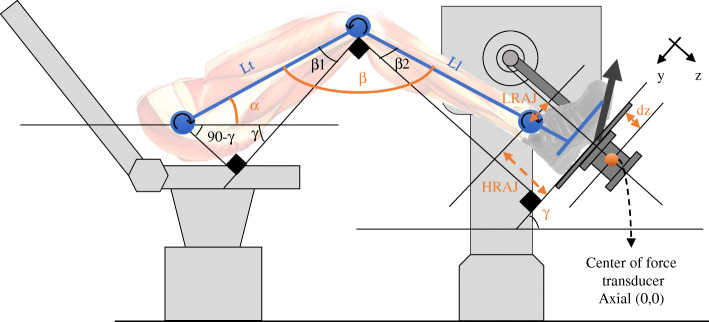
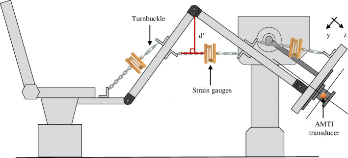
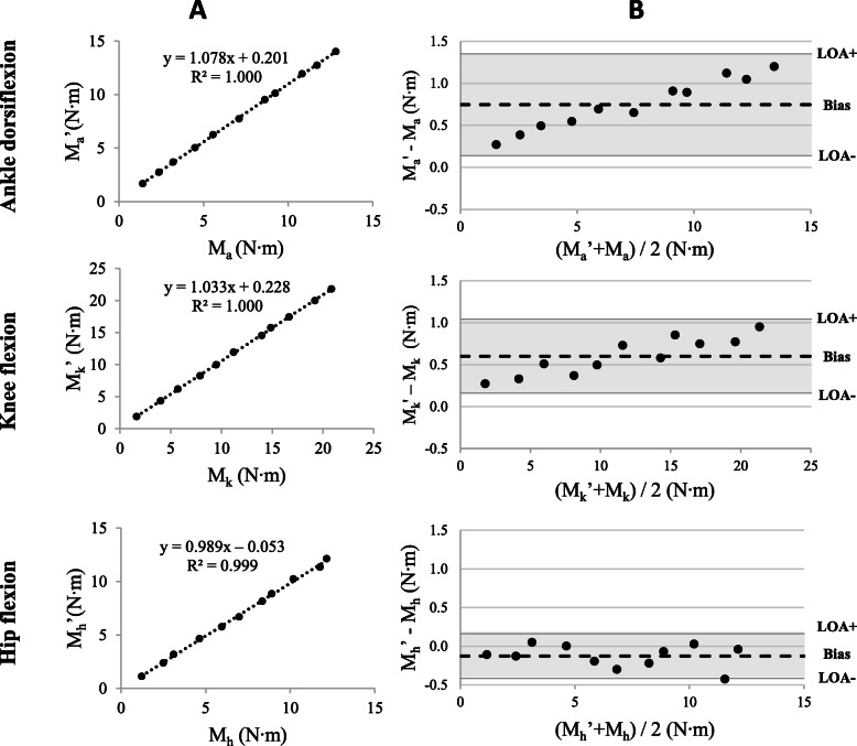
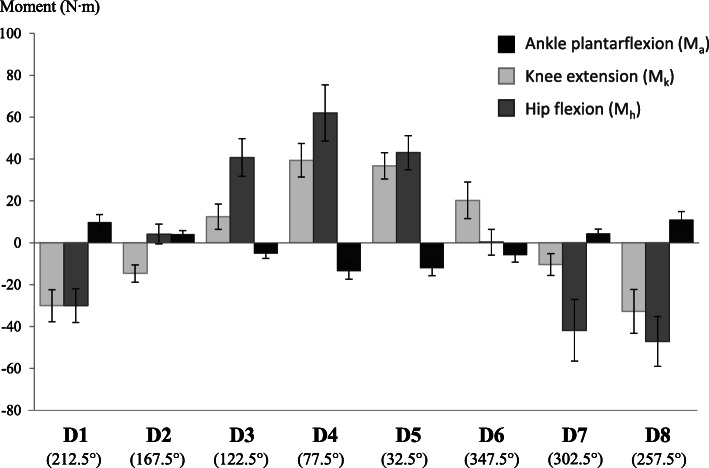
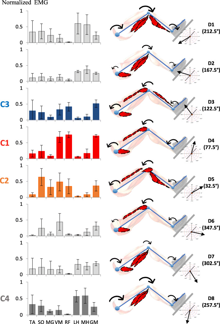
Background: Strength and coordination of lower muscle groups typically identified in healthy subjects are two prerequisites to performing functional activities. These physical qualities can be impaired following a neurological insult. A static dynamometer apparatus that measures lower limb joint moments during directional efforts at the foot was developed to recruit different patterns of muscular activity. The objectives of the present study were to 1) validate joint moments estimated by the apparatus, and 2) to characterize lower limb joint moments and muscular activity patterns of healthy subjects during progressive static efforts. Subjects were seated in a semi-reclined position with one foot attached to a force platform interfaced with a laboratory computer. Forces and moments exerted under the foot were computed using inverse dynamics, allowing for the estimation of lower limb joint moments.To achieve the study's first objective, joint moments were validated by comparing moments of various magnitudes of force applied by turnbuckles on an instrumented leg equipped with strain gauges with those estimated by the apparatus. Concurrent validity and agreement were assessed using Pearson correlation coefficients and Bland and Altman analysis, respectively. For the second objective, joint moments and muscular activity were characterized for five healthy subjects while exerting progressive effort in eight sagittal directions. Lower limb joint moments were estimated during directional efforts using inverse dynamics. Muscular activity of eight muscles of the lower limb was recorded using surface electrodes and further analyzed using normalized root mean square data.

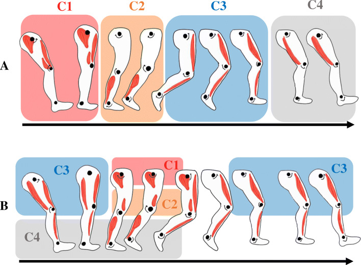
Results: The joint moments estimated with the instrumented leg were correlated (r > 0.999) with those measured by the dynamometer. Limits of agreement ranged between 8.5 and 19.2% of the average joint moment calculated by both devices. During progressive efforts on the apparatus, joint moments and patterns of muscular activity were specific to the direction of effort. Patterns of muscular activity in four directions were similar to activation patterns reported in the literature for specific portions of gait cycle.

Conclusion: This apparatus provides valid joint moments exerted at the lower limbs. It is suggested that this methodology be used to recruit muscular activity patterns impaired in neurological populations.

Keywords: (3–10) dynamometer; Electromyography; Gait; Lower limb; Muscle strength; Rehabilitation.


Conflict of interest statement

Competing interestsThe authors declare that they have no competing interests related to the presented research.

Figures

Fig. 1
Fig. 1
a) Modules of muscular activity identified by matrix factorization during gait. From 0 to 12% of the gait cycle (C1), gluteus medius, vastus medialis and rectus femoris are activated and provide body support and decelerate forward motion during early stance. From 30 to 50% of the gait cycle (C2), medial gastrocnemius and soleus are activated and provide body support and forward propulsion during stance. From 62 to 75% of the gait cycle (C3), tibialis anterior and rectus femoris ensure limb clearance during the early swing phase. From 87 to 100% of the walking cycle (C4), the semitendinosus and biceps femoris decelerate the limb during the late swing phase. b) Merging of the four muscular modules after a stroke. Compared to healthy subjects, the modules are modified during the gait cycle. Based on data presented by Clark et al. 2010
Fig. 2
Fig. 2
The subject’s foot is firmly secured on a force transducer interfaced with a laboratory computer. The location of the center of pressure in the Y axis is monitored in real time. By measuring the different angles (α, β,γ), the different lever arms (Ll, Lt, LRAJ, HRJ), and the force vector, the joint moments exerted at the different joints can be calculated
Fig. 3
Fig. 3
Experimental set-up for measuring moments exerted at the hip (Mh), knee (Mk) and ankle (Ma) from the strain gauges force and lever arm (d’). A turnbuckle was used to induce tension in the cable measured by a force transducer at one joint. The joint moment exerted was compared to the joint moment estimated by the apparatus
Fig. 4
Fig. 4
Progressive static efforts were exerted in eight directions (D1-D8) covering 360 degrees in the sagittal plane. The vector y indicates the angle of the force plate on which the foot was secured
Fig. 5
Fig. 5
a) The regression line between the joint moments calculated by the strain gauges (M’) and the joint moment estimated using the AMTI measurements (M) at each joint of the articulated leg using the set-up illustrated in Fig. 3. b) Bland and Altman plots showing the differences between joint moments as calculated by the strain gauges at the ankle (Ma’), knee (Mk’) and hip (Mh’), and estimated with the AMTI force platform at the ankle (Ma’), knee (Mk’) and hip (Mh’) against the average values (dotted line), with 95% limits of agreement (LOA; grey shadowing) for each of the eleven tests conducted for each joint
Fig. 6
Fig. 6
Joint moments for the hip, knee and ankle averaged among the 5 subjects during efforts in the eight directions (D1 to D8). Standard deviations are indicated by a bar. Positive values indicate plantarflexion of the ankle, knee extension and hip flexion
Fig. 7
Fig. 7
Normalized RMS values of the EMG during progressive efforts in the eight directions with the corresponding LL joint moment directions and predominant muscular activity. Standard deviations are indicated by a bar. Four muscle synergies were previously identified during gait: synergy 1 includes activity of the VM, RF and GM (red); synergy 2 includes activity of MG and SO (orange); synergy 3 includes activity of TA and RF (blue) and synergy 4 includes activity of LH and MH (grey)

References

    1. Ada L, Dorsch S, Canning CG. Strengthening interventions increase strength and improve activity after stroke: a systematic review. Aust J Physiother. 2006;52(4):241–248. doi: 10.1016/S0004-9514(06)70003-4. - DOI - PubMed
    1. Hunnicutt JL, Gregory CM. Skeletal muscle changes following stroke: a systematic review and comparison to healthy individuals. Top Stroke Rehabil. 2017;24(6):463–471. doi: 10.1080/10749357.2017.1292720. - DOI - PMC - PubMed
    1. Bohannon RW, Walsh S. Association of paretic lower extremity muscle strength and standing balance with stair-climbing ability in patients with stroke. J Stroke Cerebrovasc Dis. 1991;1(3):129–133. doi: 10.1016/S1052-3057(10)80004-7. - DOI - PubMed
    1. Pradon D, et al. Relationship between lower limb muscle strength and 6-minute walk test performance in stroke patients. J Rehabil Med. 2013;45(1):105–108. doi: 10.2340/16501977-1059. - DOI - PubMed
    1. Lomaglio MJ, Eng JJ. Muscle strength and weight-bearing symmetry relate to sit-to-stand performance in individuals with stroke. Gait Posture. 2005;22(2):126–131. doi: 10.1016/j.gaitpost.2004.08.002. - DOI - PMC - PubMed

LinkOut - more resources