

Human motor decoding from neural signals: a review
- PMID: 32903354
- PMCID: PMC7422484
- DOI: 10.1186/s42490-019-0022-z
Human motor decoding from neural signals: a review
Abstract
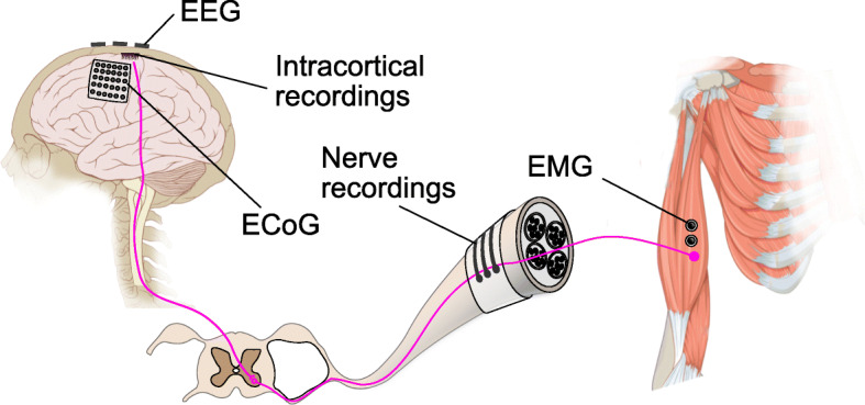
Many people suffer from movement disability due to amputation or neurological diseases. Fortunately, with modern neurotechnology now it is possible to intercept motor control signals at various points along the neural transduction pathway and use that to drive external devices for communication or control. Here we will review the latest developments in human motor decoding. We reviewed the various strategies to decode motor intention from human and their respective advantages and challenges. Neural control signals can be intercepted at various points in the neural signal transduction pathway, including the brain (electroencephalography, electrocorticography, intracortical recordings), the nerves (peripheral nerve recordings) and the muscles (electromyography). We systematically discussed the sites of signal acquisition, available neural features, signal processing techniques and decoding algorithms in each of these potential interception points. Examples of applications and the current state-of-the-art performance were also reviewed. Although great strides have been made in human motor decoding, we are still far away from achieving naturalistic and dexterous control like our native limbs. Concerted efforts from material scientists, electrical engineers, and healthcare professionals are needed to further advance the field and make the technology widely available in clinical use.
Keywords: Brain-machine interfaces; Motor decoding; Neural signal processing; Neuroprosthesis.
© The Author(s) 2019.
Conflict of interest statement
Competing interestsThe authors declare that they have no competing interests.
Figures

Similar articles
-
Computational approaches to decode grasping force and velocity level in upper-limb amputee from intraneural peripheral signals.J Neural Eng. 2021 Apr 6;18(5). doi: 10.1088/1741-2552/abef3a. J Neural Eng. 2021. PMID: 33725672
-
Validating Deep Neural Networks for Online Decoding of Motor Imagery Movements from EEG Signals.Sensors (Basel). 2019 Jan 8;19(1):210. doi: 10.3390/s19010210. Sensors (Basel). 2019. PMID: 30626132 Free PMC article.
-
Improved multi-unit decoding at the brain-machine interface using population temporal linear filtering.J Neural Eng. 2010 Aug;7(4):046012. doi: 10.1088/1741-2560/7/4/046012. Epub 2010 Jul 19. J Neural Eng. 2010. PMID: 20644245
-
Neural signal recording and processing in somatic neuroprosthetic applications. A review.J Neurosci Methods. 2020 May 1;337:108653. doi: 10.1016/j.jneumeth.2020.108653. Epub 2020 Feb 27. J Neurosci Methods. 2020. PMID: 32114143 Review.
-
Neural Decoding for Intracortical Brain-Computer Interfaces.Cyborg Bionic Syst. 2023 Jul 28;4:0044. doi: 10.34133/cbsystems.0044. eCollection 2023. Cyborg Bionic Syst. 2023. PMID: 37519930 Free PMC article. Review.
Cited by
-
Neural signal analysis in chronic stroke: advancing intracortical brain-computer interface design.Front Hum Neurosci. 2025 Feb 21;19:1544397. doi: 10.3389/fnhum.2025.1544397. eCollection 2025. Front Hum Neurosci. 2025. PMID: 40060267 Free PMC article.
-
Decoding spoken English from intracortical electrode arrays in dorsal precentral gyrus.J Neural Eng. 2020 Nov 25;17(6):066007. doi: 10.1088/1741-2552/abbfef. J Neural Eng. 2020. PMID: 33236720 Free PMC article.
-
DTCNet: finger flexion decoding with three-dimensional ECoG data.Front Comput Neurosci. 2025 Jul 9;19:1627819. doi: 10.3389/fncom.2025.1627819. eCollection 2025. Front Comput Neurosci. 2025. PMID: 40703668 Free PMC article.
-
Decoding force production of skeletal muscle from the female brain using functional near-infrared spectroscopy.BMC Res Notes. 2023 Nov 1;16(1):304. doi: 10.1186/s13104-023-06588-5. BMC Res Notes. 2023. PMID: 37915005 Free PMC article.
-
Classifying mental motor tasks from chronic ECoG-BCI recordings using phase-amplitude coupling features.Front Hum Neurosci. 2025 Mar 12;19:1521491. doi: 10.3389/fnhum.2025.1521491. eCollection 2025. Front Hum Neurosci. 2025. PMID: 40144587 Free PMC article.
References
-
- Ziegler-Graham K, MacKenzie EJ, Ephraim PL, Travison TG, Brookmeyer R. Estimating the Prevalence of Limb Loss in the United States: 2005 to 2050. Arch Phys Med Rehabil. 2008;89(3):422–9. - PubMed
-
- Adams PF, Hendershot GE, Marano MA. Current estimates from the National Health Interview Survey. Vital Health Stat. 1999;10(200):1996. - PubMed
-
- Wing-Kin T, Kai-yu T, Fei M, Shangkai Gao. A Minimal Set of Electrodes for Motor Imagery BCI to Control an Assistive Device in Chronic Stroke Subjects: A Multi-Session Study. IEEE Tran Neural Syst Rehabil Eng. 2011;19(6):617–27. - PubMed
-
- Ang KK, Chua KSG, Phua KS, Wang C, Chin ZY, Kuah CWK, Low W, Guan C. A Randomized Controlled Trial of EEG-Based Motor Imagery Brain-Computer Interface Robotic Rehabilitation for Stroke. Clin EEG Neurosci. 2015;46(4):310–20. - PubMed
-
- Bear MF, Connors BW, Paradiso MA. Neuroscience: Exploring the Brain. Philadelphia: Wolters Kluwer Health; 2015.
Publication types
LinkOut - more resources
Full Text Sources