

Human motion component and envelope characterization via wireless wearable sensors
- PMID: 32903362
- PMCID: PMC7422588
- DOI: 10.1186/s42490-020-0038-4
Human motion component and envelope characterization via wireless wearable sensors
Abstract
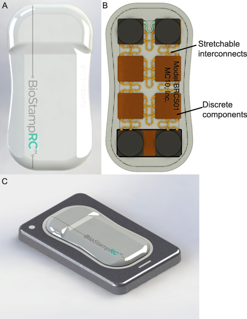
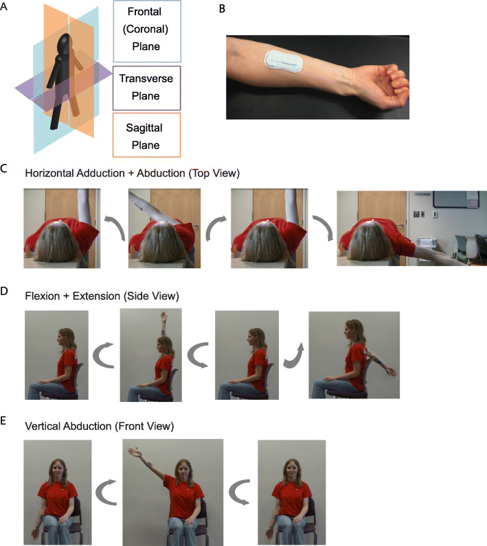
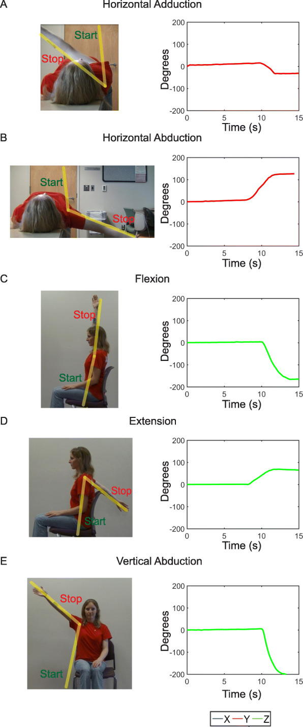
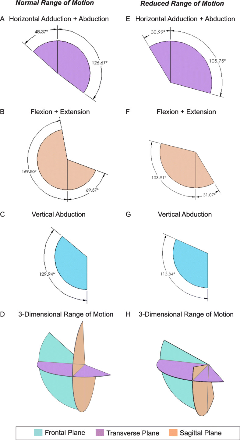
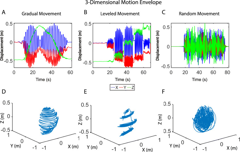
Background: The characterization of limb biomechanics has broad implications for analyzing and managing motion in aging, sports, and disease. Motion capture videography and on-body wearable sensors are powerful tools for characterizing linear and angular motions of the body, though are often cumbersome, limited in detection, and largely non-portable. Here we examine the feasibility of utilizing an advanced wearable sensor, fabricated with stretchable electronics, to characterize linear and angular movements of the human arm for clinical feedback. A wearable skin-adhesive patch with embedded accelerometer and gyroscope (BioStampRC, MC10 Inc.) was applied to the volar surface of the forearm of healthy volunteers. Arms were extended/flexed for the range of motion of three different regimes: 1) horizontal adduction/abduction 2) flexion/extension 3) vertical abduction. Data were streamed and recorded revealing the signal "pattern" of movement in three separate axes. Additional signal processing and filtering afforded the ability to visualize these motions in each plane of the body; and the 3-dimensional motion envelope of the arm.
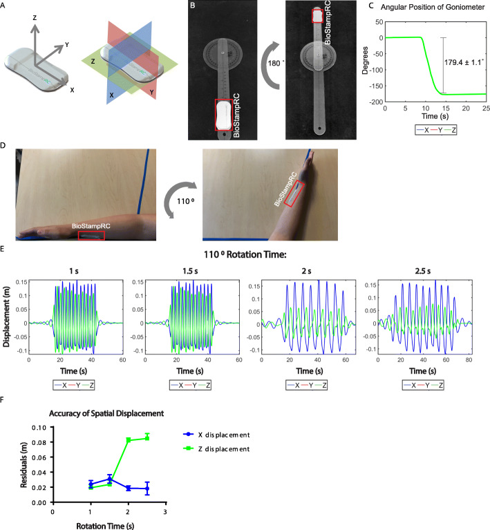
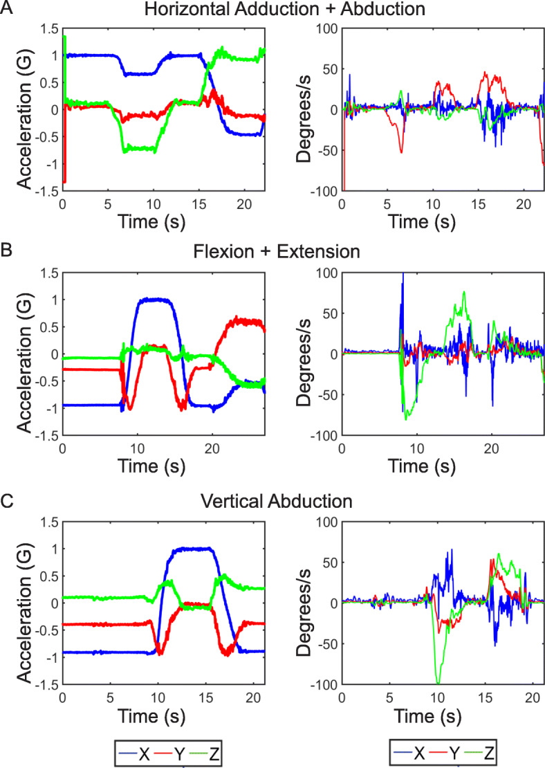
Results: Each of the three motion regimes studied had a distinct pattern - with identifiable qualitative and quantitative differences. Integration of all three movement regimes allowed construction of a "motion envelope," defining and quantifying motion (range and shape - including the outer perimeter of the extreme of motion - i.e. the envelope) of the upper extremity. The linear and rotational motion results from multiple arm motions match measurements taken with videography and benchtop goniometer.
Conclusions: A conformal, stretchable electronic motion sensor effectively captures limb motion in multiple degrees of freedom, allowing generation of characteristic signatures which may be readily recorded, stored, and analyzed. Wearable conformal skin adherent sensor patchs allow on-body, mobile, personalized determination of motion and flexibility parameters. These sensors allow motion assessment while mobile, free of a fixed laboratory environment, with utility in the field, home, or hospital. These sensors and mode of analysis hold promise for providing digital "motion biomarkers" of health and disease.
Keywords: Accelerometers; Biomechanics; Biomedical measurement; Biometrics; Biosensors; Biotechnology; Engineering in medicine and biology; Gyroscopes; Motion analysis; Wearable sensors.
© The Author(s) 2020.
Conflict of interest statement
Competing interestsAll authors report no competing interests, with exception of MJS who reports receiving equipment support (Biostamps) for research from MC10, Inc. Study was conducted at arms length from MC10, with MC10 having no role in design of the study, nor in collection, analysis, and interpretation of data or writing of the manuscript.
Figures
References
-
- WHO | International Classification of Functioning, Disability and Health (ICF). WHO [Internet]. 2017 [cited 2017 Dec 18]; Available from: http://www.who.int/classifications/icf/en/
-
- Ropper AH, Adams RD (Raymond D, Victor M, Samuels MA, Ropper AH. Adams and Victor’s principles of neurology [Internet]. McGraw-Hill Medical; 2009 [cited 2018 Feb 22]. 1572 p. Available from: https://books.google.com/books/about/Adams_and_Victor_s_Principles_of_Ne...
-
- Pearson OR, Busse ME, van Deursen RWM, Wiles CM. Quantification of walking mobility in neurological disorders. QJM [Internet]. 2004 Aug 1 [cited 2017 Dec 18];97(8):463–475. Available from: https://academic.oup.com/qjmed/article-lookup/doi/10.1093/qjmed/hch084 - DOI - PubMed
-
- Wada O, Nagai K, Hiyama Y, Nitta S, Maruno H, Mizuno K. Diabetes is a Risk Factor for Restricted Range of Motion and Poor Clinical Outcome After Total Knee Arthroplasty. J Arthroplasty [Internet]. 2016 Sep 1 [cited 2018 Aug 9];31(9):1933–1937. Available from: https://www.sciencedirect.com/science/article/pii/S0883540316001820 - PubMed
-
- Węgrzynowska-Teodorczyk Kinga, Siennicka Agnieszka, Josiak Krystian, Zymliński Robert, Kasztura Monika, Banasiak Waldemar, Ponikowski Piotr, Woźniewski Marek. Evaluation of Skeletal Muscle Function and Effects of Early Rehabilitation during Acute Heart Failure: Rationale and Study Design. BioMed Research International. 2018;2018:1–8. doi: 10.1155/2018/6982897. - DOI - PMC - PubMed
Grants and funding
LinkOut - more resources
Full Text Sources