Review
. 2020 Aug 12:11:845.
doi: 10.3389/fneur.2020.00845. eCollection 2020.

Neurological Manifestations of Severe SARS-CoV-2 Infection: Potential Mechanisms and Implications of Individualized Mechanical Ventilation Settings

Affiliations
Review

Neurological Manifestations of Severe SARS-CoV-2 Infection: Potential Mechanisms and Implications of Individualized Mechanical Ventilation Settings

Denise Battaglini et al. Front Neurol. .

Abstract

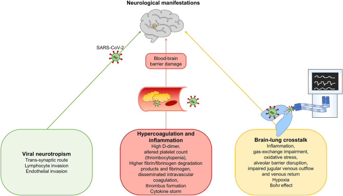
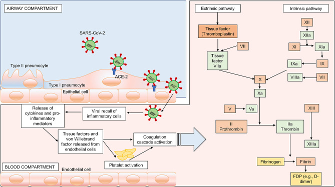
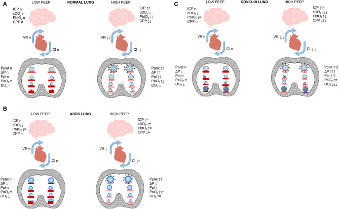
In December 2019, an outbreak of illness caused by a novel coronavirus (2019-nCoV, subsequently renamed SARS-CoV-2) was reported in Wuhan, China. Coronavirus disease 2019 (COVID-19) quickly spread worldwide to become a pandemic. Typical manifestations of COVID-19 include fever, dry cough, fatigue, and respiratory distress. In addition, both the central and peripheral nervous system can be affected by SARS-CoV-2 infection. These neurological changes may be caused by viral neurotropism, by a hyperinflammatory and hypercoagulative state, or even by mechanical ventilation-associated impairment. Hypoxia, endothelial cell damage, and the different impacts of different ventilatory strategies may all lead to increased stress and strain, potentially exacerbating the inflammatory response and leading to a complex interaction between the lungs and the brain. To date, no studies have taken into consideration the possible secondary effect of mechanical ventilation on brain recovery and outcomes. The aim of our review is to provide an updated overview of the potential pathogenic mechanisms of neurological manifestations in COVID-19, discuss the physiological issues related to brain-lung interactions, and propose strategies for optimization of respiratory support in critically ill patients with SARS-CoV-2 pneumonia.

Keywords: COVID-19; SARS-CoV-2; coronavirus; neurological manifestations; neurotropism.


Figures

Figure 1
Figure 1
Proposed mechanisms for neurological manifestations in SARS-CoV-2 infection. We hypothesize three possible mechanisms for neurological manifestations in SARS-CoV-2 infection: (1) Viral neurotropism; (2) Hypercoagulation and inflammation, and (3) Brain-lung crosstalk.
Figure 2
Figure 2
SARS-CoV-2-induced hypercoagulability. Passage of the virus from the airway to the systemic circulation is facilitated by the sluggish movement of blood within the microcirculation and subsequent binding of ACE-2 receptors, expressed on the capillary endothelium, followed by endothelial damage, enhanced inflammation, and hypercoagulability. In this figure, we represent the activation of both intrinsic and extrinsic coagulation pathways as a possible mechanism for hypercoagulability and potential brain damage. Intrinsic pathway: activation of factor (F) XIIa, followed by activation of FXIa and VIII. Extrinsic pathway: activation of FVIIa and tissue factor. Both pathways converge in the common pathway with activation of FXa, FVa, prothrombin into thrombin, fibrinogen into fibrin, and fibrin degradation products (FDP) such as D-dimer.
Figure 3
Figure 3
Bohr effect. The oxyhemoglobin dissociation curve is shifted to the left in response to respiratory alkalosis (lower PaCO2 and higher pH), with increased affinity of oxygen for the hemoglobin. Conversely, during respiratory acidosis (higher PaCO2 and lower pH), the alveolar oxygen tension and systemic saturation improve, thus reducing alveolar carbon dioxide tension, as explained by the Bohr effect: the higher the acidity, the more carbon dioxide is eliminated.
Figure 4
Figure 4
Improving oxygen delivery to the brain. Raising hemoglobin and cardiac output should be considered for improving oxygen delivery, especially in COVID-19 phenotype 1. This figure represents different delivery of oxygen (DO2) at a fixed cardiac output, by changing hemoglobin, or at fixed hemoglobin, by changing cardiac output.
Figure 5
Figure 5
(A–C) Brain–lung–heart cross talk. SARS-CoV-2 lung infection can require mechanical ventilation, which heightens the pro-inflammatory cascade. In this figure, we propose the effect of increased PEEP on the cardiovascular system and CNS in healthy subjects (A), ARDS (B), and COVID-19 (C). In normal lungs (A), high PEEP and alveolar hyperdistention cause increased plateau pressure (Pplat), driving pressure (ΔP), and pleural pressure (Ppl), with consequent reduction of venous return (VR) and cardiac index (CI) and reduced cerebral perfusion pressure (CPP) and increased intracranial pressure (ICP). This can be partially offset by the presence of preserved gas exchange. In ARDS patients (B), the increase in PEEP with recruitment of collapsed areas does not cause significant changes in hemodynamics or cerebral function, and can increase oxygen delivery (cDO2). Conversely, in COVID-19 patients (C) who do not respond to recruitment, the concomitance of alveolar hyperdistention after PEEP increase and hypoxemia can cause serious impairment of cerebral dynamics and cerebral hypoxemia (low PbtO2).

References

    1. Huang C, Wang Y, Li X, Ren L, Zhao J, Hu Y, et al. . Clinical features of patients infected with 2019 novel coronavirus in Wuhan, China. Lancet. (2020) 395:497–506. 10.1016/S0140-6736(20)30183-5 - DOI - PMC - PubMed
    1. Naming the Coronavirus Disease (COVID-19) and the Virus that Causes It Available online at: https://www.who.int/emergencies/diseases/novel-coronavirus-2019/technica... (accessed March 31, 2020).
    1. Zhu N, Zhang D, Wang W, Li X, Yang B, Song J, et al. . A novel coronavirus from patients with pneumonia in China, 2019. NEJM. (2020) 382:727–33. 10.1056/NEJMoa2001017 - DOI - PMC - PubMed
    1. Chen Y, Liu Q, Guo D. Emerging coronaviruses: genome structure, replication, and pathogenesis. J Med Virol. (2020) 92:418–23. 10.1002/jmv.25681 - DOI - PMC - PubMed
    1. Choi WJ, Lee KN, Kang EJ, Lee H. Middle east respiratory syndrome-coronavirus infection: a case report of serial computed tomographic findings in a young male patient. Korean J Radiol. (2016) 17:166–70. 10.3348/kjr.2016.17.1.166 - DOI - PMC - PubMed