

MRI Observation After Intralabyrinthine and Vestibular Schwannoma Resection and Cochlear Implantation
- PMID: 32903399
- PMCID: PMC7434924
- DOI: 10.3389/fneur.2020.00759
MRI Observation After Intralabyrinthine and Vestibular Schwannoma Resection and Cochlear Implantation
Abstract
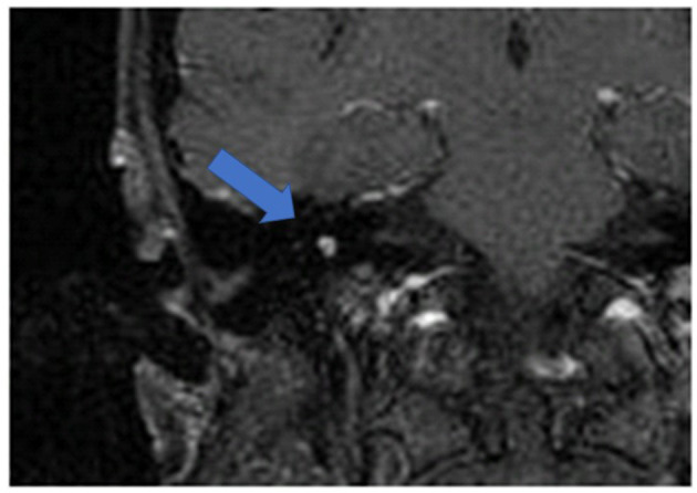
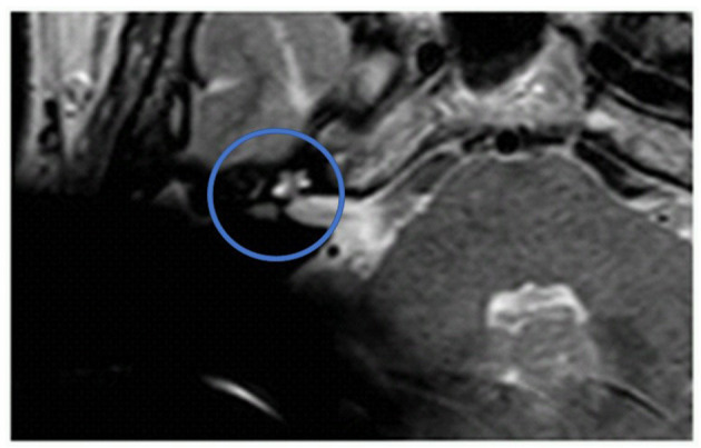
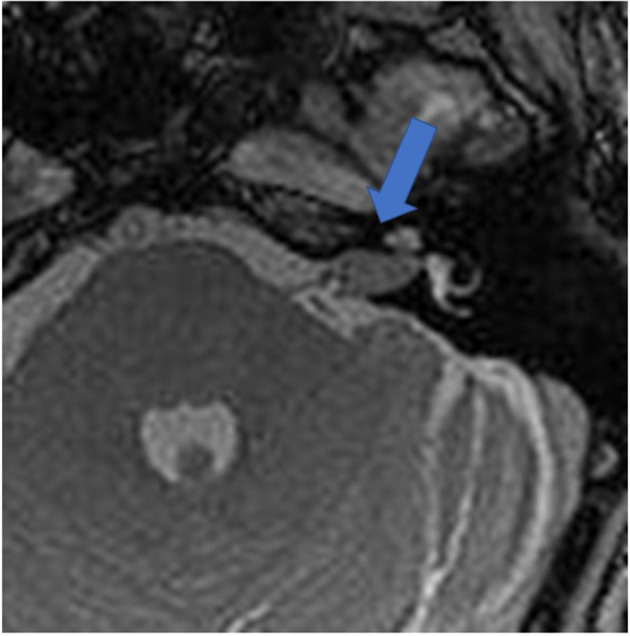
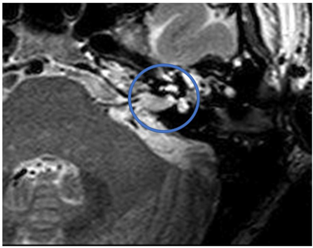
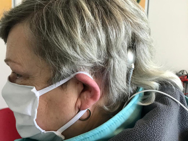
Objective: MRI observation is part of the regular follow-up after vestibular schwannoma (VS) or intralabyrinthine schwannoma (ILS) resection. Because cochlear implantation (CI) after resection is part of the audiological rehabilitation process, the magnet resonance imaging (MRI) behavior of CI systems needs to be considered. In light of recent developments in MRI artifact positioning and pain prevention, this study evaluates reproducible MRI observations after tumor resection and CI surgery as part of follow-up. Methods: In a retrospective study, we evaluated 9 patients with a T1 KM, T2 sequence MRI observation, and cone beam computed tomography (CBCT) after ILS/VS resection and CI. In all but one case, a CI with a diametrically bipolar magnet and a receiver positioned 8-9 cm behind the external auditory canal was performed. Results: In all but one case, MRI observation allowed for a pain-free visual assessment of the intralabyrinthine and internal auditory canal (IAC) regions. In one case, a painful dislodgement of the receiver magnet occurred. Conclusion: MRI follow-up after ILS and VS resection and CI is reproducibly possible. Implant choice and positioning should be considered before implantation to allow for a pain-free visual assessment afterward. This finding allows for the first time a widening of the indication into this patient group.
Keywords: MRI; cochlear implant; intralabyrinthine schwannoma; tumor follow up; vestibular schwannoma.
Copyright © 2020 Sudhoff, Gehl, Scholtz and Todt.
Figures

References
-
- Arndt S, Aschendorff A, Laszig R, Beck R, Schild C, Kroeger S, et al. . Comparison of pseudobinaural hearing to real binaural hearing rehabilitation after cochlear implantation in patients with unilateral deafness and tinnitus. Otol Neurotol. (2011) 32:39–47. 10.1097/MAO.0b013e3181fcf271 - DOI - PubMed
LinkOut - more resources
Full Text Sources