. 2020 Aug 12:11:1204.
doi: 10.3389/fphar.2020.01204. eCollection 2020.

Human α-Defensin-5 Efficiently Neutralizes Clostridioides difficile Toxins TcdA, TcdB, and CDT

Affiliations

Human α-Defensin-5 Efficiently Neutralizes Clostridioides difficile Toxins TcdA, TcdB, and CDT

Michael Korbmacher et al. Front Pharmacol. .

Abstract

Infections with the pathogenic bacterium Clostridioides (C.) difficile are coming more into focus, in particular in hospitalized patients after antibiotic treatment. C. difficile produces the exotoxins TcdA and TcdB. Since some years, hypervirulent strains are described, which produce in addition the binary actin ADP-ribosylating toxin CDT. These strains are associated with more severe clinical presentations and increased morbidity and frequency. Once in the cytosol of their target cells, the catalytic domains of TcdA and TcdB glucosylate and thereby inactivate small Rho-GTPases whereas the enzyme subunit of CDT ADP-ribosylates G-actin. Thus, enzymatic activity of the toxins leads to destruction of the cytoskeleton and breakdown of the epidermal gut barrier integrity. This causes clinical symptoms ranging from mild diarrhea to life-threatening pseudomembranous colitis. Therefore, pharmacological inhibition of the secreted toxins is of peculiar medical interest. Here, we investigated the neutralizing effect of the human antimicrobial peptide α-defensin-5 toward TcdA, TcdB, and CDT in human cells. The toxin-neutralizing effects of α-defensin-5 toward TcdA, TcdB, and CDT as well as their medically relevant combination were demonstrated by analyzing toxins-induced changes in cell morphology, intracellular substrate modification, and decrease of trans-epithelial electrical resistance. For TcdA, the underlying mode of inhibition is most likely based on the formation of inactive toxin-defensin-aggregates whereas for CDT, the binding- and transport-component might be influenced. The application of α-defensin-5 delayed intoxication of cells in a time- and concentration-dependent manner. Due to its effect on the toxins, α-defensin-5 should be considered as a candidate to treat severe C. difficile-associated diseases.

Keywords: AB-type protein toxins; C. difficile infection; binary actin ADP-ribosylating toxin; large clostridial glucosylating toxins; toxin inhibitor.


Figures

Figure 1
Figure 1
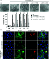
α-Defensin-5 decreases the cytotoxic effects of TcdA, TcdB and of the combination of both toxins. (A) Vero cells were either treated with TcdA (10 pM, left panel) or TcdB (10 pM, right panel) in the presence or absence of increasing concentrations of α-defensin-5 (1, 3, 6 µM), and the percentage or rounded cells was determined. For comparison, Vero cells were treated with native TcdA (10 pM, left lower panel) in the presence or absence of α-defensin-5 (6 µM). Values are given as mean ± SD (n = 3). Significance was determined using the one-way ANOVA test (n.s. = not significant, *p < 0.05, **p < 0.01, ***p < 0.001, ****p < 0.0001). (B) Vero cells (upper panel) were treated with TcdA (10 pM) and α-defensin-5 with increasing concentrations (1, 3, 6 µM) for 3.5 h. Caco-2 cells (lower panel) were treated with either TcdA (10 pM), TcdB (10 pM) or the combination of both toxins (each 10 pM) with or without α-defensin-5 (6 µM) for 8 h. Afterward, cells were lysed and subjected to Western blot analysis. Non-glucosylated Rac1 was detected using a specific antibody. Hsp90 or GAPDH were used as controls for equal protein loading. (C) Caco-2 cells were treated with the combination of TcdA (10 pM) plus TcdB (10 pM) and with/without α-defensin-5 (6 µM) for 8.5 h. For control, cells were left untreated. After incubation, cells were fixed and permeabilized. Non-glucosylated Rac1 was detected using a specific anti-Rac1-antibody, F-actin was stained with phalloidin-FITC, nuclei were stained with Hoechst33342.
Figure 2
Figure 2
α-Defensin-5 protects cells from intoxication with the binary toxin CDT. (A) Vero cells (upper left panel) were treated with CDT (1 nM/1.3 nM) and Caco-2 cells (lower left panel) were treated with CDT (4.1 nM/5.3 nM) in the presence or absence of α-defensin-5 with varying concentrations. Representative images for Vero cells (6 h) and Caco-2 cells (4.5 h) plus/minus α-defensin-5 (6 µM) are depicted. For Vero cells, the amount of rounded cells over time was determined (right panel). Values are given as mean ± SD (n = 3). Significance was determined using the one-way ANOVA test (n.s. = not significant, *p < 0.05, **p < 0.01, ***p < 0.001). (B) Caco-2 cells were treated for 4.5 h with CDT (4.1 nM/5.3 nM) with and without α-defensin-5 (6 µM). Afterward, cells were washed, lysed and subjected with 10 µM biotinylated NAD+ and 50 ng fresh CDTa. Biotin-labeled, e.g. ADP-ribosylated actin was detected by immunoblotting using the ECL system. GAPDH was stained for comparable protein loading. (C) Caco-2 cells were treated with CDT (2 nM/2.7 nM) with or without α-defensin-5 (6 µM) for 5 h. For control, cells were left untreated. Then, cells were fixed and permeabilized. Phalloidin-FITC was used to stain F-actin, Hoechst33342 was used to stain nuclei. (D) Transepithelial electrical resistance was investigated using Caco-2 cells with CDT (1.6 nM/2 nM) with or without α-defensin-5 (6 µM).
Figure 3
Figure 3
α-Defensin-5 protects cells in a time- and concentration-dependent manner from intoxication with the combination of TcdA, TcdB, and CDT. (A) Vero cells were treated with the combination of all three C. difficile toxins (TcdA: 10 pM, TcdB: 10 pM, CDT: 1 nM/1. 3 nM) and increasing concentrations of α-defensin-5 (1, 3, 6 µM). Representative images after 6 h are shown (upper panel). The amount of rounded cells was determined over time (lower panel). Values are given as mean ± SD (n=3). Significance was determined using the one-way ANOVA test (n.s. = not significant, *p < 0.05, **p < 0.01, ****p < 0.0001). (B) Caco-2 cells were treated with the combination of TcdA (10 pM), TcdB (10 pM), and CDT (2 nM/2.7 nM) in the presence or absence of α-defensin-5 (6 µM). After 5.5 h, cells were fixed, permeabilized and non-glucosylated Rac1 was stained using a specific antibody. Phalloidin-FITC was used to stain F-actin, Hoechst33342 was used to stain nuclei.
Figure 4
Figure 4
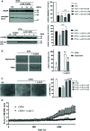
α-Defensin-5 has no influence on the enzymatic activity of TcdA and CDTa but leads to precipitation of TcdA and inhibition of the cytotoxic pore-forming activity of CDTb. (A) Caco-2 lysate (40 µg) was incubated with TcdA (300 ng) and varying concentrations of α-defensin-5 (6, 12, 24 µM) for 1 h (left panel) at 37°C. Caco-2 lysate (40 µg) was incubated with CDTa (1 ng), 10 µM biotinylated NAD+ and varying concentrations of α-defensin-5 (1, 3, 6 µM) for 30 min at 37°C. Next, samples were subjected to SDS-PAGE and Western Blotting. Non-glucosylated Rac1 was detected with a specific antibody, biotin-labeled, e.g., ADP-ribosylated actin was detected using streptavidin-peroxidase. Hsp90 and GAPDH were used to confirm equal protein loading. Values are given as mean ± SD (n = 2). Significance was determined using the one-way ANOVA test (n.s. = not significant). (B) TcdA (1 µg) was incubated with and without α-defensin-5 (6 µM) for 15 min at 37°C in serum-free medium. Afterward, samples were centrifuged and separated fractions were subjected to SDS-PAGE (left panel). Densitometric analyses from individual experiments are shown as bar graph (right panel). Values are given as mean ± SD (n = 2). Significance was determined using the one-way ANOVA test (n.s. = not significant, **p < 0.01). (C) Vero cells were treated with CDTb (5.3 nM) in the absence of CDTa with or without increasing concentrations of α-defensin-5 (1, 3, 6 µM) for 4 h at 37°C. Representative images are shown in the left panel. After the incubation time, a MTS cell viability assay was performed. Values are given as mean ± SD (n=3). Significance was determined using the one-way ANOVA test (n.s. = not significant, ****p < 0.0001). (D) Caco-2 cells were seeded in an 8-well ibidi plate and pretreated with Fura-2AM (3 µM) for 45 min. Next, baseline was measured for 2 min, and the cells were then treated with CDTb (13 nM) in the presence or absence of α-defensin-5 (6 µM) as indicated.

Similar articles

Cited by

References

    1. Aktories K. (2011). Bacterial protein toxins that modify host regulatory GTPases. Nat. Rev. Microbiol. 9, 487–498. 10.1038/nrmicro2592 - DOI - PubMed
    1. Alpha Defensin - an overview | ScienceDirect Topics Available at: https://www.sciencedirect.com/topics/neuroscience/alpha-defensin (Accessed March 19, 2020).
    1. Bachmeyer C., Benz R., Barth H., Aktories K., Gilbert M., Popoff M. R. (2001). Interaction of Clostridium botulinum C2 toxin with lipid bilayer membranes and Vero cells: inhibition of channel function by chloroquine and related compounds in vitro and intoxification in vivo. FASEB J. 15, 1658–1660. 10.1096/fj.00-0671fje - DOI - PubMed
    1. Bachmeyer C., Orlik F., Barth H., Aktories K., Benz R. (2003). Mechanism of C2-toxin inhibition by Fluphenazine and related compounds: investigation of their binding kinetics to the C2II-channel using the current noise analysis. J. Mol. Biol. 333, 527–540. 10.1016/j.jmb.2003.08.044 - DOI - PubMed
    1. Carter G. P., Rood J., II, Lyras D. (2012). The role of toxin A and toxin B in the virulence of Clostridium difficile. Trends Microbiol. 20, 21–29. 10.1016/j.tim.2011.11.003 - DOI - PubMed

LinkOut - more resources