. 2020 Aug 12:11:1786.
doi: 10.3389/fimmu.2020.01786. eCollection 2020.

Polymicrobial Sepsis Impairs Antigen-Specific Memory CD4 T Cell-Mediated Immunity

Affiliations

Polymicrobial Sepsis Impairs Antigen-Specific Memory CD4 T Cell-Mediated Immunity

Frances V Sjaastad et al. Front Immunol. .

Abstract

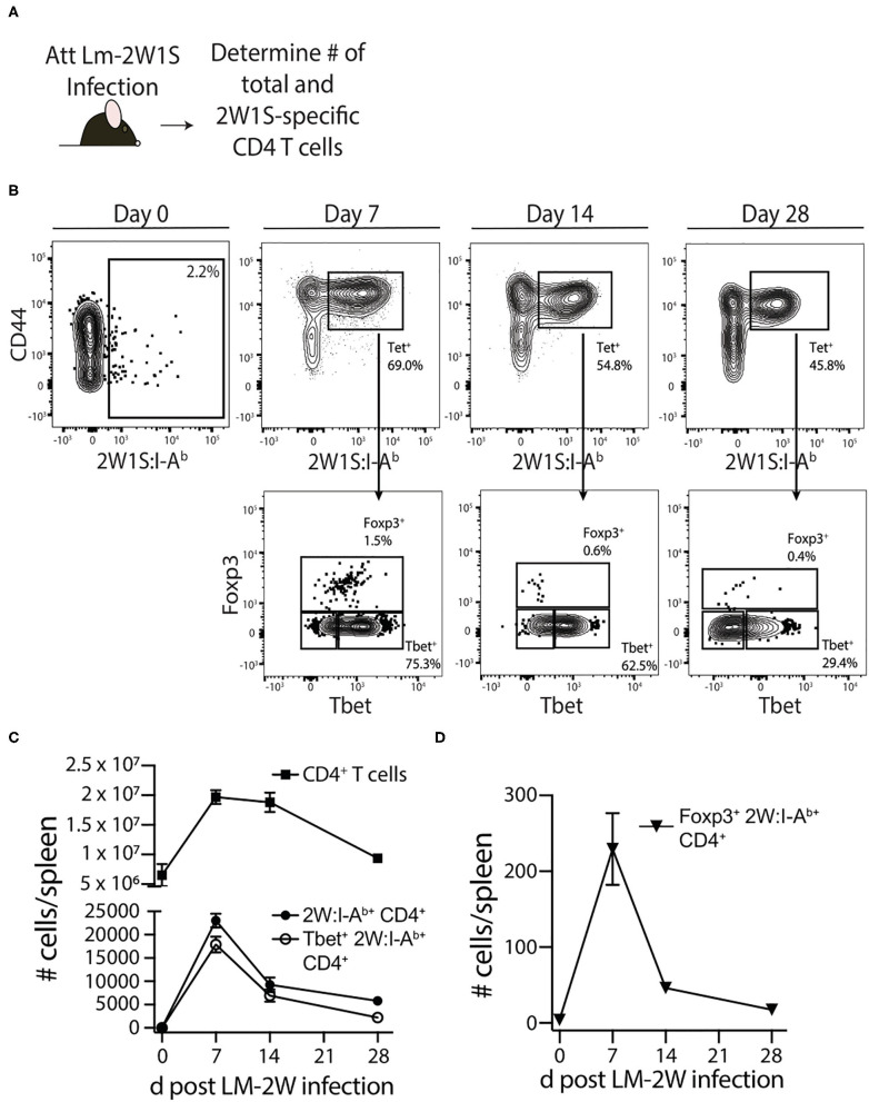
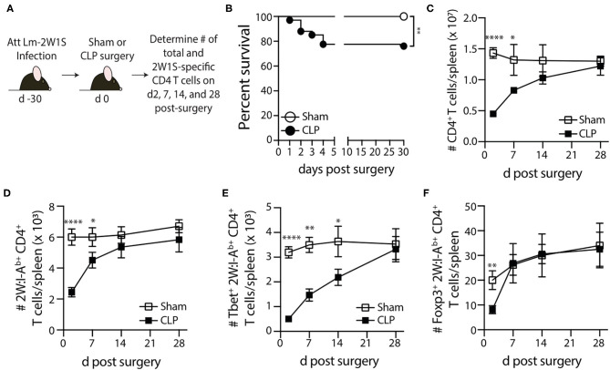
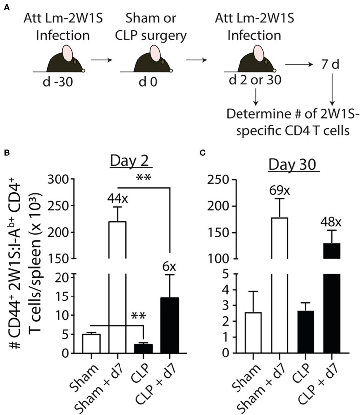
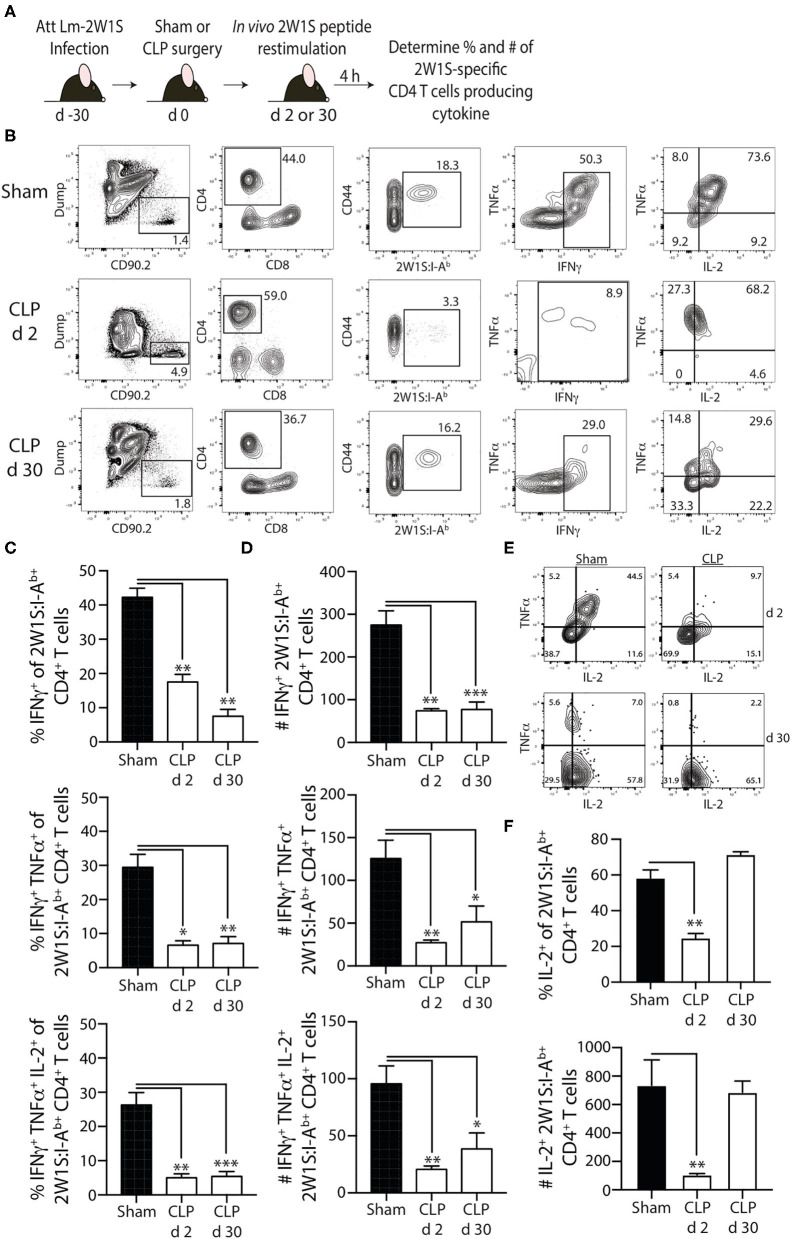
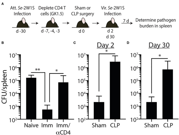
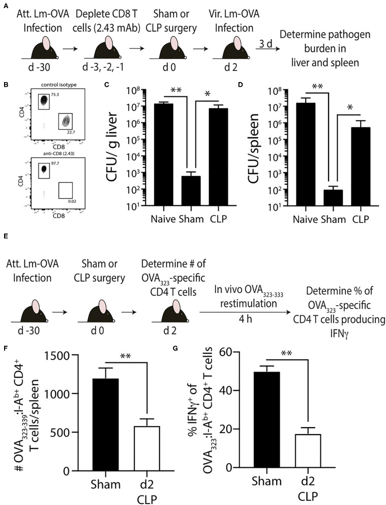
Patients who survive sepsis display prolonged immune dysfunction and heightened risk of secondary infection. CD4 T cells support a variety of cells required for protective immunity, and perturbations to the CD4 T cell compartment can decrease overall immune system fitness. Using the cecal ligation and puncture (CLP) mouse model of sepsis, we investigated the impact of sepsis on endogenous Ag-specific memory CD4 T cells generated in C57BL/6 (B6) mice infected with attenuated Listeria monocytogenes (Lm) expressing the I-Ab-restricted 2W1S epitope (Lm-2W). The number of 2W1S-specific memory CD4 T cells was significantly reduced on day 2 after sepsis induction, but recovered by day 14. In contrast to the transient numerical change, the 2W1S-specific memory CD4 T cells displayed prolonged functional impairment after sepsis, evidenced by a reduced recall response (proliferation and effector cytokine production) after restimulation with cognate Ag. To define the extent to which the observed functional impairments in the memory CD4 T cells impacts protection to secondary infection, B6 mice were infected with attenuated Salmonella enterica-2W (Se-2W) 30 days before sham or CLP surgery, and then challenged with virulent Se-2W after surgery. Pathogen burden was significantly higher in the CLP-treated mice compared to shams. Similar reductions in functional capacity and protection were noted for the endogenous OVA323-specific memory CD4 T cell population in sepsis survivors upon Lm-OVA challenge. Our data collectively show CLP-induced sepsis alters the number and function of Ag-specific memory CD4 T cells, which contributes (in part) to the characteristic long-lasting immunoparalysis seen after sepsis.

Keywords: CD4 T cells; IFN-gamma; immune suppression; memory; sepsis.


Figures

Figure 1
Figure 1
Generation of Ag-specific memory CD4 T cells following attenuated Listeria monocytogenes-2W1S infection. (A) Experimental design—B6 mice were immunized with attenuated Listeria monocytogenes-2W1S (107 CFU i.v.). The number of 2W1S-specific CD4 T cells were determined before and after infection. (B) Representative flow plots show the gating strategy used to identify 2W1S:I-Ab+ cells first identified as being CD3+ and CD4+. From the tetramer+ gate, cells expressing the transcription factors Tbet (Th1 phenotype) or Foxp3 (regulatory T cell phenotype) were then identified. Positive and negative gating determined using FMO controls. The number of (C) total CD4 T cells, 2W1S-specific CD4 T cells, Tbet+ 2W1S-specific CD4 T cells, and (D) Foxp3+ 2W1S-specific CD4 T cells in the spleen was determined 7, 14, and 28 days after attenuated LM-2W1S infection. Data shown are representative from 3 independent experiments, with at least 3 mice/group/time point in each experiment.
Figure 2
Figure 2
Loss and recovery of 2W1S-specific memory CD4 T cells following CLP-induced sepsis. (A) Experimental design—B6 mice were immunized with attenuated Listeria monocytogenes-2W1S (107 CFU i.v.) 30 days before sham or CLP surgery. The number of 2W1S-specific CD4 T cells were determined after surgery. (B) Survival of LM-2W-infected B6 mice after sham and CLP surgery (n = 29 sham; n = 67 CLP). The number of (C) total CD4 T cells and (D) 2W1S-specific CD4 T cells in the spleen was determined 2, 7, 14, and 28 days after sham or CLP surgery by flow cytometry. In addition, the 2W1S-specific CD4 T cells were subtyped based on (E) Tbet (Th1 phenotype) and (F) Foxp3 (regulatory T cell phenotype) expression. Data shown are cumulative from 3 independent experiments, with at least 3 mice/group/time point in each experiment. *p ≤ 0.05, **p ≤ 0.01, ***p ≤ 0.005, and ****p ≤ 0.001.
Figure 3
Figure 3
Sepsis induces transient loss in number of pre-existing “Ag-experienced” CD4 T cells in microbially-experienced “dirty” mice. (A) Experimental design—SPF B6 mice were cohoused with pet store mice for 60 days to permit microbe transfer and immune system maturation. (B) Age-matched SPF and cohoused (CoH) mice were bled prior to and at 2, 4, 6, and 8 weeks after cohousing to determine the frequency of CD44hi CD4 T cells. (C–G) Sham or CLP surgery was performed on cohoused B6 mice. The number of total, CD44hi, CD11a+CD49d+ “Ag-experienced,” and CD11aCD49d naive CD4 T cells in the spleen was determined 2 and 30 days post-surgery by flow cytometry. (E) Representative flow plots show gating strategy. Positive and negative gating determined using FMO controls. The number of (C) total, (D) CD11a+CD49d+, and (E) CD11aCD49d CD4 T cells was determined. Data shown are representative of 2 independent experiments, with at least 4 mice/group in each experiment. *p ≤ 0.05 and **p ≤ 0.01.
Figure 4
Figure 4
Sepsis impairs the recall response by pre-existing 2W1S-specific memory CD4 T cells to cognate Ag. (A) Experimental design—B6 mice were immunized with attenuated L. monocytogenes-2W1S (107 CFU i.v.) 30 days before sham or CLP surgery. The mice were given a second infection with attenuated L. monocytogenes-2W1S (107 CFU i.v.) (B) 2 or (C) 30 days after surgery. Total number of 2W1S-specific CD4 T cells in the spleen was determined the day of and 7 days after the second LM-2W1S infection (107 CFU i.v.). The fold increase in cell numbers at day 7 post-secondary infection is indicated. Data shown are representative of 2 independent experiments, with 4 mice/group in each experiment. **p ≤ 0.01.
Figure 5
Figure 5
Sepsis impairs the ability of 2W1S-specific memory CD4 T cells to produce effector cytokines after in vivo cognate Ag restimulation. (A) Experimental design—B6 mice were immunized with attenuated L. monocytogenes-2W1S (107 CFU i.v.) 30 days before sham or CLP surgery. Mice were injected with 2W1S peptide (100 μg i.v.) 2 or 30 days after surgery to restimulate the 2W1S-specific memory CD4 T cells. Spleens were harvested 4 h later, and the frequency and number of IFNγ+, IFNγ+TNFα+, and IFNγ+TNFα+IL-2+ 2W1S-specific CD4 T cells was determined by flow cytometry. (B) Representative flow plots of intracellular IFNγ, TNFα, and IL-2 detection in CD44hi2W1S:I-Ab+ CD4 T cells after in vivo peptide restimulation. Plots show cells gated from 2W:I-Ab-enriched CD4 T cells from sham- or CLP-treated mice. Positive and negative gating determined using FMO controls. Frequency (C) and number (D) of CD44hi2W1S:I-Ab+-specific CD4 T cells in the spleen producing IFNγ, IFNγ/TNFα, and IFNγ/TNFα/IL-2. (E) Representative flow plots of intracellular IL-2 detection in CD44hi2W1S:I-Ab+ CD4 T cells after in vivo peptide restimulation. Plots show cells gated from 2W:I-Ab-enriched CD4 T cells from sham- or CLP-treated mice. Positive and negative gating determined using FMO controls. (F) Frequency and number of CD44hi2W1S:I-Ab+-specific CD4 T cells in the spleen producing IL-2. Data shown are representative of 2 independent experiments, with at least 4 mice/group in each experiment. *p ≤ 0.05, **p ≤ 0.01, and ***p ≤ 0.005.
Figure 6
Figure 6
Impaired 2W1S-specific memory CD4 T cell-mediated immunity to secondary Salmonella-2W1S infection after CLP-induced sepsis. (A) Experimental design—B6 mice were immunized with attenuated Salmonella enterica strain BRD509-2W1S (Se-2W1S; AroA; 106 CFU i.v.) 30 d before sham or CLP surgery. (B) One group of mice (Imm/αCD4) was depleted of CD4 T cells by injecting anti-CD4 mAb GK1.5 i.v. (800 μg on day -7, and 400 μg on days −4 and −3) before second infection with virulent Salmonella-2W1S (103 CFU i.v.). Bacterial titers in the spleen were determined 7 days later. (C–D) In separate cohorts of attenuated Se-2W1S infected mice, sham or CLP surgery was performed. These mice were then challenged with virulent Se-2W1S (103 CFU i.v.) (C) 2 or (D) 30 days after surgery. Bacterial titers in the spleen were determined 7 days later. Data shown are representative of 3 independent experiments, with at least 5 mice/group in each experiment. *p ≤ 0.05, **p ≤ 0.01.
Figure 7
Figure 7
Effect of sepsis on OVA323−339-specific memory CD4 T cells. (A) Experimental design—B6 mice were infected with attenuated L. monocytogenes-OVA (LM-OVA; 107 CFU i.v.) 30 d before sham or CLP surgery. Mice in the naïve, sham, and CLP groups were depleted of CD8 T cells by injecting 100 μg anti-CD8 mAb (clone 2.43) i.v. 3, 2, and 1 days prior to surgery. (B) A small amount of blood was collected from the anti-CD8 mAb-treated mice on the day of surgery and staining for CD4 and CD8 T cells. Representative flow plots show the extent of CD8 T cell depletion compared to a reference mouse injected with a control isotype mAb. (C,D) The mice were infected with virulent LM-OVA (104 CFU i.v.) 2 days after surgery. Bacterial titers in the liver and spleen were determined 3 days post-vir LM-OVA infection. Data shown are representative of 2 independent experiments, with at least 5 mice/group in each experiment. *p ≤ 0.05 and **p < 0.01. (E) Experimental design—B6 mice were infected with attenuated L. monocytogenes-OVA (LM-OVA; 107 CFU i.v.) 30 d before sham or CLP surgery. (F) On day 2 post-surgery, the number of OVA323−339-specific memory CD4 T cells in the spleen were determined. (G) A separate cohort of mice were injected with OVA323−339 peptide (100 μg i.v.) 2 days after surgery to restimulate the OVA323−339-specific memory CD4 T cells. Spleens were harvested 4 h later, and the frequency of IFNγ+ OVA323−339-specific CD4 T cells was determined by flow cytometry. Data shown are representative of 2 independent experiments, with at least 5 mice/group in each experiment. **p < 0.01.

Similar articles

Cited by

References

    1. Shankar-Hari M, Phillips GS, Levy ML, Seymour CW, Liu VX, Deutschman CS, et al. Developing a new definition and assessing new clinical criteria for septic shock: for the third international consensus definitions for sepsis and septic shock (Sepsis-3). JAMA. (2016) 315:775–87. 10.1001/jama.2016.0289 - DOI - PMC - PubMed
    1. Brun-Buisson C, Doyon F, Carlet J, Dellamonica P, Gouin F, Lepoutre A, et al. . Incidence, risk factors, and outcome of severe sepsis and septic shock in adults. A multicenter prospective study in intensive care units French ICU Group for Severe Sepsis. JAMA. (1995) 274:968–74. 10.1001/jama.1995.03530120060042 - DOI - PubMed
    1. Levy MM, Fink MP, Marshall JC, Abraham E, Angus D, Cook D, et al. . 2001 SCCM/ESICM/ACCP/ATS/SIS international sepsis definitions conference. Intensive Care Med. (2001) 29:530–8. 10.1007/s00134-003-1662-x - DOI - PubMed
    1. Hotchkiss RS, Nicholson DW. Apoptosis and caspases regulate death and inflammation in sepsis. Nat Rev Immunol. (2006) 6:813–22. 10.1038/nri1943 - DOI - PubMed
    1. Rittirsch D, Flierl MA, Ward PA. Harmful molecular mechanisms in sepsis. Nat Rev Immunol. (2008) 8:776–87. 10.1038/nri2402 - DOI - PMC - PubMed

Publication types

MeSH terms