. 2020 Aug 13:11:927.
doi: 10.3389/fphys.2020.00927. eCollection 2020.

A Guard Cell Abscisic Acid (ABA) Network Model That Captures the Stomatal Resting State

Affiliations

A Guard Cell Abscisic Acid (ABA) Network Model That Captures the Stomatal Resting State

Parul Maheshwari et al. Front Physiol. .

Abstract

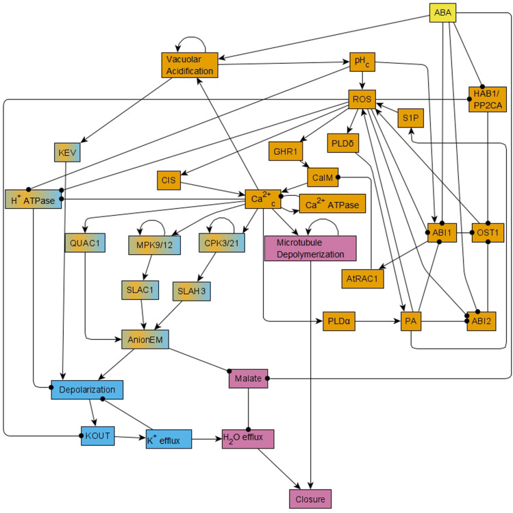
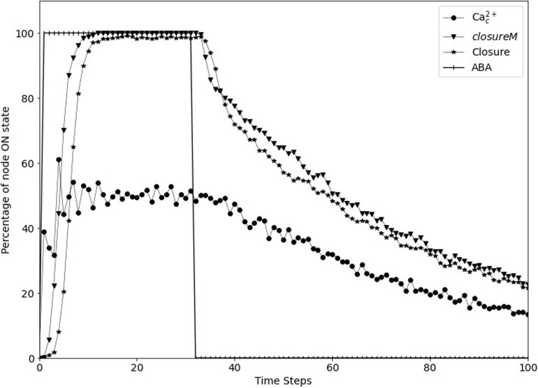
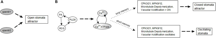
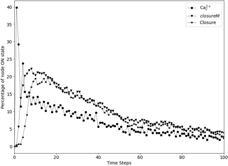
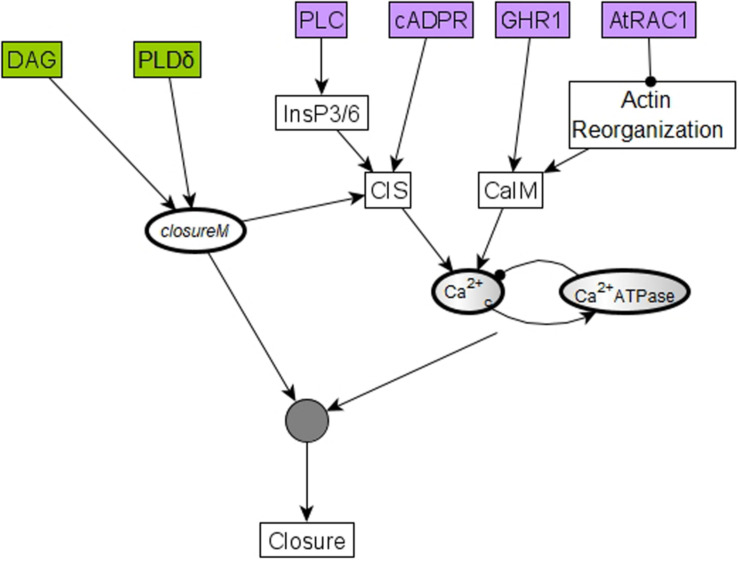
Stomatal pores play a central role in the control of carbon assimilation and plant water status. The guard cell pair that borders each pore integrates information from environmental and endogenous signals and accordingly swells or deflates, thereby increasing or decreasing the stomatal aperture. Prior research shows that there is a complex cellular network underlying this process. We have previously constructed a signal transduction network and a Boolean dynamic model describing stomatal closure in response to signals including the plant hormone abscisic acid (ABA), calcium or reactive oxygen species (ROS). Here, we improve the Boolean network model such that it captures the biologically expected response of the guard cell in the absence or following the removal of a closure-inducing signal such as ABA or external Ca2+. The expectation from the biological system is reversibility, i.e., the stomata should reopen after the closing signal is removed. We find that the model's reversibility is obstructed by the previously assumed persistent activity of four nodes. By introducing time-dependent Boolean functions for these nodes, the model recapitulates stomatal reopening following the removal of a signal. The previous version of the model predicts ∼20% closure in the absence of any signal due to uncertainty regarding the initial conditions of multiple network nodes. We systematically test and adjust these initial conditions to find the minimally restrictive combinations that appropriately result in open stomata in the absence of a closure signal. We support these results by an analysis of the successive stabilization of feedback motifs in the network, illuminating the system's dynamic progression toward the open or closed stomata state. This analysis particularly highlights the role of cytosolic calcium oscillations in causing and maintaining stomatal closure. Overall, we illustrate the strength of the Boolean network modeling framework to efficiently capture cellular phenotypes as emergent outcomes of intracellular biological processes.

Keywords: Boolean model; Boolean network; guard cell; memory; signal transduction; stomatal closure.


Figures

FIGURE 1
FIGURE 1
Simplified version of the network that forms the basis of the Boolean model of ABA induced stomatal closure. This network is reduced from the 49-node network in Maheshwari et al. (2019) using methods of causal logic reduction (Maheshwari and Albert, 2017) and binary transitive reduction (Albert et al., 2007); it preserves all the relationships among nodes of the 49-node network via edges or paths. Each edge that terminates in an arrow indicates an activating relationship and each edge that terminates in a black circle indicates an inhibitory relationship. This reduced network is presented here just for ease in visualization; all the analysis in this work was conducted on the 49-node network presented in Figure 1 of Maheshwari et al. (2019). This network contains positive feedback loops, i.e., cycles of directed edges that contain no or an even number of inhibitory edges, for example ROS – • ABI1 – • OST1→ ROS. It also contains negative feedback loops, i.e., cycles of directed edges that contain an odd number of inhibitory edges, for example Depolarization → KOUT → K+ efflux – • Depolarization. This network has two strongly connected components (SCCs) i.e., subnetworks in which every pair of nodes is connected by at least two paths of opposite direction. These two SCCs are represented in orange and light blue colors. Nodes of the orange SCC can reach the nodes of the light blue SCC via paths; the nodes linking these two SCCs are shown in a mix of orange and light blue colors. The signal ABA reaches all the nodes of the network. The out-component of this network leads from the two SCCs to the Closure node and is represented in pink color.
FIGURE 2
FIGURE 2
Stable and oscillating motifs observed in the absence of ABA. (A) Stable motif associated with open stomata attractors; we refer to this motif as openM1. (B) Stable motif associated with open stomata attractors; we refer to this motif as openM2. (C) Conditionally stable motif associated with the closed stomata attractor A0; we refer to this motif as closureM. The condition for this conditionally stable motif is the ON state of Vacuolar Acidification. In panels (A–C), the white background indicates the ON state of the node and the gray background means OFF state of the node. (D) Conditional oscillatory motif associated with closed stomata. This motif plays a role in both the presence and absence of ABA. The condition for the activation of this motif is the ON state of either of CIS or CaIM, and it yields a sustained oscillation of Ca2+c and Ca2+ ATPase (represented as a gray-white background). (E) Conditional oscillatory motif, K+ oscillation, that exists in the absence of any signal. The condition for the activation of this motif is KEV = ON. Since Vacuolar Acidification is sufficient for KEV, the ON state of Vacuolar Acidification is sufficient to establish this conditional oscillating motif, leading to sustained oscillation of its constituent nodes.
FIGURE 3
FIGURE 3
Motif succession diagrams of Model1 in the absence of ABA and any other closure signal. Stable motifs are shown with oval symbols and attractors are indicated by rectangles. Each dashed directed line between two motifs indicates that the system states in which the first motif has established and any nodes driven by it have stabilized admit the second motif as next to activate. The dashed directed line converging into an attractor symbol indicates that the succession of stable motifs ensures the system’s convergence into the respective attractor. (A) shows a subset of the succession diagram that converges to the attractor A0, corresponding to closure of the stomata in the absence of ABA (see Supplementary Table S4). The stable motif characteristic to this attractor is the conditionally stable motif closureM. (B,C) show a subset of the succession diagram corresponding to a sample of the 16 attractors that describe open stomata in the absence of ABA (see Supplementary Table S4). Each possible trajectory in this case contains exactly one of the stable motifs openM1 and openM2. (B) describes some of the trajectories containing openM1 while C describes some of the trajectories containing openM2. (A) also indicates the existence of bifurcations in the system’s trajectory due to the mutually exclusive motifs closureM and openM1. If closureM stabilizes the system converges into the attractor A0, and if openM1 stabilizes the system converges into attractor A1. The diagram encodes node states into the background color of the stable motif symbols. When referring to single nodes, white background indicates the ON state of the node, gray background means the OFF state of the node, and gray-white background represents oscillating nodes. Since in the openM1 and openM2 stable motifs all nodes are OFF (see Figure 2), we use a gray background color for these stable motifs. We use white background to represent the locking in of the closureM motif and represent the oscillating nature of the K+ oscillation motif by a gray-white background. The conditionally stable motifs are marked by thick boundaries.
FIGURE 4
FIGURE 4
Motif succession diagram of Model1 in the presence of a closure inducing signal. As in Figure 3, stable motifs are shown with oval symbols and attractors are indicated by rectangles. Each dashed directed line between two motifs indicates that the system states in which the first motif has established and any nodes driven by it have stabilized admit the second motif as next to activate. The dashed directed line converging into an attractor symbol indicates that the succession of stable motifs ensures the system’s convergence into the respective attractor. White background indicates the ON state of the corresponding node and white-gray background indicates oscillating nodes. (A) Motif succession diagram in the sustained presence of ABA when the first motif to stabilize is the PA = 1, PLDδ = 1, ROS = 1 stable motif. The top trajectory is the case when the oscillatory motif stabilizes into oscillations after the PA-PLDδ-ROS motif stabilizes; as a result of the Ca2+c – Ca2+ ATPase oscillations, the motifs CPK3/21 = 1, MPK9/12 = 1, and Microtubule Depolymerization = 1 stabilize directly and hence the system stabilizes in the closed stomata attractor. The middle and bottom trajectories represent the case when the second motif to stabilize is CPK3/21 = 1. The complete succession diagram covers all possible trajectories that start with stabilization of one or more of the four stable motifs (in any order), followed by the activation of the oscillatory motif, after which the system always stabilizes into the closed stomata attractor. (B) Motif succession diagram when external Ca2+ is simulated as the fixed ON state of the CaIM node when the first three motifs to stabilize are Vacuolar Acidification = ON, MPK9/12 = ON, and CPK3/21 = ON.
FIGURE 5
FIGURE 5
Simulated stomatal closure is maintained after removal of ABA. The state of the node ABA (vertical line symbols) is ON for 30 time-steps and is then OFF. The closureM conditionally stable motif (downward pointing triangles) establishes in less than 15 timesteps and remains stable despite the loss of ABA. The percentage of closure (star symbols) increases to 100% and stays at this value even after the signal is removed. The circles represent the percentage of the ON state of Ca2+c which after a fast increase fluctuates around 50% since Ca2+c oscillates with approximately equal ON and OFF time periods. The biological expectation is that after the signal is removed the states of ROS, Closure, and Ca2+c should go to OFF eventually.
FIGURE 6
FIGURE 6
Considering memory of the states of regulator nodes instead of persistent activity. (A) In Maheshwari et al. (2019), the nodes CPK3/21, MPK9/12, Vacuolar Acidification, and Microtubule Depolymerization have a persistence term in their update function to maintain them in a fixed ON state when Ca2+c oscillates. (B) Replacing persistent activity with short-term memory. The update function of each of the four nodes combines with an “OR” function the states of their respective regulator (Ca2+c, TCTP, or V-ATPase) at the current and previous two time-steps.
FIGURE 7
FIGURE 7
Stomatal re-opening after removal of the signal in the updated Model1. The signal, ABA (vertical lines) is set to fixed ON state for the first 30 time-steps and then set to fixed OFF state for 70 time-steps. The closureM conditionally stable motif (downward triangles) is stabilized within the first 10 time-steps and it slowly destabilizes after ABA is set to the OFF state. The state of the node Closure, depicted by the star symbols, shows a similar behavior i.e., it starts decreasing after the signal is removed. The state of the node Ca2+c, depicted by the circle symbols, is oscillating in the presence of ABA and it slowly transitions to the OFF state after ABA is fixed to OFF state.
FIGURE 8
FIGURE 8
Succession diagram for Model1 with short term memory. (A) Stable motif succession diagram in the absence of ABA and any other closure-inducing signal. Regardless of the duration of the memory, this case always leads to an open stomata attractor. (B) Motif succession diagram in the presence of ABA. The system can reach two different attractors depending on whether the memory duration is large enough. When the memory duration is large enough, sustained Ca2+c oscillations can sustain the ON state of the nodes CPK3/21, MPK9/12, Microtubule Depolymerization, and Vacuolar Acidification. With small memory duration these nodes oscillate instead of stabilizing; as a result, the node corresponding to stomatal closure also oscillates. See Supplementary Text S3 for details on the sufficient memory durations and Supplementary Table S4 for these attractors. The dashed edges denote logic succession with certainty while the dotted edges denote the variant outcomes depending on the memory duration.
FIGURE 9
FIGURE 9
Transient closure observed in Model1 in the absence of ABA when using short-term memory instead of node persistence. Due to the random initial conditions of the 17 nodes, the closureM motif temporarily stabilizes in ∼29% of the simulations (downward triangles) and there is a non-zero level of Ca2+c oscillations, which in turn lead to up to ∼20% transient closure. Eventually, the percentage of closure reduces to less than 5%.
FIGURE 10
FIGURE 10
The initial condition of six nodes can lead to temporary closure by helping establish Ca2+c oscillations and the closureM conditionally stable motif. Four of these six nodes, cADPR, GHR1, AtRAC1, and PLC, represented in purple, regulate one of the two processes (CIS or CaIM) that yield Ca2+c elevation. For example, the two-edge path between AtRAC1 and CaIM indicates that AtRAC1 inhibits the reorganization of the actin cytoskeleton that otherwise would contribute to Ca2+ influx (CaIM). Ca2+c elevation, in turn, has some probability of causing Ca2+c oscillations due to the negative feedback loop formed by Ca2+c and Ca2+c ATPase. These oscillations have a potential to drive the network to the closed stomata attractor. The remaining two nodes, PLDδ and DAG, represented in green, affect the closureM motif, indicated by the edges that start from PLDδ and DAG, respectively, and end in the node that stands for the closureM motif. Both nodes are direct regulators of PA, which is an internal driver of the motif, thus their activity has a chance of locking in the closureM motif. The stabilization of the closureM motif is sufficient for sustained Ca2+c oscillations, indicated by the path mediated by CIS. The cumulative effect of sustained Ca2+c oscillations and of the closureM motif leads to stomatal closure (see Section “Stable Motif Succession Diagrams of the Stomatal Closure Model Versions”). Hence, initiating any of these six nodes in their states corresponding to stomatal closure leads to a non-zero percentage of stomatal closure, at least transiently.

Similar articles

Cited by

References

    1. Ache P., Becker D., Ivashikina N., Dietrich P., Roelfsema M. R. G., Hedrich R. (2000). GORK, a delayed outward rectifier expressed in guard Cells of Arabidopsis thaliana, Is a K+-Selective, K+-Sensing ion channel. Febs Lett. 486 93–98. 10.1016/s0014-5793(00)02248-1 - DOI - PubMed
    1. Albert I., Thakar J., Li S., Zhang R., Albert R. (2008). Boolean network simulations for life scientists. Source Code Biol. Med. 3:16. - PMC - PubMed
    1. Albert R., Acharya B. R., Jeon B. W., Zañudo J. G., Zhu M., Osman K., et al. (2017). A new discrete dynamic model of ABA-induced stomatal closure predicts key feedback loops. PLoS Biol. 15:e2003451. 10.1371/journal.pbio.2003451 - DOI - PMC - PubMed
    1. Albert R., DasGupta B., Dondi R., Kachalo S., Sontag E., Zelikovsky A., et al. (2007). A novel method for signal transduction network inference from indirect experimental evidence. J. Comput. Biol. 14 927–949. 10.1089/cmb.2007.0015 - DOI - PubMed
    1. Allen G. J., Kuchitsu K., Chu S. P., Murata Y., Schroeder J. I. (1999). Arabidopsis ABI1-1 and ABI2-1 phosphatase mutations reduce abscisic acid–induced cytoplasmic calcium rises in guard cells. Plant Cell 11 1785–1798. 10.1105/tpc.11.9.1785 - DOI - PMC - PubMed