. 2020 Aug 13:11:1658.
doi: 10.3389/fimmu.2020.01658. eCollection 2020.

Conventional Dendritic Cells and Slan+ Monocytes During HIV-2 Infection

Affiliations

Conventional Dendritic Cells and Slan+ Monocytes During HIV-2 Infection

Marco Iannetta et al. Front Immunol. .

Abstract

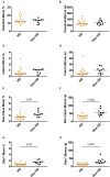
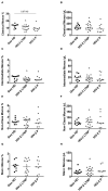
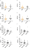
HIV-2 infection is characterized by low viremia and slow disease progression as compared to HIV-1 infection. Circulating CD14++CD16+ monocytes were found to accumulate and CD11c+ conventional dendritic cells (cDC) to be depleted in a Portuguese cohort of people living with HIV-2 (PLWHIV-2), compared to blood bank healthy donors (HD). We studied more precisely classical monocytes; CD16+ inflammatory (intermediate, non-classical and slan+ monocytes, known to accumulate during viremic HIV-1 infection); cDC1, important for cross-presentation, and cDC2, both depleted during HIV-1 infection. We analyzed by flow cytometry these PBMC subsets from Paris area residents: 29 asymptomatic, untreated PLWHIV-2 from the IMMUNOVIR-2 study, part of the ANRS-CO5 HIV-2 cohort: 19 long-term non-progressors (LTNP; infection ≥8 years, undetectable viral load, stable CD4 counts≥500/μL; 17 of West-African origin -WA), and 10 non-LTNP (P; progressive infection; 9 WA); and 30 age-and sex-matched controls: 16 blood bank HD with unknown geographical origin, and 10 HD of WA origin (GeoHD). We measured plasma bacterial translocation markers by ELISA. Non-classical monocyte counts were higher in GeoHD than in HD (54 vs. 32 cells/μL, p = 0.0002). Slan+ monocyte counts were twice as high in GeoHD than in HD (WA: 28 vs. 13 cells/μL, p = 0.0002). Thus cell counts were compared only between participants of WA origin. They were similar in LTNP, P and GeoHD, indicating that there were no HIV-2 related differences. cDC counts did not show major differences between the groups. Interestingly, inflammatory monocyte counts correlated with plasma sCD14 and LBP only in PLWHIV-2, especially LTNP, and not in GeoHD. In conclusion, in LTNP PLWHIV-2, inflammatory monocyte counts correlated with LBP or sCD14 plasma levels, indicating a potential innate immune response to subclinical bacterial translocation. As GeoHD had higher inflammatory monocyte counts than HD, our data also show that specific controls are important to refine innate immunity studies.

Keywords: HIV-2; cDC1; cDC2; controls; dendritic cells; monocytes; slan+ monocytes.


Figures

Figure 1
Figure 1
Monocyte subset percentages among CD45+ PBMC and absolute counts in controls from the blood bank (HD) compared to controls of West-African origin (GeoHD). (A) Classical monocyte (CD14++CD16) percentages among CD45+ PBMC and (B) absolute counts/μL blood, (C) intermediate monocyte CD14+CD16+ percentages and (D) absolute counts / μL blood, (E) non-classical monocyte (CD14±CD16+) percentages and (F) absolute counts, (G) slan+ monocyte percentages and (H) absolute counts, in the two groups of age- and sex- matched healthy donors residing in the Paris area, one from the French Blood Bank (EFS) (HD) and the other of West-African origin (GeoHD). Orange: unknown origin, Black: West-African origin. Horizontal bars represent median values. Mann-Whitney's post-test p are represented above the horizontal line connecting the compared groups. Kruskall-Wallis tests and descriptive statistics can be found in Table 2 (in italics for West-African participants).
Figure 2
Figure 2
Monocyte subset percentages among CD45+ PBMC and absolute counts in HIV-2-infected participants (LTNP and P) and uninfected controls (GeoHD), all of West African origin. (A) Classical monocyte (CD14++CD16) percentages among CD45+ PBMC and (B) absolute counts/μL blood, (C) intermediate monocyte CD14+CD16+ percentages and (D) absolute counts/μL blood, (E) non-classical monocyte (CD14±CD16+) percentages and (F) absolute counts, (G) slan+ monocyte percentages and (H) absolute counts, in GeoHD, HIV-2 LTNP and P groups, all of West-African origin. The data for the GeoHD group are the same as in Figure 1 Dunn's post-test p is represented above the horizontal line connecting the compared groups. Horizontal bars represent median values. Kruskall-Wallis tests and descriptive statistics can be found in Table 2, in italics.
Figure 3
Figure 3
Conventional cDC1 and cDC2 percentages among CD45+ PBMC and absolute counts. (A) Conventional DC1 percentages among CD45+ PBMC and (B) absolute counts/μL blood, (C) cDC2 percentages and (D) absolute counts/μL blood, in HD and GeoHD. Orange: unknown origin, Black: West-African origin. (E) cDC1 percentages and (F) absolute counts, (G) cDC2 percentages and (H) absolute counts, in GeoHD, HIV-2 LTNP and P, all of West-African origin. Horizontal bars represent median values. Mann-Whitney tests, Kruskall-Wallis tests and descriptive statistics can be found in Table 3 (in italics for West-African participants).
Figure 4
Figure 4
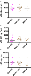
Plasma concentrations of sCD14, sCD163 and LBP in HIV-2-infected participants and uninfected controls (GeoHD), all of West African origin. Plasma concentrations of (A) sCD14, (B) sCD163, and (C) LBP. Horizontal bars represent median values. Kruskall-Wallis test and descriptive statistics can be found in Table 4.
Figure 5
Figure 5
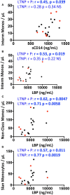
Correlations between monocyte counts and plasma concentrations of sCD14, sCD163, and LBP in HIV-2 infected participants and uninfected controls (GeoHD), all of West-African origin. Data from PLWHIV-2 from Figure 2 were correlated to data from Figure 4 by Spearman's test. The significant correlations are depicted. (A) Intermediate monocytes/μL vs. sCD14 plasma concentration. (B–D) Intermediate, non-classical or slan monocytes/μL vs. LBP. Correlations are given for all PLWHIV-2 (LTNP + P) or for LTNP alone. Progressor PLWHIV-2 (P) are indicated in red.

References

    1. Brun-Vezinet F, Rey MA, Katlama C, Girard PM, Roulot D, Yeni P, et al. . Lymphadenopathy-associated virus type 2 in AIDS and AIDS-related complex. Clinical and virological features in four patients. Lancet. (1987) 1:128–32. 10.1016/S0140-6736(87)91967-2 - DOI - PubMed
    1. Nyamweya S, Hegedus A, Jaye A, Rowland-Jones S, Flanagan KL, Macallan DC. Comparing HIV-1 and HIV-2 infection: lessons for viral immunopathogenesis. Rev Med Virol. (2013) 23:221–40. 10.1002/rmv.1739 - DOI - PubMed
    1. Tchounga B, Ekouevi DK, Balestre E, Dabis F. Mortality and survival patterns of people living with HIV-2. Curr Opin HIV AIDS. (2016) 11:537–44. 10.1097/COH.0000000000000299 - DOI - PMC - PubMed
    1. Thiebaut R, Matheron S, Taieb A, Brun-Vezinet F, Chene G, Autran B, et al. . Long-term nonprogressors and elite controllers in the ANRS CO5 HIV-2 cohort. AIDS. (2011) 25:865–7. 10.1097/QAD.0b013e328344892e - DOI - PubMed
    1. Esbjornsson J, Mansson F, Kvist A, da Silva ZJ, Andersson S, Fenyo EM, et al. . Long-term follow-up of HIV-2-related AIDS and mortality in Guinea-Bissau: a prospective open cohort study. Lancet HIV. (2018). 10.1016/S2352-3018(18)30254-6 - DOI - PubMed

Publication types

MeSH terms