

Frequency-Dependent Changes in Interhemispheric Functional Connectivity Measured by Resting-State fMRI in Children With Idiopathic Generalized Epilepsy
- PMID: 32903710
- PMCID: PMC7438858
- DOI: 10.3389/fneur.2020.00645
Frequency-Dependent Changes in Interhemispheric Functional Connectivity Measured by Resting-State fMRI in Children With Idiopathic Generalized Epilepsy
Abstract
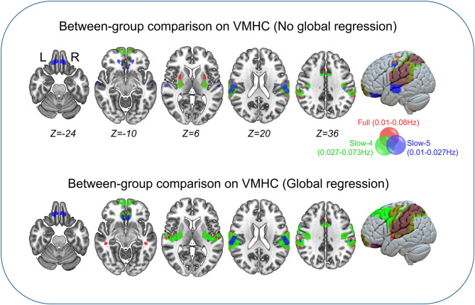
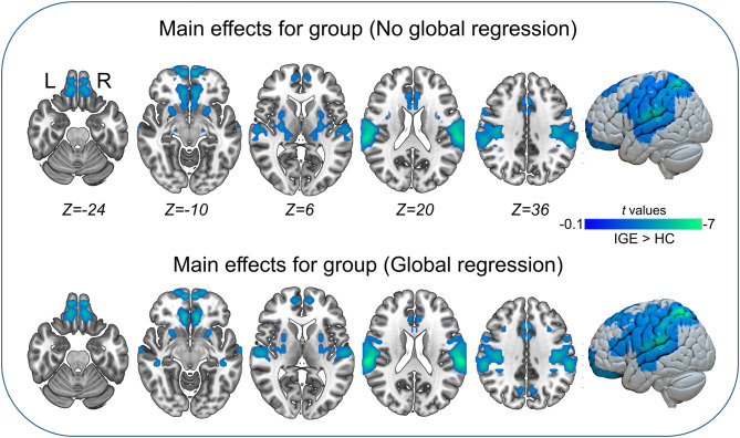
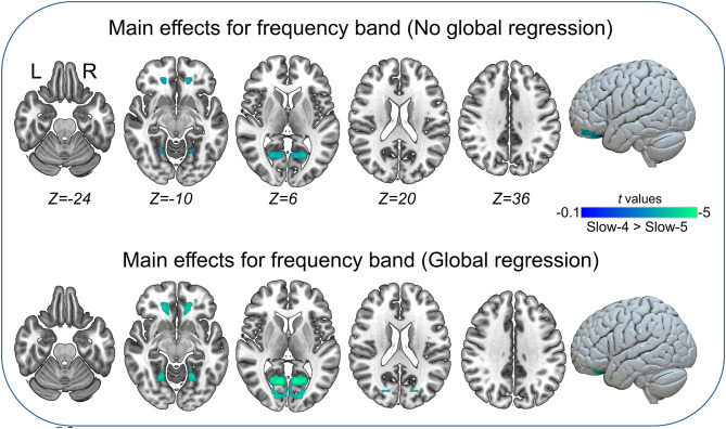
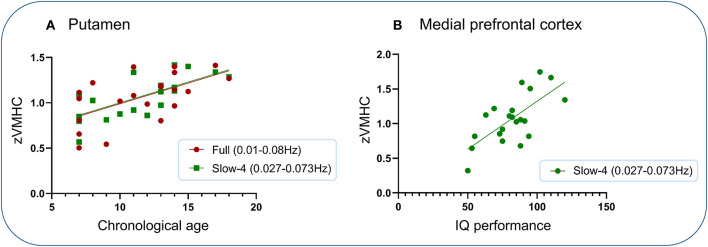
Epilepsy is associated with abnormal spatiotemporal changes in resting-state brain connectivity, but how these changes are characterized in interhemispheric coupling remains unclear. This study aimed to characterize frequency-dependent alterations in voxel-wise mirrored homotopic connectivity (VMHC) measured by resting-state functional magnetic resonance imaging (rs-fMRI) in children with idiopathic generalized epilepsy (IGE). Rs-fMRI data were collected in 21 children with IGE and 22 demographically matched children with typical development. We used three resting-state frequency bands (full, 0.01-0.08 Hz; slow-4, 0.027-0.073 Hz; slow-5, 0.01-0.027 Hz) to compute VMHC and locate the significant foci. Voxel-wise p <0.001 and cluster-level p <0.05 cluster-level family-wise error correction was applied. In between-group comparisons, we identified that the full and higher frequency (slow-4) bands showed similar reductions in VMHC including Rolandic operculum, putamen, superior frontal, lateral parietal, middle cingulate, and precuneus in children with IGE. In the lower frequency band (slow-5), we identified specific reductions in VMHC in orbitofrontal and middle temporal gyri in children with IGE. Further analyses on main effects and interaction between group and frequency band suggested significant frequency-dependent changes in VMHC, and no significant interaction was found. The results were generally similar with global brain signal regression. Additional association analysis showed that VMHC in the putamen within the full and slow-4 bands was significantly positively correlated with chronological age in children with IGE, and the same analysis was non-significant in the controls; VMHC in the medial prefrontal region in the slow-4 band was significantly positively correlated with IQ performance sub-score. Our findings suggest that IGE children show frequency-dependent changes in interhemispheric integration that spans regions and systems involving cortical-subcortical, language, and visuomotor processing. Decreased functional coupling within the dorsal striatum may reflect atypical development in children with IGE.
Keywords: IGE; fMRI; frequency-dependent; functional connectivity; interhemispheric.
Copyright © 2020 Jiang, Ma, Li, Luo, Zhang, Wang and Zhang.
Figures
Similar articles
-
Increased interhemispheric resting-state in idiopathic generalized epilepsy with generalized tonic-clonic seizures: a resting-state fMRI study.Epilepsy Res. 2014 Oct;108(8):1299-305. doi: 10.1016/j.eplepsyres.2014.06.010. Epub 2014 Jul 7. Epilepsy Res. 2014. PMID: 25043752
-
Decreased resting-state interhemispheric coordination in first-episode, drug-naive paranoid schizophrenia.Prog Neuropsychopharmacol Biol Psychiatry. 2014 Jan 3;48:14-9. doi: 10.1016/j.pnpbp.2013.09.012. Epub 2013 Sep 27. Prog Neuropsychopharmacol Biol Psychiatry. 2014. PMID: 24075897
-
Frequency-Specific Abnormalities Of Functional Homotopy In Alcohol Dependence: A Resting-State Functional Magnetic Resonance Imaging Study.Neuropsychiatr Dis Treat. 2019 Nov 19;15:3231-3245. doi: 10.2147/NDT.S221010. eCollection 2019. Neuropsychiatr Dis Treat. 2019. PMID: 31819451 Free PMC article.
-
Interhemispheric resting-state functional connectivity abnormalities in type 2 diabetes patients.Ann Palliat Med. 2021 Jul;10(7):8123-8133. doi: 10.21037/apm-21-1655. Ann Palliat Med. 2021. PMID: 34353097
-
Alternations of interhemispheric functional connectivity in patients with acute acquired concomitant esotropia: a resting state fMRI study using voxel-mirrored homotopic connectivity.Front Neurosci. 2025 Jan 6;18:1515675. doi: 10.3389/fnins.2024.1515675. eCollection 2024. Front Neurosci. 2025. PMID: 39834696 Free PMC article.
Cited by
-
Well-Being Is Associated With Local to Remote Cortical Connectivity.Front Behav Neurosci. 2022 Mar 11;16:737121. doi: 10.3389/fnbeh.2022.737121. eCollection 2022. Front Behav Neurosci. 2022. PMID: 35368310 Free PMC article.
-
Frequency-dependent alterations in functional connectivity in patients with Alzheimer's Disease spectrum disorders.Front Aging Neurosci. 2024 Mar 28;16:1375836. doi: 10.3389/fnagi.2024.1375836. eCollection 2024. Front Aging Neurosci. 2024. PMID: 38605859 Free PMC article.
-
Altered neurovascular coupling in children with idiopathic generalized epilepsy.CNS Neurosci Ther. 2023 Feb;29(2):609-618. doi: 10.1111/cns.14039. Epub 2022 Dec 8. CNS Neurosci Ther. 2023. PMID: 36480481 Free PMC article.
-
MOGAD-related epilepsy: a systematic characterization of age-dependent clinical, fluid, imaging and neurophysiological features.J Neurol. 2025 Jul 14;272(8):508. doi: 10.1007/s00415-025-13245-3. J Neurol. 2025. PMID: 40659898 Review.
References
-
- Sone D, Watanabe M, Ota M, Kimura Y, Sugiyama A, Maekawa T, et al. . Thalamic hypoperfusion and disrupted cerebral blood flow networks in idiopathic generalized epilepsy: arterial spin labeling and graph theoretical analysis. Epilepsy Res. (2017) 129:95–100. 10.1016/j.eplepsyres.2016.12.009 - DOI - PubMed
-
- Yacubian EMT, de Araújo Filho GM. Management issues for patients with idiopathic generalized epilepsies. Epileptology. (2013) 1:1–10. 10.1016/j.epilep.2012.11.001 - DOI
-
- Global Accelerated Action for the Health of Adolescents (AA-HA!): Guidance to Support Country Implementation (2017). Available online at: http://apps.who.int/iris/bitstream/10665/255415/1/9789241512343-eng.pdf?... (accessed October 16, 2019).
LinkOut - more resources
Full Text Sources