Review
. 2020 Aug 13:7:139.
doi: 10.3389/fcvm.2020.00139. eCollection 2020.

Transcatheter Tricuspid Valve Intervention: Coaptation Devices

Affiliations
Review

Transcatheter Tricuspid Valve Intervention: Coaptation Devices

Guillem Muntané-Carol et al. Front Cardiovasc Med. .

Abstract

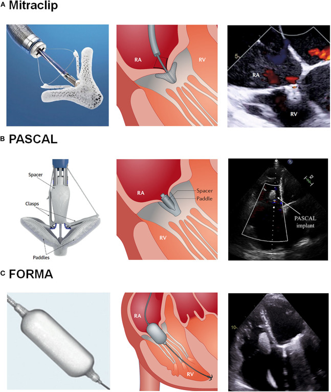
Transcatheter tricuspid valve intervention (TTVI) has recently emerged as an alternative for the treatment of severe tricuspid regurgitation (TR). Multiple percutaneous devices have been developed in the last decade with promising early results. Among them, the coaptation devices are designed to reduce TR severity by valve leaflet plication or occupying the regurgitant orifice with a spacer. To date, the MitraClip/TriClip devices (Abbott, Santa Clara, CA, USA), the PASCAL system (Edwards Lifesciencies, Irvine, CA, USA), and the FORMA device (Edwards Lifesciencies, Irvine, CA, USA) have been used as coaptation devices for treating severe TR. The present document aimed to review the clinical evidence on coaptation devices in the field of TTVI, describing its design characteristics, main procedural steps, and early and mid-term outcomes.

Keywords: FORMA; MitraClip device; PASCAL; transcatheter tricuspid intervention; tricuspid regurgitation (TR).


Figures

Figure 1
Figure 1
Transcatheter Tricuspid Valve Intervention: Coaptation Devices. (A) Illustration of the Mitraclip system and echocardiography after grasping. (B) Illustration of the Pascal system and echocardiography after grasping. (C) Illustration of the Mitraclip system and echocardiography after device implantation. RA, Right atrium; RV, Right ventricle. Reproduced with permission from Asmarats et al. (1) and Fam et al. (6) and modified for the authors.
Figure 2
Figure 2
TEE protocol to patient selection for MitraClip/TriClip procedures. (A) Transgastric basal short-axis view. ATL, anterior tricuspid leaflet; PPM, posterior papillary muscle; PTL, posterior tricuspid leaflet; RA, right atrium; RV, right ventricle; STL, septal tricuspid leaflet; TV, tricuspid valve. (B) Mid-esophageal TEE view. The left view is orientated along the anteroseptal coaptation line usually at 80–90°. The right image is obtained by a sweep from the aorta to the valve center. Ao, aorta; ATL, anterior tricuspid leaflet; RA, right atrium; RV, right ventricle; STL, septal tricuspid leaflet; TV, tricuspid valve. (C) 3D-TEE imaging view. Ao, aorta; ATL, anterior tricuspid leaflet; PTL, posterior tricuspid leaflet; STL, septal tricuspid leaflet. Reproduced with permission from Hausleiter et al. (9).
Figure 3
Figure 3
Intraprocedural main fluoroscopic views in transcatheter tricuspid valve intervention using the Mitraclip system. (A–D) Left anterior oblique projection. Once the CDS is straddled, turning the SGC clockwise steers away the MitraClip from the interatrial septum toward the TV. In that case, the position of the prior MitraClip placed at the TV can serve as a reference. (E,F) Right anterior oblique projection. Steering of the MitraClip toward the TV. The existing MitraClip is marked with white arrows. RAO, Right anterior oblique; LAO, Left anterior oblique. Reproduced with permission from Tang (21).
Figure 4
Figure 4
Intraprocedural fluoroscopic images in transcatheter tricuspid valve intervention using the FORMA system. (A) Right ventriculography to locate the tricuspid annular plane and identify the ideal anchor location (red asterisk) on fluoroscopy. (B) Right ventricular anchoring via a steerable delivery catheter. (C) Device positioning in the tricuspid annular plane. (D) Final right ventriculography showing the device in correct position (red arrow) and reduction of tricuspid regurgitation from baseline. Reproduced with permission from Campelo-Parada et al. (33).

References

    1. Asmarats L, Taramasso M, Rodés-Cabau J. Tricuspid valve disease: diagnosis, prognosis and management of a rapidly evolving field. Nat Rev Cardiol. (2019) 16:538–54. 10.1038/s41569-019-0186-1 - DOI - PubMed
    1. Alqahtani F, Berzingi CO, Aljohani S, Hijazi M, Al-Hallak A, Alkhouli M. Contemporary trends in the use and outcomes of surgical treatment of tricuspid regurgitation. J Am Heart Assoc. (2017) 6:e007597. 10.1161/JAHA.117.007597 - DOI - PMC - PubMed
    1. Lau GT, Tan HC, Kritharides L. Type of liver dysfunction in heart failure and its relation to the severity of tricuspid regurgitation. Am J Cardiol. (2002) 90:1405–9. 10.1016/S0002-9149(02)02886-2 - DOI - PubMed
    1. Maeder MT, Holst DP, Kaye DM. Tricuspid regurgitation contributes to renal dysfunction in patients with heart failure. J Cardiac Fail. (2008) 14:824–30. 10.1016/j.cardfail.2008.07.236 - DOI - PubMed
    1. Asmarats L, Puri R, Latib A, Navia JL, Rodés-Cabau J. Transcatheter tricuspid valve interventions: landscape, challenges, and future directions. J Am Coll Cardiol. (2018) 71:2935–56. 10.1016/j.jacc.2018.04.031 - DOI - PubMed

LinkOut - more resources