. 2020 Aug 13:8:681.
doi: 10.3389/fcell.2020.00681. eCollection 2020.

Reg-1α Promotes Differentiation of Cortical Progenitors via Its N-Terminal Active Domain

Affiliations

Reg-1α Promotes Differentiation of Cortical Progenitors via Its N-Terminal Active Domain

Marjorie Varilh et al. Front Cell Dev Biol. .

Abstract

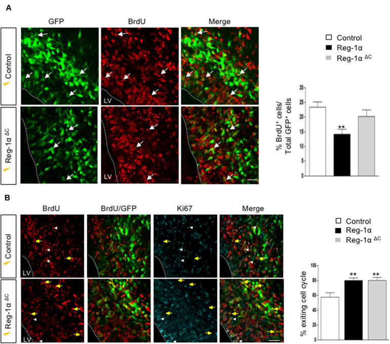
Reg-1α belongs to the Reg family of small, secreted proteins expressed in both pancreas and nervous system. Reg-1α is composed of two domains, an insoluble C-type lectin domain and a short soluble N-terminal peptide, which is released from the molecule upon proteolytic N-terminal processing, although the biological significance of this proteolysis remains unclear. We have previously shown that binding of Reg-1α to its receptor Extl3 stimulates axonal outgrowth. Reg-1α and Extl3 genes are expressed in the developing cortex but their expression decreases in adulthood, pointing to a possible function of this signaling system at the early developmental stages. Here, we demonstrate that recombinant Reg-1α increases migration and differentiation of cultured embryonic rat telencephalic progenitors via the activation of GSK-3β activity. In vivo overexpression of Reg-1α by in utero electroporation, has a similar effect, favoring premature differentiation of cortical progenitors. Notably, the N-terminal soluble domain, but not the C-type lectin domain, is largely responsible for Reg-1α effects on cortical neuronal differentiation. We thus conclude that Reg-1α via its proteolytically generated N-terminal domain is required for basic development processes.

Keywords: Extl3; GSK-3β; Reg-1α; axon elongation; differentiation; neural progenitors.


Figures

FIGURE 1
FIGURE 1
Age-dependent expression of Reg-1α protein and Extl3 in brain tissues. (A) Levels of Reg-1α protein were determined using western blotting of cortical tissue extracts prepared from rats of the following ages: E13.5 and E17.5 (n > 10 per group), P1, P15, and P30 (n > 4 per group) and 2, 10 months-old (2M, 10M) (n > 3 per group). Extract of ileum from 2 months-old animals was used as positive control. Relative Reg-1α protein levels were normalized with β-actin values and plotted according to the age group. (B) qPCR analysis of Reg-1α and Extl3 gene expression in brain homogenates from the same developmental stages used above. Values were normalized to cyclophilin expression. (A,B) Bars represent the mean from independent embryos per group. One-way ANOVA followed by Bonferroni’s multiple comparison test. Statistical significance: *p < 0.05, **p < 0.01, ***p < 0.001. (C) Coronal cryosections of E17.5 cortex immunostained for Reg-1α, NeuN and Calbindin. Note that Reg-1α colocalizes with NeuN in the dorso-lateral (a, boxed area 1) and ventral cortex (a, boxed area 2) and with Calbindin in interneurons of the lateral and piriform cortex (a, boxed areas 3–4). High magnification pictures in (b,c) correspond to the boxed areas in (a). (D) Double immunostaining for Reg-1α/NeuN in the parietal cortex of 2 months-old rats shows double-positive cells scattered through all cortical layers (a, boxed areas 5-6-7). High magnification of the boxed areas are illustrated in (b). Cx, cortex; Lge, Lateral ganglionic eminence. Scale bars, 80 μm.
FIGURE 2
FIGURE 2
Neurospheres of telencephalic embryonic precursor cells express Reg-1α and Extl3. (A) Bright field images of neurospheres at 2 and 48 h of culture on non-adherent and adherent substrate. The graph shows the quantification of neurosphere size (pixel areas). Scale bar, 100 μm. (B) Adherent neurospheres after 48 h of culture immunostained for β3-Tubulin and Nestin. β3-Tubulin-positive differentiated cells are predominant outside the neurosphere whereas Nestin-positive cells localize inside and at the edge of the neurosphere. Scale bar, 50 μm. (C) RT-qPCR analysis of Reg-1α and Extl3 mRNA expression in neurospheres grown for 48 h on an adherent substrate (left panel). Values were normalized to those of cyclophilin expression. Western blots of cellular extracts and culture medium from neurospheres grown on an adherent substrate for 48 h (right panel). Reg-1α and Extl3 are expressed in the cells. Reg-1α is secreted in the medium. (D) Immunofluorescence analysis of Reg-1α, Nestin, and β3-Tubulin expression in neurospheres. Merged images show that Reg-1α localizes to Nestin-positive progenitors and in β3-Tubulin-positive post-mitotic neurons in both cell bodies and neuritic extensions. Scale bar, 10 μM.
FIGURE 3
FIGURE 3
Reg-1α induces differentiation, migration and processes elongation growth of telencephalic precursor cells. (A) Neurospheres grown on an adherent substrate in proliferation medium in absence (Co) or presence of 10– 7 M recombinant Reg-1α for 48 h (Reg-1α). DAPI and β3-Tubulin immunostaining shows the effect of Reg-1α. Scale bar, 50 μM. (B) Quantification of neurosphere size (a), number of migrated cells (b), cell distance from the neurosphere edge (c), β3-Tubulin staining intensity outside the neurosphere (d) and length of the longest neurite in β3-Tubulin-positive cells (e) in the two experimental conditions. Reg-1α treatment promotes cell differentiation of neurospheres. (C) Dissociated neural precursor cells immunostained for β3-Tubulin in presence or absence of 10– 7M recombinant Reg-1α. Scale bar, 25 μM. (D) Reg-1α significantly increases the length of the longest neurite (a) and the total neuritic length per cell (b) but not the number of neurites per cell (c). Results represent three independent experiments Student’s t-test (mean ± SEM) *p < 0.05; **p < 0.01; ***p < 0.001.
FIGURE 4
FIGURE 4
Treatment with anti-Reg-1α antibodies neutralizes the effect of Reg-1α. (A,B) Quantitative analysis of the neutralizing effect of anti-Reg-1α polyclonal antibody on the differentiative activity of Reg-1α on neurosphere cultures. Neurosphere size (Aa), the number of migrating cells (Ab), the distance from the neurosphere edge (Ac) and the length of the longest neurite (Bd) were measured. Flow cytometry analysis of the number of β3-Tubulin-positive cells in neurosphere grown in control conditions (proliferation medium alone, white bars), or in the presence of Reg-1α (10–7 M; black bars) or recombinant Reg-1α (10–7 M) + anti-Reg-1α antibody (10–6 M; gray bars) (Be). The histograms show the quantification of three independent experiments (mean ± SEM). p < 0.05; ∗∗∗p < 0.001 relative to control and ##p < 0.01; ###p < 0.001 relative to Reg-1α treated cells based on the Student’s t-test. #p ¡ 0.05.
FIGURE 5
FIGURE 5
Reg-1α leads to changes in GSK-3β phosphorylation and β-catenin subcellular localization. Western blot assays of the levels of pSer9GSK-3β (A), pY216GSK-3β (B), total GSK-3β, and cytosolic β-catenin (C) from homogenates of neurospheres grown in proliferation medium alone (Co, white bars) or with the addition of Reg-1α (10–7 M; black bars). Note that Reg-1α enhances pSer9GSK-3β/GSK-3β total and decreases pY216GSK-3β/GSK-3β total ratios. Student’s t-test p < 0.05; ∗∗p < 0.01.
FIGURE 6
FIGURE 6
In vivo overexpression of Reg-1α induces a premature cortical migration. (A) Schematic representation of the experimental procedures used to electroporate in utero constructs encoding human or mouse Reg-1α cDNAs in E14.5 telencephalon. A GFP expressing plasmid was co-electroporated as tracer. Embryos were analyzed at E17.5. (B) Frontal sections of the telencephalon immunostained for GFP and Tbr1, a differentiation marker. Sections were counterstained with DAPI. The number of GFP+ cells present in the different layers were quantified and plotted against the total number of GFP+ present in the entire thickness of the section (graphs on the right). Bars represent the mean from a minimum of 3 independent embryos. Mann–Whitney test; *p < 0.05, **p < 0.01. Scale bar, 100 μm. (C) Representative confocal images of GFP+ cells in the CP after Reg-1α transfected neural cells. Sections were immunostained for Reg-1α. Note the presence of the protein on the neuronal surface. LV, lateral ventricle, CP, cortical plate, VZ/IZ, ventricular zone/intermediate zone. White lines indicate boundaries of the VZ or the meninges (mn). Scale bars, 10 μm.
FIGURE 7
FIGURE 7
Reg-1α induces telencephalic progenitors to exit the cell cycle. (A) Timeline of human Reg-1α/GFP in utero electroporation and BrdU labeling of embryonic telencephalon. (B) Frontal sections of harvested brains immunostained for BrdU and GFP (arrows). The graph on the right shows the percentage of BrdU/GFP+ over the total number of GFP+ cells in the VZ/IZ. Reg-1α significantly decreases the number of BrdU+/GFP+ cellsScale bar, 50 μm. (C) Frontal sections of E16.5 embryos immunostained for BrdU and the cell-cycle marker Ki67. GFP/BrdU/Ki67 positive cells (white arrowheads) are actively cycling. GFP/BrdU positive cells (yellow arrows) have exited the cell cycle. The graph on the right shows the percentage of cells exiting the cell cycle, measured as the percentage of the GFP+BrdU+KI67- over the total number of GFP+BrdU+ cells. Bars represent the mean from a minimum of three independent embryos. Mann–Whitney test; **p < 0.01, ***p < 0.001. LV, lateral ventricle. Scale bar, 80 μm.
FIGURE 8
FIGURE 8
The N-terminal domain of Reg-1α mimics the effect of the full length protein. (A) Schematic representation of the different deleted versions of Reg-1α. (B) Confocal images of frontal sections from E17.5 embryos electroporated with GFP, and the deleted versions of Reg-1α as indicated in the panels. Sections were immunostained for GFP and Tbr1 and counterstained with DAPI. Quantification of GFP+ cells in the VZ/IZ and CP in the different conditions is plotted in the graphs on the right. Each bar represents the mean from at least three independent embryos. Mann–Whitney test (*p < 0.05, versus the control group). LV, lateral ventricle, CP, cortical plate, VZ/IZ, ventricular zone/intermediate zone. Dotted white lines indicate VZ or meninge (mn) boundaries. Scale bar, 100 μm.
FIGURE 9
FIGURE 9
Reg-1αΔ C promotes exit from the cell cycle. (A) Confocal images of frontal sections from E16.5 cortex electroporated at E14.5 with a Reg-1αΔ C (n = 4) or Reg-1α (n = 3) and GFP (n = 5) plasmids. 24 h later, BrdU was injected intraperitoneally at E15.5. Sections were double labeled for BrdU/GFP. The graph shows the percentage of BrdU/GFP+ cells in the VZ/IZ. Scale bar, 50 μm. (B) Confocal images of frontal sections immunostained for GFP, BrdU and Ki67. Both Reg-1α and Reg-1αΔ C increase the percentage of GFP + BrdU + KI67–/GFP + BrdU+. Bars represent the mean from 3 independent embryos. ANOVA followed by Dunnett’s multiple comparison test (**p < 0.01 versus the control group). LV, lateral ventricle. Scale bar, 80 μm.

Similar articles

Cited by

References

    1. Acquatella-Tran Van Ba I., Marchal S., François F., Silhol M., Lleres C., Michel B., et al. (2012). Regenerating islet-derived 1α (Reg-1α) protein is new neuronal secreted factor that stimulates neurite outgrowth via exostosin Tumor-like 3 (EXTL3) receptor. J. Biol. Chem. 287 4726–4739. 10.1074/jbc.m111.260349 - DOI - PMC - PubMed
    1. Armando S., Lebrun A., Hugnot J.-P., Ripoll C., Saunier M., Simonneau L. (2007). Neurosphere-derived neural cells show region-specific behaviour in vitro. Neuroreport 18 1539–1542. 10.1097/wnr.0b013e3282f03d54 - DOI - PubMed
    1. Cavard C., Terris B., Grimber G., Christa L., Audard V., Radenen-Bussiere B., et al. (2006). Overexpression of regenerating islet-derived 1 alpha and 3 alpha genes in human primary liver tumors with beta-catenin mutations. Oncogene 25 599–608. 10.1038/sj.onc.1208860 - DOI - PubMed
    1. Cerini C., Peyrot V., Garnier C., Duplan L., Veesler S., Caer J.-P. L., et al. (1999). Biophysical characterization of lithostathine evidences for A polymeric structure at physiological Ph and A proteolysis mechanism leading to the formation of fibrils. J. Biol. Chem. 274 22266–22274. 10.1074/jbc.274.32.22266 - DOI - PubMed
    1. Chen Z., Downing S., Tzanakakis E. S. (2019). Four decades after the discovery of regenerating islet-derived (Reg) proteins: current understanding and challenges. Front. Cell Dev. Biol. 7:235. 10.3389/fcell.2019.00235 - DOI - PMC - PubMed