

MiR-3194-3p Inhibits Breast Cancer Progression by Targeting Aquaporin1
- PMID: 32903818
- PMCID: PMC7438898
- DOI: 10.3389/fonc.2020.01513
MiR-3194-3p Inhibits Breast Cancer Progression by Targeting Aquaporin1
Abstract
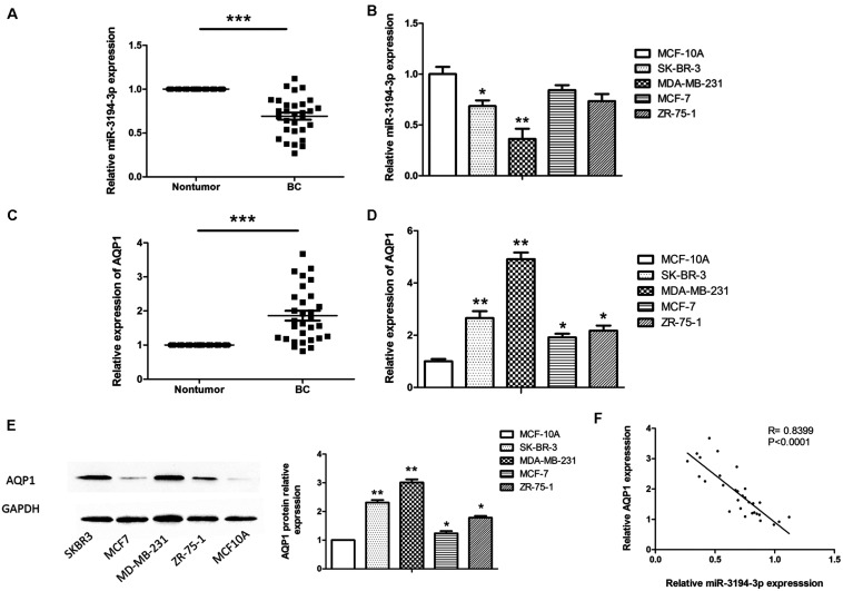
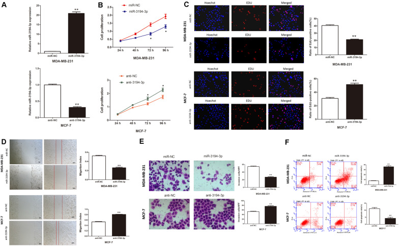
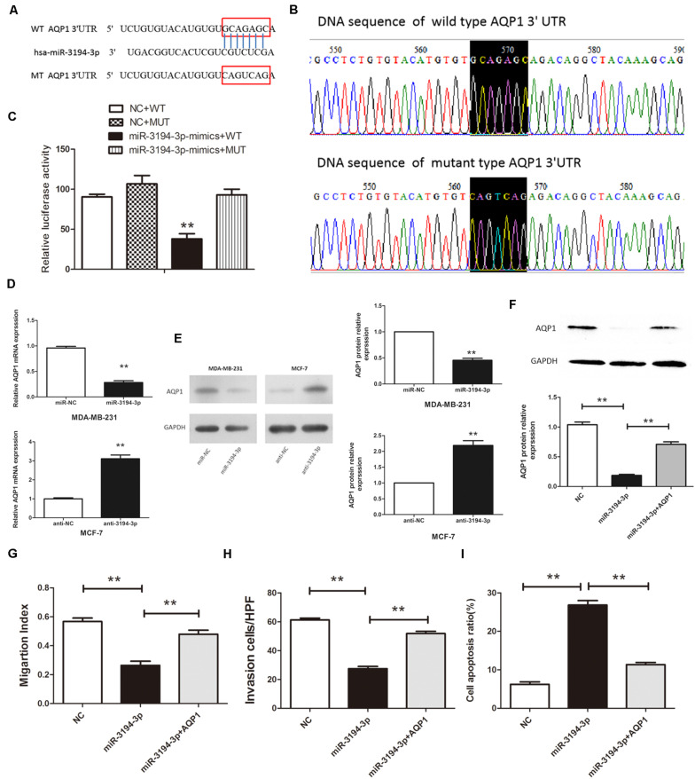
Increasing evidence indicates that the Aquaporin1 (AQP1) aberrant expression may be related to a wide variety of human cancers, including breast cancer (BC). In the present study, we explore the effects and possible mechanism of miR-3194-3p on the biological behaviors of BC. At first, miR-3194-3p is found to modulate AQP1 expression targeting the 3'-UTR using miRNA target prediction algorithms. MiR-3194-3p expression is markedly downregulated, and AQP1 expression is upregulated in BC tissues compared with adjacent normal breast tissues. Moreover, the differential expression of miR-3194-3p and AQP1 are observed in four BC cells with different malignancy degree. Meanwhile, a significant negative correlation between AQP1 and miR-3194-3p expressions in tumor tissues from 30 BC patients is revealed. miR-3194-3p mimic remarkably inhibits cell proliferation, migration, and invasion as well as promotes apoptosis in MDA-MB-231 cells while miR-3194-3p inhibitors exert an opposite role in MCF-7 cells. Dual-luciferase reporter system demonstrates that AQP1 is a direct target gene of miR-3194-3p. Overexpression of AQP1 by pBABE-puro-AQP1 vector partially abrogates the effect of miR-3194-3p mimic in MDA-MB-231 cells. In short, our results suggest that miR-3194-3p suppresses BC cell proliferation, migration, and invasion by targeting AQP1, providing a novel insight into BC tumorigenesis and treatment.
Keywords: AQP1; apoptosis; breast cancer; invasion; miR-3194-3p; migration; proliferation.
Copyright © 2020 Wei, Yu, Cai, Gao, Liu and Zhu.
Figures
Similar articles
-
MiR-1301-3p inhibits human breast cancer cell proliferation by regulating cell cycle progression and apoptosis through directly targeting ICT1.Breast Cancer. 2018 Nov;25(6):742-752. doi: 10.1007/s12282-018-0881-5. Epub 2018 Jun 27. Breast Cancer. 2018. PMID: 29951881
-
MicroRNA-133a-3p inhibits cell proliferation, migration and invasion in colorectal cancer by targeting AQP1.Oncol Lett. 2021 Sep;22(3):649. doi: 10.3892/ol.2021.12910. Epub 2021 Jul 9. Oncol Lett. 2021. Retraction in: Oncol Lett. 2024 Nov 20;29(2):66. doi: 10.3892/ol.2024.14812. PMID: 34386071 Free PMC article. Retracted.
-
MicroRNA-409-3p regulates cell invasion and metastasis by targeting ZEB1 in breast cancer.IUBMB Life. 2016 May;68(5):394-402. doi: 10.1002/iub.1494. Epub 2016 Apr 15. IUBMB Life. 2016. PMID: 27079864
-
miR-758-3p suppresses human bladder cancer cell proliferation, migration and invasion by targeting NOTCH2.Exp Ther Med. 2019 May;17(5):4273-4278. doi: 10.3892/etm.2019.7400. Epub 2019 Mar 18. Exp Ther Med. 2019. PMID: 30988799 Free PMC article.
-
miR-3614-3p suppresses cell aggressiveness of human breast cancer by targeting AKT3 and HDAC1 expression.Transl Cancer Res. 2022 Jun;11(6):1565-1575. doi: 10.21037/tcr-21-2419. Transl Cancer Res. 2022. PMID: 35836520 Free PMC article.
Cited by
-
Peroxiporins in Triple-Negative Breast Cancer: Biomarker Potential and Therapeutic Perspectives.Int J Mol Sci. 2024 Jun 17;25(12):6658. doi: 10.3390/ijms25126658. Int J Mol Sci. 2024. PMID: 38928364 Free PMC article. Review.
-
Identification of a dysregulated CircRNA-associated gene signature for predicting prognosis, immune landscape, and drug candidates in bladder cancer.Front Oncol. 2022 Oct 10;12:1018285. doi: 10.3389/fonc.2022.1018285. eCollection 2022. Front Oncol. 2022. PMID: 36300085 Free PMC article.
-
Leptin Promotes Vasculogenic Mimicry in Breast Cancer Cells by Regulating Aquaporin-1.Int J Mol Sci. 2024 May 10;25(10):5215. doi: 10.3390/ijms25105215. Int J Mol Sci. 2024. PMID: 38791252 Free PMC article.
-
A critical ETV4/Twist1/Vimentin axis in Ha-RAS-induced aggressive breast cancer.Cancer Gene Ther. 2022 Nov;29(11):1590-1599. doi: 10.1038/s41417-022-00471-4. Epub 2022 Apr 27. Cancer Gene Ther. 2022. PMID: 35477769
-
Inhibition of aquaporins as a potential adjunct to breast cancer cryotherapy.Oncol Lett. 2021 Jun;21(6):458. doi: 10.3892/ol.2021.12719. Epub 2021 Apr 8. Oncol Lett. 2021. PMID: 33907568 Free PMC article.
References
LinkOut - more resources
Full Text Sources
Miscellaneous