. 2020 Aug 21:15:6279-6294.
doi: 10.2147/IJN.S254969. eCollection 2020.

TiO2 Nanoparticles Caused DNA Damage in Lung and Extra-Pulmonary Organs Through ROS-Activated FOXO3a Signaling Pathway After Intratracheal Administration in Rats

Affiliations

TiO2 Nanoparticles Caused DNA Damage in Lung and Extra-Pulmonary Organs Through ROS-Activated FOXO3a Signaling Pathway After Intratracheal Administration in Rats

Bin Han et al. Int J Nanomedicine. .

Abstract

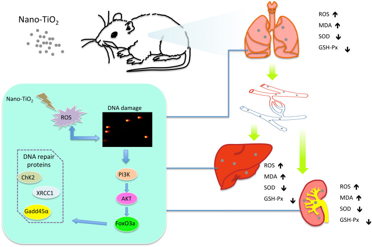
Introduction: Because of the increased production and application of manufactured Nano-TiO2 in the past several years, it is important to investigate its potential hazards. TiO2 is classified by IARC as a possible human carcinogen; however, the potential mechanism of carcinogenesis has not been studied clearly. The present study aimed to investigate the mechanism of DNA damage in rat lung and extra-pulmonary organs caused by TiO2nanoparticles.

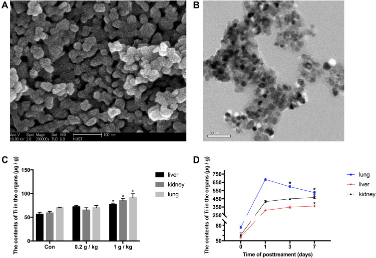
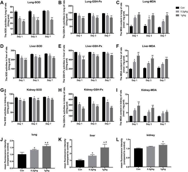
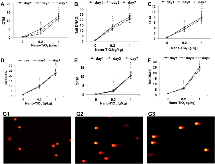
Methods: In the present study, SD rats were exposed to Nano-TiO2 by intratracheal injection at a dose of 0, 0.2, or 1 g/kg body weight. The titanium levels in tissues were detected by ICP-MS. Western blot was used to detect the protein expression levels. The DNA damage and oxidative stress were detected by comet assay and ROS, MDA, SOD, and GSH-Px levels, respectively.

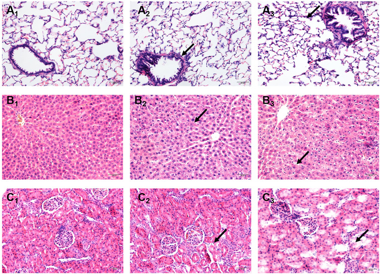
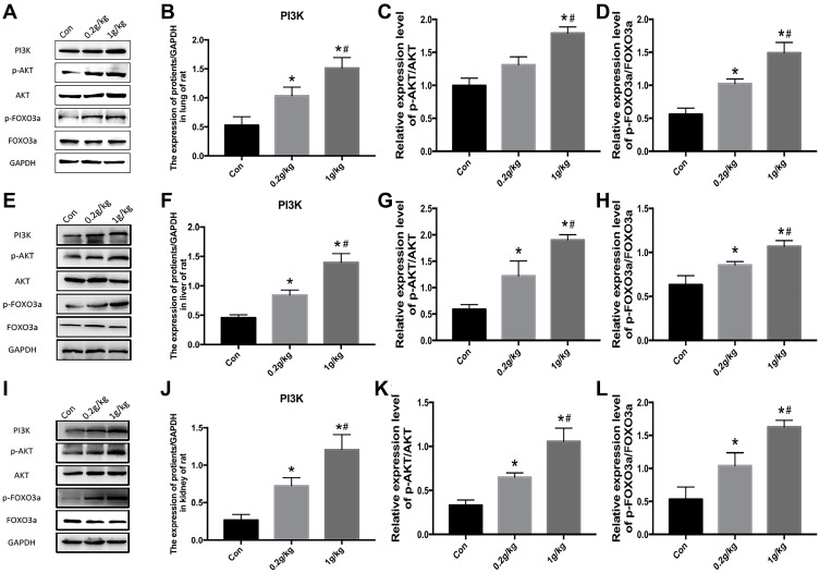
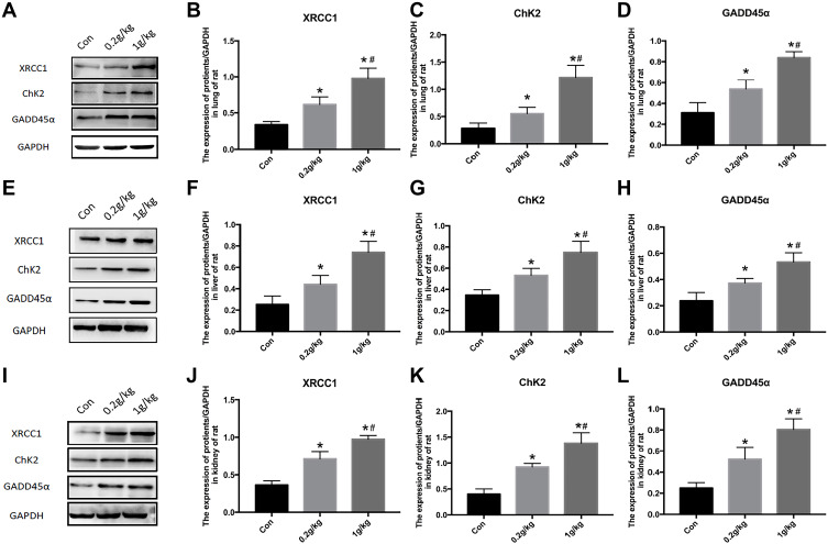
Results: The titanium levels of the 1 g/kg group on day-3 and day-7 were significantly increased in liver and kidney as well as significantly decreased in lung compared to day-1. ROS and MDA levels were statistically increased, whereas SOD and GSH-Px levels were statistically decreased in tissues of rats in dose-dependent manners after Nano-TiO2 treatment. PI3K, p-AKT/AKT, and p-FOXO3a/FOXO3a in lung, liver, and kidney activated in dose-dependent manners. The levels of DNA damage in liver, kidney, and lung in each Nano-TiO2 treatment group were significantly increased and could not recover within 7 days. GADD45α, ChK2, and XRCC1 in liver, kidney, and lung of rats exposed to Nano-TiO2 statistically increased, which triggered DNA repair.

Conclusion: This work demonstrated that Ti could deposit in lung and enter extra-pulmonary organs of rats and cause oxidative stress, then trigger DNA damage through activating the PI3K-AKT-FOXO3a pathway and then promoting GADD45α, ChK2, and XRCC1 to process the DNA repair.

Keywords: DNA damage; DNA repair; GADD45α/ChK2/XRCC1 signaling pathway; Nano-TiO2; PI3K/AKT/FOXO3a signaling pathway.


Conflict of interest statement

We declare that we do not have any commercial or associative interest that represents a conflict of interest in connection with the work submitted. The authors report no conflicts of interest for this work.

Figures

Figure 1
Figure 1
Characterization of Ti and the contents of Ti in organs. (A and B) Scanning electron microscopy and transmission electron microscopy images of Nano-TiO2. The powder was deposited on 200-mesh copper grids. The particle primary structure was 10–30 nm and globular. (C) On the 7th day after instillation, the contents of Ti in tissues of rats in the 0, 0.2, and 1 g/kg Nano-TiO2 treatment groups. N=5, *P<0.05, in comparison to the control group. (D) The contents of Ti in tissues of rats after 1 g/kg Nano-TiO2 treatment for 1, 3, and 7 days. N=5, *P<0.05, in comparison to the 1-day group.
Figure 2
Figure 2
Effects of Nano-TiO2 exposure on the histopathological changes in the lung, liver, and kidney at 7 days after treatment. (A) The representative images of lung tissue of rats stained with HE. (B) The representative images of liver tissue of rats stained with HE. (C) The representative images of kidney tissue of rats stained with HE. Scale bar represents 50 μm. The arrow in A2 indicated irregular luminal inflammatory cell infiltration; The arrow in A3indicated the alveolar wall thickening. The arrow in B2 indicated cell disorder. The arrow in B3 indicated the spotty necrosis. The arrow in C2 indicated the gap increases. The arrow in C3 indicated necrosis. 1: Control group, 2: 0.2 g/kg group, 3: 1 g/kg group.
Figure 3
Figure 3
The oxidative stress levels in the lung, liver, and kidney of rats after Nano-TiO2 treatment. (AC) The SOD, GSH-Px activities, and MDA content in lung of rats. (DF) The SOD, GSH-Px activities, and MDA content in liver of rats. (GI) The SOD, GSH-Px activities, and MDA content in kidney of rats. (JL) ROS levels induced by Nano-TiO2 in organs of rats. N=5, *P<0.05, in comparison to the control group; #P<0.05, in comparison to the 0.2 g/kg group. Abbreviations: SOD, superoxide dismutase; GSH-Px, glutathione peroxidase; MDA, malonaldehyde.
Figure 4
Figure 4
The DNA damage levels in the lung, liver, and kidney of rats after Nano-TiO2 treatment. (A and B): The OTM value and Tail DNA% value changed in the lung after Nano-TiO2 treatment at different times. (C and D) The OTM value and Tail DNA% value changed in the liver after Nano-TiO2 treatment at different times. (E and F) The OTM value and Tail DNA% value changed in the lung after Nano-TiO2 treatment at different times. (G) Representative images of DNA damage in the lung of rats after Nano-TiO2 treatment for 7 days (400x). (G1: control group; G2: 0.2 g/kg Nano-TiO2 treatment group; G3: 1 g/kg Nano-TiO2 treatment group). The result was the average of at least three independent experiments. N=5, *P<0.05: day 1 in comparison to the control group; #P<0.05: day 3 in comparison to the control group; P<0.05: day 7 in comparison to the control group.
Figure 5
Figure 5
Activation of the PI3K-AKT-FOXO3a signal pathway in the lung, liver, and kidney of rats after Nano-TiO2 treatment for 7 days. (A) The represented protein bands of PI3K, AKT, FOXO3a, p-AKT, and p-FOXO3a in the lung. (BD) The fold changes of PI3K, p-AKT/AKT, and p-FOXO3a/FOXO3a levels in the lung. (EH) The represented protein bands and fold changes of PI3K, AKT, FOXO3a, p-AKT, and p-FOXO3a in the liver. (IL) The represented protein bands and fold changes of PI3K, AKT, FOXO3a, p-AKT, and p-FOXO3a in the kidney. N=5, *P<0.05, in comparison to the control group. #P<0.05, in comparison to the 0.2 g/kg group. Abbreviations: PI3K, phosphatidyl inositol 3-kinase; AKT, protein kinase B; FOXO3a, the Fork head box class O 3a.
Figure 6
Figure 6
Changes of DNA repair proteins in the lung, liver, and kidney of rats exposed to Nano-TiO2 for 7 days. (AD) The represented protein bands and fold changes of XRCC1, ChK2, and GADD45α in the lung. (EH) The represented protein bands and fold changes of XRCC1, ChK2, and GADD45α in the liver. (IL) The represented protein bands and fold changes of XRCC1, ChK2, and GADD45α in the kidney. N=5, *P<0.05, in comparison to the control group. #P<0.05, in comparison to the 0.2 g/kg group. Abbreviations: XRCC1, x-ray repair cross complementing gene 1; ChK2, checkpoint kinase 2; GADD45α, growth arrest and DNA damage α.
Figure 7
Figure 7
A proposed action model for the PI3K/AKT/FoxO3a pathway via ROS regulating DNA damage in the lung, liver, and kidney induced by Nano-TiO2 intratracheal administration.

Similar articles

Cited by

References

    1. Pelclova D, Zdimal V, Kacer P, et al. Markers of lipid oxidative damage in the exhaled breath condensate of nano TiO2 production workers. Nanotoxicology. 2017;11(1):52–63. doi:10.1080/17435390.2016.1262921 - DOI - PubMed
    1. Bhattacharya K, Davoren M, Boertz J, Schins RP, Hoffmann E, Dopp E. Titanium dioxide nanoparticles induce oxidative stress and DNA-adduct formation but not DNA-breakage in human lung cells. Part Fibre Toxicol. 2009;6:17. doi:10.1186/1743-8977-6-17 - DOI - PMC - PubMed
    1. Brezová V, Gabčová S, Dvoranová D, Staško A. Reactive oxygen species produced upon photoexcitation of sunscreens containing titanium dioxide (an EPR study). J Photochem Photobiol B. 2005;79(2):121–134. doi:10.1016/j.jphotobiol.2004.12.006 - DOI - PubMed
    1. Dunford R, Cai LSN, Horikoshi S, Hidaka HKJ, Salinaro A. Chemical oxidation and DNA damage catalysed by inorganic sunscreen ingredients. FEBS Lett. 1997;418(2):87–90. doi:10.1016/S0014-5793(97)01356-2 - DOI - PubMed
    1. Yu KN, Sung JH, Lee S, et al. Inhalation of titanium dioxide induces endoplasmic reticulum stress-mediated autophagy and inflammation in mice. Food Chem Toxicol. 2015;85:106–113. doi:10.1016/j.fct.2015.08.001 - DOI - PubMed

MeSH terms