

Sareomycetes cl. nov.: A new proposal for placement of the resinicolous genus Sarea (Ascomycota, Pezizomycotina)
- PMID: 32904095
- PMCID: PMC7451776
- DOI: 10.3114/fuse.2020.06.02
Sareomycetes cl. nov.: A new proposal for placement of the resinicolous genus Sarea (Ascomycota, Pezizomycotina)
Abstract
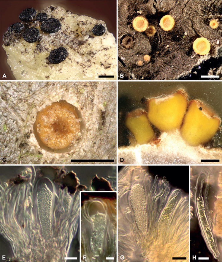
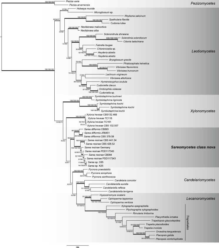
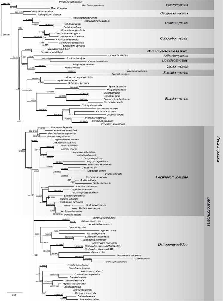
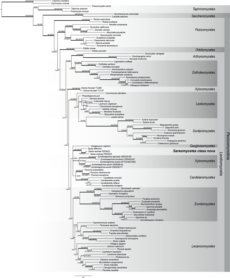
Resinicolous fungi constitute a heterogeneous assemblage of fungi that live on fresh and solidified plant resins. The genus Sarea includes, according to current knowledge, two species, S. resinae and S. difformis. In contrast to other resinicolous discomycetes, which are placed in genera also including non-resinicolous species, Sarea species only ever fruit on resin. The taxonomic classification of Sarea has proven to be difficult and currently the genus, provisionally and based only on morphological features, has been assigned to the Trapeliales (Lecanoromycetes). In contrast, molecular studies have noted a possible affinity to the Leotiomycetes. Here we review the taxonomic placement of Sarea using sequence data from seven phylogenetically informative DNA regions including ribosomal (ITS, nucSSU, mtSSU, nucLSU) and protein-coding (rpb1, rpb2, mcm7) regions. We combined available and new sequence data with sequences from major Pezizomycotina classes, especially Lecanoromycetes and Leotiomycetes, and assembled three different taxon samplings in order to place the genus Sarea within the Pezizomycotina. Based on our data, none of the applied phylogenetic approaches (Bayesian Inference, Maximum Likelihood and Maximum Parsimony) supported the placement of Sarea in the Trapeliales or any other order in the Lecanoromycetes. A placement of Sarea within the Leotiomycetes is similarly unsupported. Based on our data, Sarea forms an isolated and highly supported phylogenetic lineage within the "Leotiomyceta". From the results of our multilocus phylogenetic analyses we propose here a new class, order, and family, Sareomycetes, Sareales and Sareaceae in the Ascomycota to accommodate the genus Sarea. The genetic variability within the newly proposed class suggests that it is a larger group that requires further infrageneric classification.
Keywords: Ascomycota; Sarea difformis; Sarea resinae; Sareaceae fam. nov.; Sareales ord. nov.; Sareomycetes cl. nov.; new taxa; resinicolous fungi; taxonomy.
© 2020 Westerdijk Fungal Biodiversity Institute.
Figures
Similar articles
-
Sareomycetes: more diverse than meets the eye.IMA Fungus. 2021 Mar 16;12(1):6. doi: 10.1186/s43008-021-00056-0. IMA Fungus. 2021. PMID: 33726866 Free PMC article.
-
Revision of Xylonaceae (Xylonales, Xylonomycetes) to include Sarea and Tromera.Mycoscience. 2021 Jan 20;62(1):47-63. doi: 10.47371/mycosci.2020.11.001. eCollection 2021. Mycoscience. 2021. PMID: 37090019 Free PMC article.
-
Phylogenetic comparison of protein-coding versus ribosomal RNA-coding sequence data: a case study of the Lecanoromycetes (Ascomycota).Mol Phylogenet Evol. 2007 Jul;44(1):412-26. doi: 10.1016/j.ympev.2006.10.016. Epub 2006 Oct 25. Mol Phylogenet Evol. 2007. PMID: 17207641
-
A five-gene phylogeny of Pezizomycotina.Mycologia. 2006 Nov-Dec;98(6):1018-28. doi: 10.3852/mycologia.98.6.1018. Mycologia. 2006. PMID: 17486977
-
A multigene phylogenetic synthesis for the class Lecanoromycetes (Ascomycota): 1307 fungi representing 1139 infrageneric taxa, 317 genera and 66 families.Mol Phylogenet Evol. 2014 Oct;79:132-68. doi: 10.1016/j.ympev.2014.04.003. Epub 2014 Apr 18. Mol Phylogenet Evol. 2014. PMID: 24747130 Free PMC article.
Cited by
-
Chaetocapnodiummagnum and Chaetocapnodiumpolonicum from conifer resins disclose an unknown lifestyle in the Capnodiales (Dothideomycetes).MycoKeys. 2025 Jul 14;119:315-333. doi: 10.3897/mycokeys.119.159094. eCollection 2025. MycoKeys. 2025. PMID: 40697982 Free PMC article.
-
Sareomycetes: more diverse than meets the eye.IMA Fungus. 2021 Mar 16;12(1):6. doi: 10.1186/s43008-021-00056-0. IMA Fungus. 2021. PMID: 33726866 Free PMC article.
-
Revision of Xylonaceae (Xylonales, Xylonomycetes) to include Sarea and Tromera.Mycoscience. 2021 Jan 20;62(1):47-63. doi: 10.47371/mycosci.2020.11.001. eCollection 2021. Mycoscience. 2021. PMID: 37090019 Free PMC article.
-
Discovery of the first resinicolous fungus in Mycosphaerellales (Dothideomycetes): Resinomelaniacommunis from conifer resins in Poland.MycoKeys. 2025 Jul 29;120:119-138. doi: 10.3897/mycokeys.120.154464. eCollection 2025. MycoKeys. 2025. PMID: 40778229 Free PMC article.
-
Resin outpourings on conifers are inhabited by more members of Nectriaceae (Hypocreales, Sordariomycetes) than previously thought.MycoKeys. 2025 Feb 12;113:337-358. doi: 10.3897/mycokeys.113.140446. eCollection 2025. MycoKeys. 2025. PMID: 39980719 Free PMC article.
References
-
- Aptroot A, Schumm F. (2012). The genus Melanophloea, an example of convergent evolution towards polyspory. The Lichenologist 44: 501–509.
-
- Ayers TT. (1941). Biatorella resinae: The Perfect Stage of Zythia resinae. Mycologia 33: 130–135.
-
- Baral HO, Marson G. (2012). Deltopyxis triangulispora gen. et sp. nov., a polysporous Tromeropsis-like discomycete of unclear relationship. Andrias 19: 175–183.
-
- Baral HO, Weber E, Marson G, et al. , (2018). A new connection between wood saprobism and beetle endosymbiosis: the rarely reported saprobic discomycete Tromeropsis is congeneric with the symbiotic yeast Symbiotaphrina (Symbiotaphrinales, Xylonomycetes) and two asexual morphs misplaced in Hyphozyma. Mycological Progress 17: 215–254.
LinkOut - more resources
Full Text Sources