. 2020 Apr 9:96:185-308.
doi: 10.1016/j.simyco.2020.03.001. eCollection 2020 Jun.

Venturiales

Affiliations

Venturiales

M Shen et al. Stud Mycol. .

Abstract

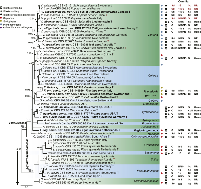
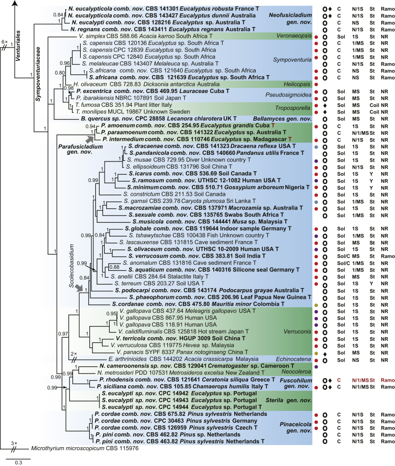
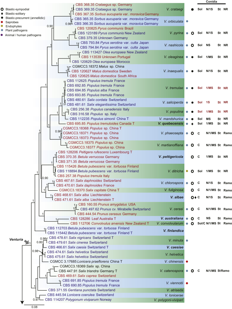
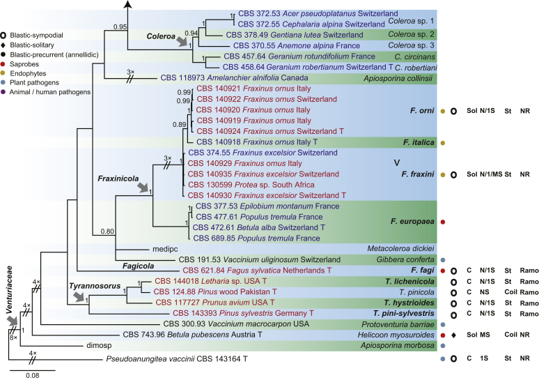
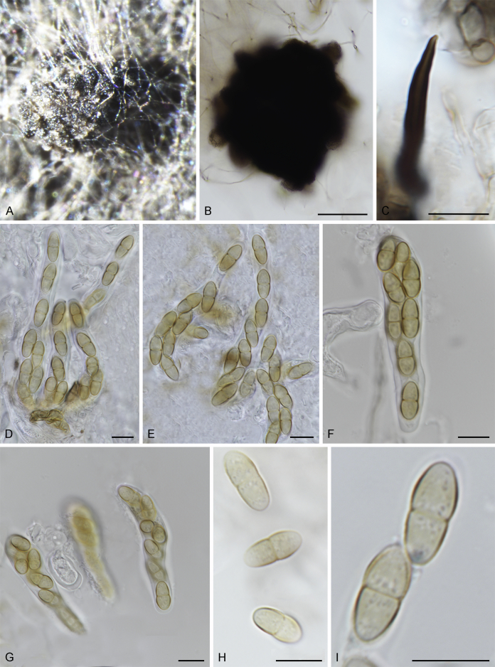
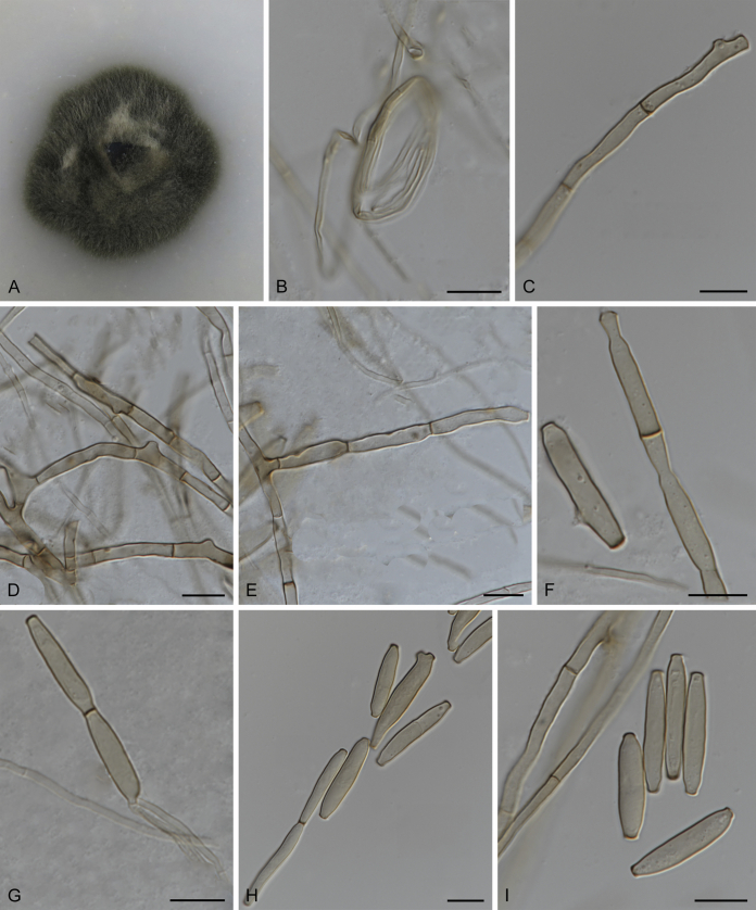
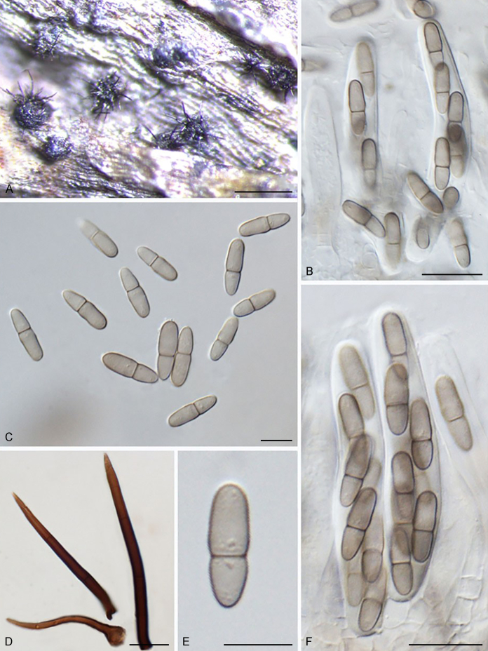
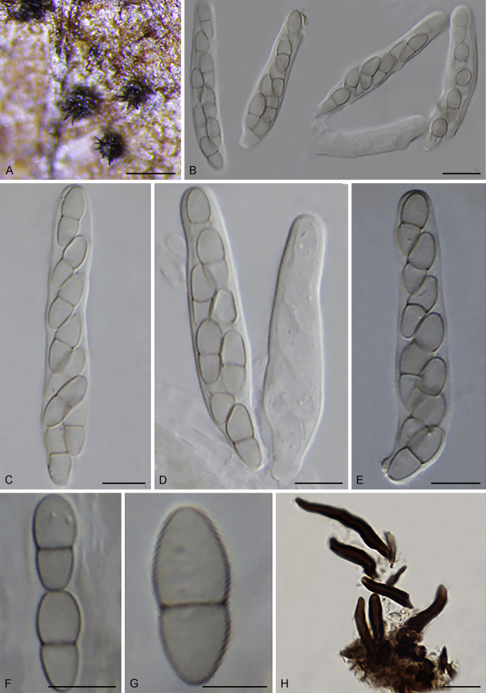
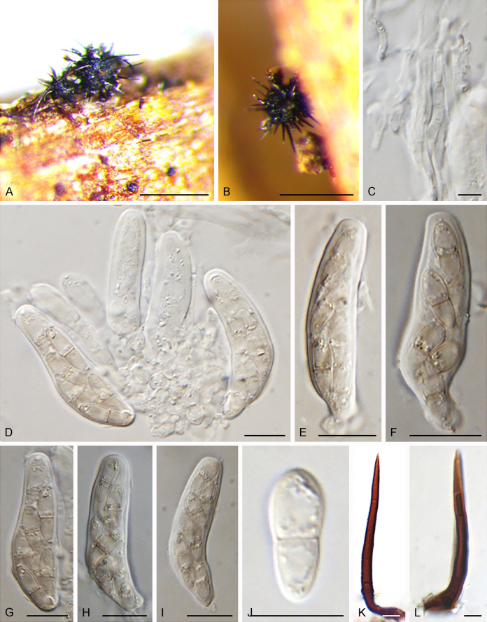
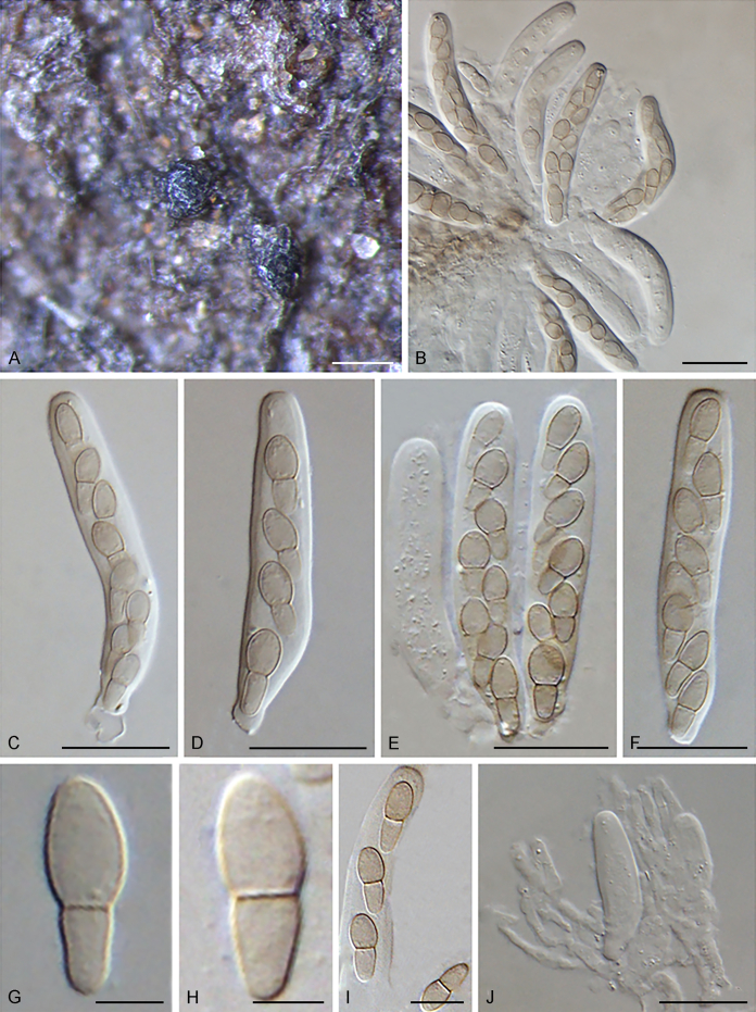
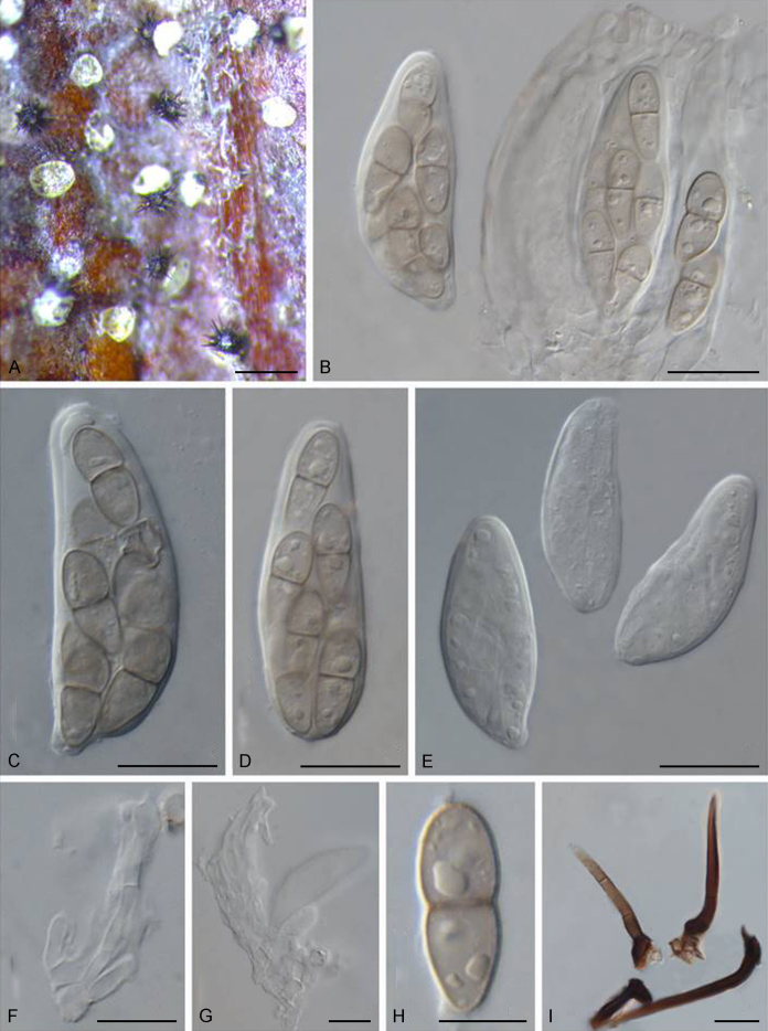
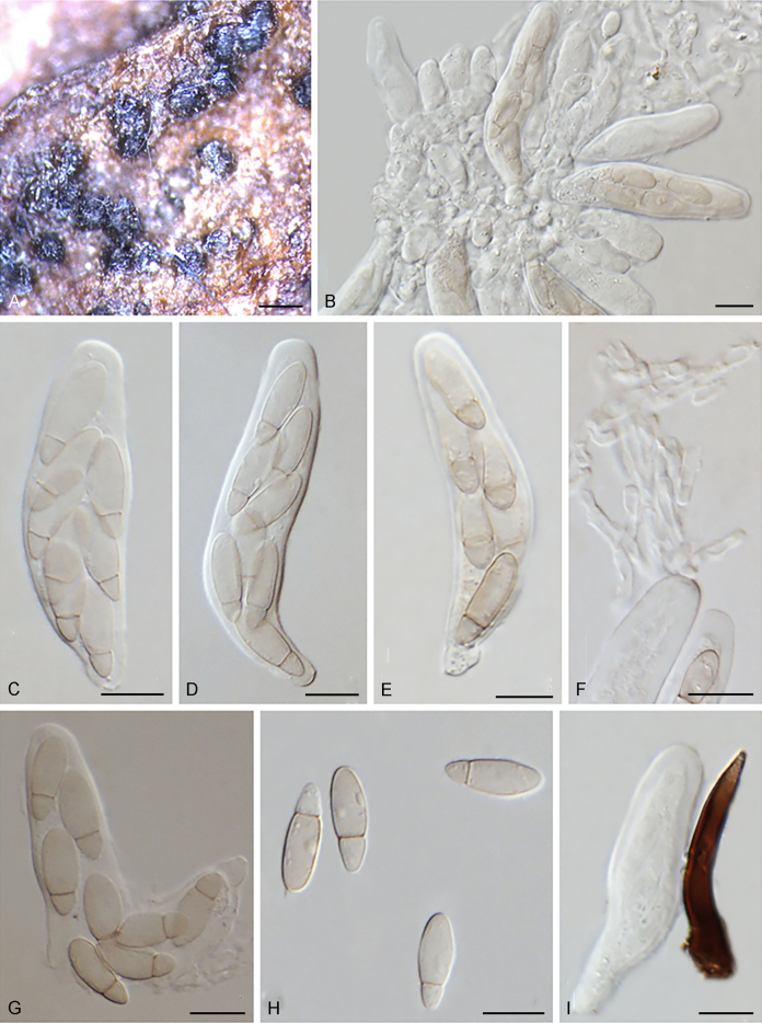
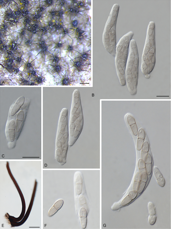
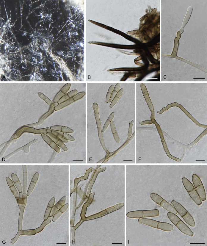
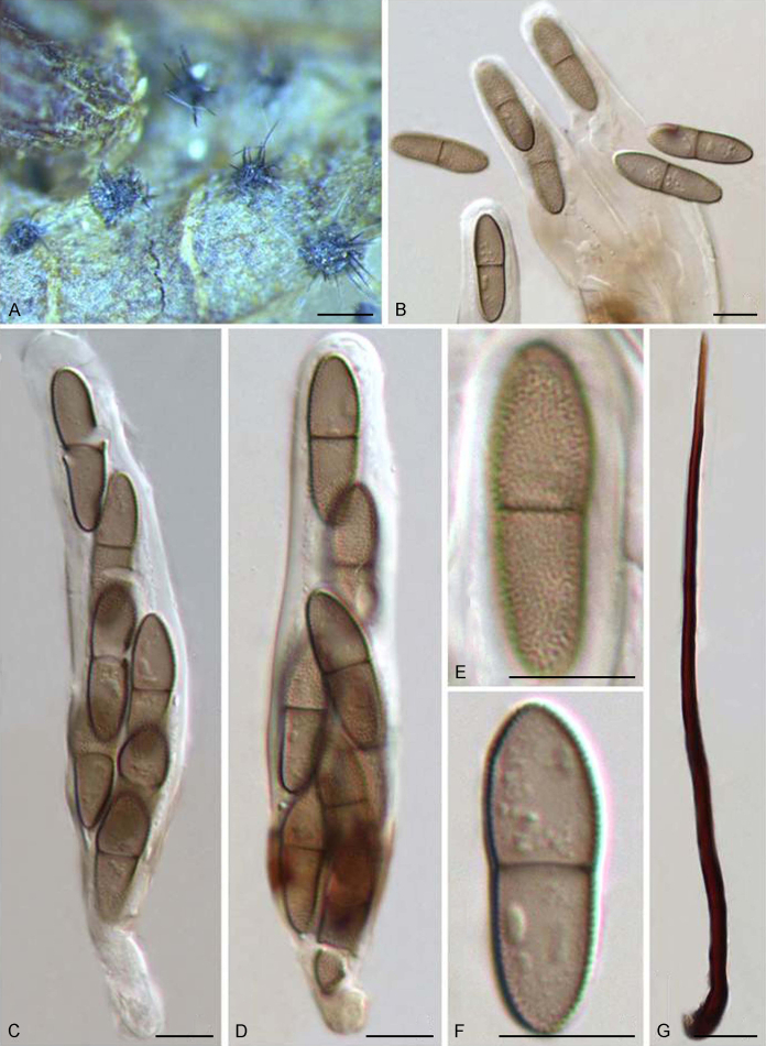
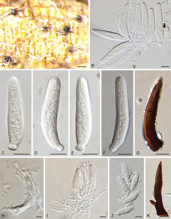
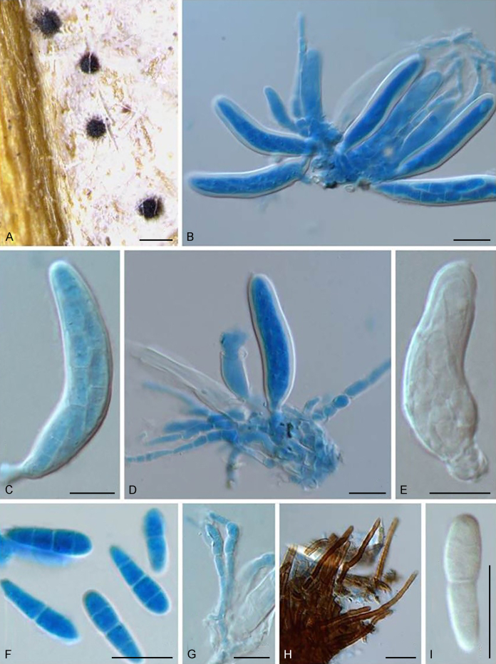
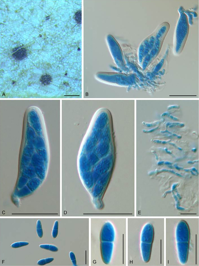
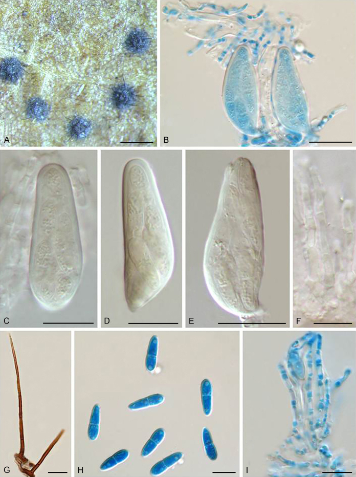
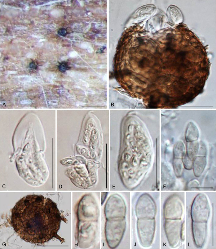
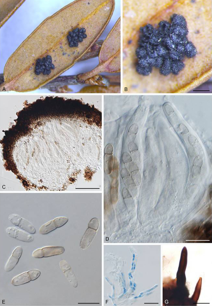
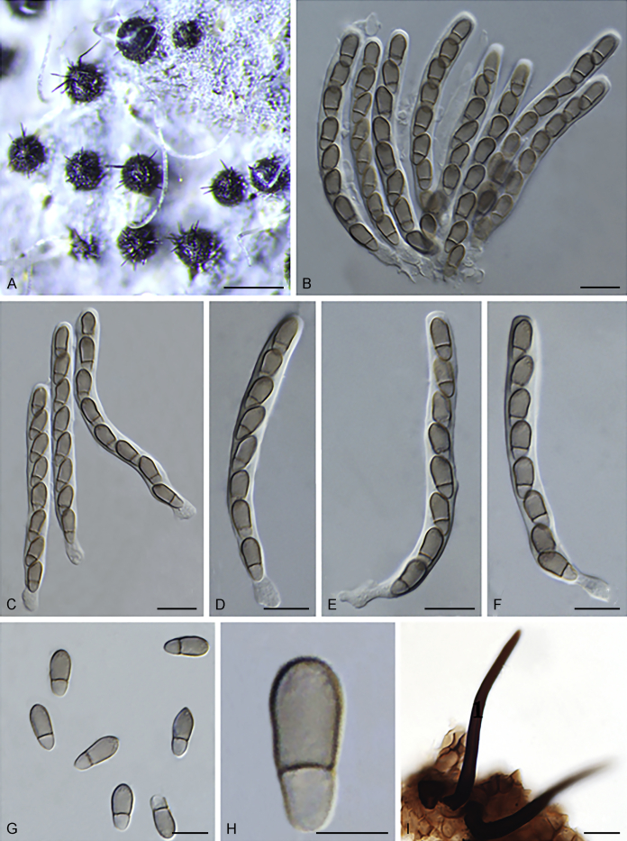
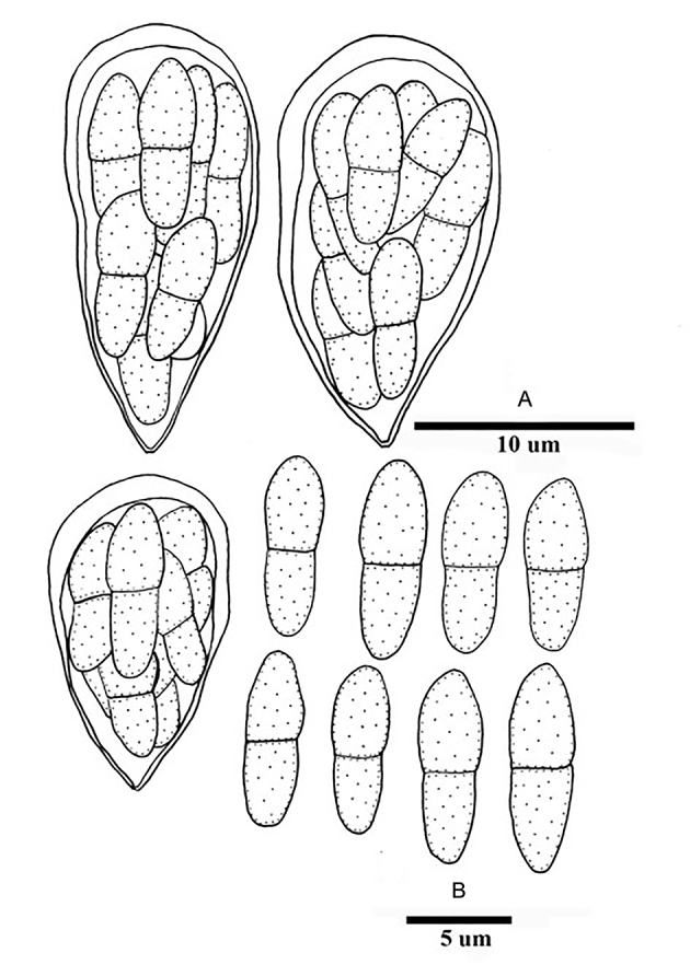
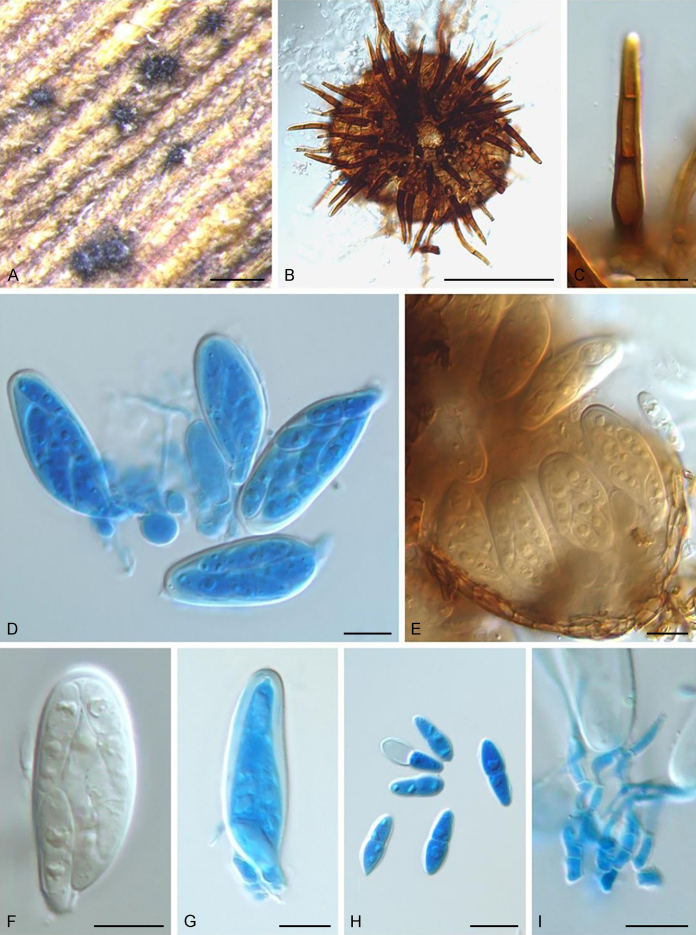
Members of Venturiales (Dothideomycetes) are widely distributed, and comprise saprobes, as well as plant, human and animal pathogens. In spite of their economic importance, the general lack of cultures and DNA data has resulted in taxa being poorly resolved. In the present study five loci, ITS, LSU rDNA, tef1, tub2 and rpb2 are used for analysing 115 venturialean taxa representing 30 genera in three families in the current classification of Venturiales. Based on the multigene phylogenetic analysis, morphological and ecological characteristics, one new family, Cylindrosympodiaceae, and eight new genera are described, namely Bellamyces, Fagicola, Fraxinicola, Fuscohilum, Neofusicladium, Parafusicladium, Pinaceicola and Sterila. In addition, 12 species are described as new to science, and 41 new combinations are proposed. The taxonomic status of 153 species have been re-evaluated with 20 species excluded from Venturiales. Based on this revision of Venturiales, morphological characteristics such as conidial arrangement (solitary or in chains) or conidiogenesis (blastic-solitary, sympodial or annellidic), proved to be significant at generic level. Venturia as currently defined represents a generic complex. Furthermore, plant pathogens appear more terminal in phylogenetic analyses within Venturiaceae and Sympoventuriaceae, suggesting that the ancestral state of Venturiales is most likely saprobic.

Keywords: Bellamyces Crous, Coppins & U. Braun; Bellamyces quercus Crous, Coppins & U. Braun; Cylindrosympodiaceae Crous, M. Shen & Y. Zhang ter; Fagicola Crous, M. Shen & Y. Zhang ter; Fagicola fagi (Crous & de Hoog) Crous, M. Shen & Y. Zhang ter; Fraxinicola Crous, M. Shen & Y. Zhang ter; Fraxinicola europaea Crous, M. Shen & Y. Zhang ter; Fraxinicola fraxini (Aderh.) Crous, M. Shen & Y. Zhang ter; Fraxinicola italica Crous, M. Shen & Y. Zhang ter; Fraxinicola orni (M. Ibrahim et al.) Crous, M. Shen & Y. Zhang ter; Fuscohil`um Crous, M. Shen & Y. Zhang ter; Fuscohilum Crous, M. Shen & Y. Zhang ter; Fuscohilum rhodensis (Crous & M.J. Wingf.) Crous, M. Shen & Y. Zhang ter, Fuscohilum siciliana (Koukol) Crous, M. Shen & Y. Zhang ter; Multigene analysis; Neocoleroa cameroonensis Crous, M. Shen & Y. Zhang ter; Neofusicladium Crous, M. Shen & Y. Zhang ter; Neofusicladium eucalypti (Crous & R.G. Shivas) Crous, M. Shen & Y. Zhang ter; Neofusicladium eucalypticola (Crous & M.J. Wingf.) Crous, M. Shen & Y. Zhang ter; Neofusicladium regnans (Crous) Crous, M. Shen & Y. Zhang ter; New taxa; Niesslia iridicola (M.E. Barr) Crous, M. Shen & Y. Zhang ter; Niesslia parasitica (Ellis & Everh.) M. Shen & Y. Zhang ter; Niesslia vaccinii (Ellis & Everh.) Crous, M. Shen & Y. Zhang ter; Parafusicladium Crous, M. Shen & Y. Zhang ter; Parafusicladium amoenum (R.F. Castañeda & Dugan) Crous, M. Shen & Y. Zhang ter; Parafusicladium intermedium (Crous & W.B. Kendr.) Crous, M. Shen & Y. Zhang ter; Parafusicladium paraamoenum (Crous et al.) Crous, M. Shen & Y. Zhang ter; Pinaceicola Crous, M. Shen & Y. Zhang ter; Pinaceicola cordae (Koukol) Crous, M. Shen & Y. Zhang ter; Pinaceicola pini(Crous & de Hoog) Crous, M. Shen & Y. Zhang ter; Pseudosigmoidea excentrica (R.F. Castañeda et al.) Crous, M. Shen & Y. Zhang ter; Scab disease; Scolecobasidium aquaticum (Samerp. et al.) Crous, M. Shen & Y. Zhang ter; Scolecobasidium atlanticuum (A.M. Wellman) Crous, M. Shen & Y. Zhang ter; Scolecobasidium bacilliforme (Samerp. et al.) Crous, M. Shen & Y. Zhang ter; Scolecobasidium capsici (Crous & Cheew.) Crous, M. Shen & Y. Zhang ter; Scolecobasidium cordanae (Samerp. et al.) Crous, M. Shen & Y. Zhang ter; Scolecobasidium dracaenae (Crous) Crous, M. Shen & Y. Zhang ter; Scolecobasidium globale (Samerp. et al.) Crous, M. Shen & Y. Zhang ter; Scolecobasidium icarus (Samerp. et al.) Crous, M. Shen & Y. Zhang ter; Scolecobasidium macrozamiae (Crous & R.G. Shivas) Crous, M. Shen & Y. Zhang ter; Scolecobasidium minimum (Fassat.) Crous, M. Shen & Y. Zhang ter; Scolecobasidium musicola (Crous) Crous, M. Shen & Y. Zhang ter; Scolecobasidium olivaceum (A. Giraldo et al.) Crous, M. Shen & Y. Zhang ter; Scolecobasidium pandanicola (Crous & M.J. Wingf.) Crous, M. Shen & Y. Zhang ter; Scolecobasidium phaeophorum (Samerp. et al.) Crous, M. Shen & Y. Zhang ter; Scolecobasidium podocarpi (Crous) Crous, M. Shen & Y. Zhang ter; Scolecobasidium ramosum (A. Giraldo et al.) Crous, M. Shen & Y. Zhang ter; Scolecobasidium robustum (Samerp. et al.) Crous, M. Shen & Y. Zhang ter; Scolecobasidium sexuale (Samerp. et al.) Crous, M. Shen & Y. Zhang ter; Scolecobasidium verrucosum (Zachariah et al.) Crous, M. Shen & Y. Zhang ter; Sterila Crous, M. Shen & Y. Zhang ter; Sterila eucalypti Crous, M. Shen & Y. Zhang ter; Sympoventuria africana (Crous) Crous, M. Shen & Y. Zhang ter; Systematics; Tyrannosorus hanlinianus (U. Braun & Feiler) Crous, M. Shen & Y. Zhang ter; Tyrannosorus hystrioides (Dugan et al.) Crous, M. Shen & Y. Zhang ter; Tyrannosorus lichenicola Crous, M. Shen & Y. Zhang ter; Tyrannosorus pini-sylvestris Crous & R.K. Schumach.; Venturia; Venturia albae Crous, M. Shen & Y. Zhang ter; Venturia australiana Crous, M. Shen & Y. Zhang ter; Venturia caesiae Crous, M. Shen & Y. Zhang ter; Venturia finlandica Crous, M. Shen & Y. Zhang ter; Venturia peltigericola (Crous & Diederich) Crous, M. Shen & Y. Zhang ter; Venturia quebecensis Crous, M. Shen & Y. Zhang ter; Verruconis terricola (J. Ren et al.) Crous, M. Shen & Y. Zhang ter.


Figures

Fig 1
Fig 1
Consensus phylogram (50 % majority rule) of 691 952 trees resulting from a Bayesian analysis of the combined alignment of ITS, LSU, tef1, tub2 and rpb2 sequences of Venturiales. Bayesian posterior probabilities (PP) > 0.80 are shown at the nodes and the scale bar represents the expected changes per site. Some branches were shortened to facilitate layout. The tree was rooted with Microthyrium microscopicum (CBS 115976). Culture collection numbers, substrates and countries are indicated behind the species names. Those highlighted in bold are new taxa or new combinations proposed in this study, and type strains are marked with “T” (ex-type in black, ex-epitype in red). Relevant morphological characteristics plotted are abbreviated as follows: Sol – conidia solitary, C – conidia in chains, NS – aseptate conidia, 1S – 1-septate conidia, MS – multi-septate conidia (septa ≥ 2), St – straight or slightly curved conidia, Coil – coiled conidia, Y – Y-shaped conidia; Ramo – ramoconidia present, NR – ramoconidia not observed; ? – asexual morphology not available (either from references or from sporulation induced in this study); and morphological characters plotted in red means strains failed to sporulate in this study and plotted values are taken from the original description, observation of this study or related references. Other characteristics are explained in the legend.
Fig 1
Fig 1
Consensus phylogram (50 % majority rule) of 691 952 trees resulting from a Bayesian analysis of the combined alignment of ITS, LSU, tef1, tub2 and rpb2 sequences of Venturiales. Bayesian posterior probabilities (PP) > 0.80 are shown at the nodes and the scale bar represents the expected changes per site. Some branches were shortened to facilitate layout. The tree was rooted with Microthyrium microscopicum (CBS 115976). Culture collection numbers, substrates and countries are indicated behind the species names. Those highlighted in bold are new taxa or new combinations proposed in this study, and type strains are marked with “T” (ex-type in black, ex-epitype in red). Relevant morphological characteristics plotted are abbreviated as follows: Sol – conidia solitary, C – conidia in chains, NS – aseptate conidia, 1S – 1-septate conidia, MS – multi-septate conidia (septa ≥ 2), St – straight or slightly curved conidia, Coil – coiled conidia, Y – Y-shaped conidia; Ramo – ramoconidia present, NR – ramoconidia not observed; ? – asexual morphology not available (either from references or from sporulation induced in this study); and morphological characters plotted in red means strains failed to sporulate in this study and plotted values are taken from the original description, observation of this study or related references. Other characteristics are explained in the legend.
Fig 2
Fig 2
Consensus phylogram (50 % majority rule) of 42 902 trees resulting from a Bayesian analysis of the combined alignment of ITS, LSU, tef1, tub2 and rpb2 sequences of Venturiaceae. Bayesian posterior probabilities (PP) > 0.80 are shown at the nodes and the scale bar represents the expected changes per site. Some branches were shortened to facilitate layout. The tree was rooted with Pseudoanungitea vaccinii (CBS 143164). See title of Fig. 1 for an explanation of the characters plotted on the tree. Strains in red text sporulated in this study, while those in blue text failed to sporulate and those in black text were not studied.
Fig 2
Fig 2
Consensus phylogram (50 % majority rule) of 42 902 trees resulting from a Bayesian analysis of the combined alignment of ITS, LSU, tef1, tub2 and rpb2 sequences of Venturiaceae. Bayesian posterior probabilities (PP) > 0.80 are shown at the nodes and the scale bar represents the expected changes per site. Some branches were shortened to facilitate layout. The tree was rooted with Pseudoanungitea vaccinii (CBS 143164). See title of Fig. 1 for an explanation of the characters plotted on the tree. Strains in red text sporulated in this study, while those in blue text failed to sporulate and those in black text were not studied.
Fig. 3
Fig. 3
Bellamyces quercus (culture ex-type CPC 28858) asexual morph. A. Colony on OA. B–E. Conidiogenous cells producing conidia. F. Multi-septate conidia. Scale bars: B–F = 10 μm.
Fig. 4
Fig. 4
Fuscohilum siciliana (culture ex-type CBS 105.85) asexual morph. A. Colony on OA. B, C. Conidia arising from hyphae. D–H. Cylindrical and subcylindrical conidia in chains. Scale bars: B–H = 10 μm.
Fig. 5
Fig. 5
Neofusicladium eucalypti (culture ex-type CBS 128216) asexual morph. A. Colony on OA. B–D. Concatenated conidia arising from hypha. E. Pale brown, fusiform and aseptate conidia. Scale bars: B–E = 10 μm.
Fig. 6
Fig. 6
Neofusicladium eucalypticola (culture ex-type CBS 141301) asexual morph. A. Colony on OA. B–E. Concatenated conidia arising from hypha. F, G. Pale brown and aseptate or 1-septate conidia. Scale bars: B–G = 10 μm.
Fig. 7
Fig. 7
Parafusicladium amoenum (culture ex-type CBS 254.95) asexual morph. A. Colony on OA. B–F. Long conidiophores reduced to sympodial conidiogenous cells. G, H. Pale brown and 1-septate conidia. Scale bars: B–H = 10 μm.
Fig. 8
Fig. 8
Parafusicladium intermedium (culture ex-epitype CBS 110746) asexual morph. A. Colony on OA. B–E. Brown conidiophores with sympodial conidiogenous loci. F–H. Subhyaline and cylindrical conidia. Scale bars: B–H = 10 μm.
Fig. 9
Fig. 9
Parafusicladium paraamoenum (culture ex-type CBS 141322) asexual morph. A. Colony on OA. B. Hyphal coil. C–E. Conidia arising from sympodial conidiogenous cells. F. Pale brown, aseptate or 1-septate conidia. Scale bars: B–F = 10 μm.
Fig. 10
Fig. 10
Pinaceicola cordae (culture CBS 675.82) asexual morph. A. Colony on OA. B, C. Conidia arising from conidiogenous cells. D–F. Pale brown, aseptate or 1-septate conidia in branched chains. Scale bars: B–F = 10 μm.
Fig. 11
Fig. 11
Pinaceicola pini (culture ex-type CBS 463.82) asexual morph. A. Colony on OA. B. Hyphal coil. C–H. Sympodial conidiogenous loci and concatenated conidia arising from conidiogenous cells. Scale bars: B–H = 10 μm.
Fig. 12
Fig. 12
Fagicola fagi (culture ex-type CBS 621.84) asexual morph. A. Colony on OA. B–E. Conidia in simple or branched chains arising from conidiogenous cells. F, G. Brown, aseptate or 1-septate conidia. Scale bars: B–G = 10 μm.
Fig. 13
Fig. 13
Fraxinicola fraxini (culture CBS 140930) asexual morph. A. Colony on OA. B–F. Sympodial conidiogenous cells producing conidia. G–J. Brown, 1–3-septate, tapering conidia. Scale bars: B–J = 10 μm.
Fig. 14
Fig. 14
Fraxinicola orni (culture CBS 140920) asexual morph. A, B. Sporodochia produced on OA. C–E. Sympodial conidiogenous cells producing conidia. F, G. Medium brown, 1-septate and asymmetrical conidia. Scale bars: A, C = 20 μm; B, D–G = 10 μm.
Fig. 15
Fig. 15
Gibbera kalmiae (holotype NYSf1621) sexual morph. A. Ascomata scattered on the host surface. B, D, E. Clavate, asci. C. Released, pale brown, 1-septate ascospores. F. Evanesent, cellular pseudoparaphyses. G. Setae. Scale bars: A = 200 μm; B–G = 10 μm.
Fig. 16
Fig. 16
Tyrannosorus hystrioides (culture ex-type CBS 117727) asexual morph. A. Colony on OA. B. Hyphal coil. C–F. Concatenated conidia arising from conidiogenous cells. G, H. Aseptate or 1-septate conidia. Scale bars: B–H = 10 μm.
Fig. 17
Fig. 17
Tyrannosorus lichenicola (culture ex-type CBS 144018) asexual morph A. Colony on OA. B. Hyphal coil. C. Conidium arising from conidiogenous locus. D–H. Aseptate or 1-septate conidia in chains. Scale bars: B–H = 10 μm.
Fig. 18
Fig. 18
Tyrannosorus pinicola (culture ex-type CBS 124.88) asexual morph. A. Colony on OA. B. Conidiogenous loci on hypha. C–F. Conidia in chains forming three-dimensional helix. G. Straight or curved conidia. Scale bars: B–G = 10 μm.
Fig. 19
Fig. 19
Tyrannosorus pini-sylvestris (culture ex-type CBS 143393) asexual morph. A. Colony on OA. B–E. Conidia in penicillate-like chains. Scale bars: B applies to B, C, E = 10 μm; G = 10 μm.
Fig. 20
Fig. 20
Venturia acerina (holotype CUP 029477) sexual morph. A, B. Ascomata scattered on the host surface. C–E, M, N. Cylindrical asci. F, H, I. Setae. G. Evanescent pseudoparaphyses. J–L. Ascospores. Scale bars: A, B = 200 μm; C applies to C–F, M, N = 10 μm; G applies to G–I = 10 μm; J applies to J–L = 5 μm.
Fig. 21
Fig. 21
Venturia aesculi (isotype K(M) 189168) sexual morph. A. Ascomata on the host surface. B. Section of an ascoma (showing the asci). C, D. Broadly cylindrical asci. E–H. Released, olivaceous brown ascospores. Scale bars: A = 300 μm; B–E, H = 10 μm; F, G = 5 μm.
Fig. 22
Fig. 22
Venturia alaskensis (type NY 00914426) sexual morph. A. Ascomata scattered on the host surface. B, C. Evanescent pseudoparaphyses. D–F, I. Subcylindrical asci. G, H. Released, pale brown, 1-septate ascospores. J, K. Dark brown setae. Scale bars: A = 200 μm; B–C = 5 μm; D–F, I = 20 μm; G, H, J, K = 10 μm.
Fig. 23
Fig. 23
Venturia albae (culture ex-type CBS 471.61) sexual morph. A, B. Ascomata on OA. C. Dark brown seta. D–G. Cylindrical, narrowly cylindrical or obclavate asci. H, I. Released, yellowish to brown ascospores. Scale bars: B = 100 μm; C–I = 10 μm.
Fig. 24
Fig. 24
Venturia albae (culture ex-type CBS 471.61) asexual morph. A. Colony on OA. B. Hyphal coil. C, D. Conidiophores with conidiogenous loci. E. Conidiogenous cell giving rise to conidia. F, G. Conidia in chains. H, I. Ramoconidium and conidia. Scale bars: B–I = 10 μm.
Fig. 25
Fig. 25
Venturia antherici (NY) sexual morph. A. Ascomata on the host surface. B, F. Broadly cylindrical asci. C, E. Released, broadly cylindrical, pale brown ascospores. D. Dark brown setae. Scale bars: A = 200 μm; B, D, F = 20 μm; C, E = 10 μm.
Fig. 26
Fig. 26
Venturia asperata (holotype PDD 31846) sexual morph. A. Ascomata scattered on the host surface. B–E. Broadly cylindrical to somewhat obclavate asci. F, G. Ellipsoid, pale brown ascospores. H. Dark brown setae. Scale bars: A = 200 μm; B–F, H = 10 μm; G = 5 μm.
Fig. 27
Fig. 27
Venturia asperata (holotype PDD 31846) asexual morph. A. Colony growing on OA. B–H. Conidiophores reduced to conidiogenous cells. I, J. Fusiform conidia. K. Subcylindrical ramoconidium. L. Germinating conidia. Scale bars: B–L = 5 μm.
Fig. 28
Fig. 28
Venturia atriseda (isotype K(M) 189232) sexual morph. A. Ascomata scattered on the host surface. B. Dark brown setae. C–E, H. Broadly cylindrical to somewhat obclavate asci. F, G. Released, fusiform, pale brown ascospores. Scale bars: A = 200 μm; B, F, G = 10 μm; C–E, H = 20 μm.
Fig. 29
Fig. 29
Venturiaaustraliana (culture ex-type CBS 128286) asexual morph. A. Colony on OA. B–D. Conidiogenous cells giving rise to conidia. E–G. Ramoconidia and conidia in chains. Scale bars: B–G = 10 μm.
Fig. 30
Fig. 30
Venturia bistortae (isotype K(M) 189233) sexual morph. A. Ascomata scattered on the host surface. B–E. Broadly obclavate asci. F–I. Released, pale brown ascospores. Scale bars: A = 200 μm; B–E = 20 μm; F–I = 10 μm.
Fig. 31
Fig. 31
Venturia canadensis (type NY 00914436) sexual morph. A, B. Ascomata scattered on the host surface. C. Evanescent pseudoparaphyses. D–I. Broadly cylindrical to somewhat obclavate asci. J. Released, fusiform, pale brown ascospores. K, L. Dark brown setae. Scale bars: A, B = 200 μm; C–L = 10 μm.
Fig. 32
Fig. 32
Venturia carpophila (isotype K(M) 189234 and PDD 32688) sexual morph. A. Ascomata scattered on the host surface. B–F. Clavate or cylindrical asci. G–I. Clavate, pale brown ascospores. J. Evanescent pseudoparaphyses. Scale bars: A = 200 μm; B–F, J = 20 μm; G–I = 10 μm.
Fig. 33
Fig. 33
Venturia cassandrae (holotype NYSf672) sexual morph. A. Ascomata scattered on the host surface. B–E. Broadly obclavate asci. F, G. Evanescent pseudoparaphyses. H. Released, pale brown ascospores. I. Setae. Scale bars: A = 200 μm; B–E, I = 20 μm; F–H = 10 μm.
Fig. 34
Fig. 34
Venturia cephalariae (type K(M) 189236) sexual morph. A. Ascomata scattered on the host surface. B–E. Broadly cylindrical asci. F. Evanescent pseudoparaphyses. G, H. Released, pale brown, 1-septate, apiosporous ascospores. I. Dark brown setae. Scale bars: A = 200 μm; B–I = 10 μm.
Fig. 35
Fig. 35
Venturiacerasi (CBS 160.55) asexual morph. A. Colony on OA. B–F. Conidiogenous cells giving rise to conidia. Scale bars: B–F = 10 μm.
Fig. 36
Fig. 36
Venturia chamaemori (isotype K(M) 189238) sexual morph. A. Ascomata scattered on the host surface. B. Evanescent pseudoparaphyses. C. Dark brown setae. D. Pale brown ascospores. Scale bars: A = 100 μm; B, D = 10 μm; C = 20 μm.
Fig. 37
Fig. 37
Venturia chlorospora (culture CBS 467.61) asexual morph. A. Colony on OA. B, C. Hypha with conidiogenous loci. D. Long, branched conidial chains. E, F. Conidiophores giving rise to concatenate conidia. G, H. Ramoconidium and conidia in chains. I. Germinating conidia. Scale bars: B–I = 10 μm.
Fig. 38
Fig. 38
Venturia crataegi (MICH 15147) sexual morph. A. Ascomata scattered on the host surface. B, C. Squash mounts with a large number of asci (C in cotton blue). D–F. Cylindrical asci (in cotton blue). G, H. Released, colourless to pale brown ascospores (in cotton blue). Scale bars: A = 100 μm; B–F = 10 μm; G, H = 5 μm.
Fig. 39
Fig. 39
Venturia crataegi (culture CBS 367.35) asexual morph. A. Colonies on OA. B, C. Conidiophores giving rise to conidia. D. Conidiogenous cells with sympodial proliferation. E–G. Germinating conidia. Scale bars: B–G = 10 μm.
Fig. 40
Fig. 40
Venturia curviseta (holotype NYSf 925) sexual morph. A. Ascoma scattered on the host surface. B, D, G. Broadly clavate to somewhat obclavate asci. C, F. Pale brown ascospores. E. Dark brown setae. Scale bars: A = 100 μm; B applies to B, D = 10 μm; C applies to C, F, G = 10 μm; E = 10 μm.
Fig. 41
Fig. 41
Venturia ditricha (culture CBS 115426) sexual / asexual morph. A. Ascomata scattered on OA. B. Dark brown setae on ascoma. C–H. Sympodial conidiogenous cells giving rise to conidia. I. Fusiform to subcylindrical conidia. Scale bars: B–I = 10 μm.
Fig. 42
Fig. 42
Venturia elegantula (type NY 00914439) sexual morph. A. Globose ascomata scattered on the host surface. B, E, F. Released, ellipsoid, olivaceous or brown ascospores. C, D. Somewhat obclavate asci with short pedicels. G. Long, dark brown seta. Scale bars: A = 200 μm; B–G = 10 μm.
Fig. 43
Fig. 43
Venturia fagi (holotype NY 00914440) sexual morph. A. Ascomata scattered on the host surface. B–F. Broadly cylindrical to subcylindrical asci with short, knob-like pedicels. G, K. Dark brown setae. H, I. Evanescent pseudoparaphyses and immature asci. J. Colourless ascospores. Scale bars: A = 100 μm; B–G, K = 10 μm; H–J = 5 μm.
Fig. 44
Fig. 44
Venturia frangulae (syntype MICH 15149) sexual morph. A. Ascomata scattered on the host surface. B–F. Asci and immature asci. G. Dark brown seta. H. Released, pale brown ascospores. I. Evanescent pseudoparaphyses and with mature and immature asci. Scale bars: A = 200 μm; B = 20 μm; C–I = 10 μm.
Fig. 45
Fig. 45
Gibbera gaultheriae (isotype MICH 15150) sexual morph. A. Ascomata scattered on the host surface. B. Section of an ascoma. C–E. Pale brown, broadly cylindrical to somewhat obclavate asci. F. Brown seta. G–I. Released, fusiform, pale brown ascospores. Scale bars: A = 100 μm; B = 20 μm; C–F = 10 μm; G–I = 5 μm.
Fig. 46
Fig. 46
Venturia inaequalis (NY 00914442) sexual morph. A. Ascomata scattered on the host surface. B, C. Evanescent pseudoparaphyses. D–H. Broadly cylindrical to somewhat obclavate asci. I–L. Olivaceous brown, asymmetrical ascospores. M, N. Dark brown setae. Scale bars: A = 200 μm; B–N = 10 μm.
Fig. 47
Fig. 47
Venturia inaequalis (CGMCC 3.18372) asexual morph. A, B. Leaf spots caused by Venturia inaequalis (from herbarium specimen). C. Conidiophores. D–F. Annellidic conidiogenous cells giving rise to conidia. G, H. Released subpyriform conidia. Scale bars: C, G, H = 20 μm; D–F = 10 μm.
Fig. 48
Fig. 48
Venturia maculans (holotype NYSf1816) sexual morph. A. Ascomata scattered on the host surface. B–E. Broadly subcylindrical to obclavate asci. F. Evanescent pseudoparaphyses. G. Released, brown ascospores. H. Dark brown setae. Scale bars: A = 200 μm; B–H = 10 μm.
Fig. 49
Fig. 49
Venturia maculiformis (HMAS 243785) sexual morph. A. Ascomata scattered on the host surface. B–I. Oblong to obclavate asci. J–O. Released, pale brown to olivaceous brown, 1-septate ascospores. Scale bars: A = 200 μm; B–I = 20 μm; J–O = 10 μm.
Fig. 50
Fig. 50
Venturia martianoffiana (BJFU 150828-1) asexual morph. A, B. Leaf spots caused by V. martianoffiana (from herbarium specimen). C–F, H. Sympodial conidiophores and conidiogenous cells. G. Fusiform, non-septate or 1-septate conidia. I. Conidia in chains. Scale bars: C–I = 10 μm.
Fig. 51
Fig. 51
Venturia martianoffiana (culture CGMCC 3.18375) asexual morph. A. Colony growing on MEA. B. Hyphae and conidial chains. C–G. Conidiophores with conidiogenous loci. H. Geniculate-sinuous hyphae. I, L, M. Conidiophore with branching conidial chains. J. Ramoconidia and conidia. K. Conidiophores reduced to conidiogenous cells. Scale bars: B–E, H, J, K = 20 μm; F, G, I, L, M = 10 μm.
Fig. 52
Fig. 52
Venturia oleaginea (culture CBS 113539) asexual morph. A. Colony on OA. B, C, E. Conidiogenous cells giving rise to conidia. D. Hypha with conidiogenous loci and conidia. FH. Released conidia. Scale bars: B–H = 10 μm.
Fig. 53
Fig. 53
Venturia orbicula (lectotype NYSf 2176) sexual morph. A. Ascomata scattered on the host surface. B, D–F. Oblong to somewhat obclavate asci. C. Evanescent pseudoparaphyses. G, H. Released, fusiform, 1-septate ascospores. I. Dark brown setae. Scale bars: A = 200 μm; B–I = 10 μm.
Fig. 54
Fig. 54
Venturia peltigericola (culture CBS 370.35) asexual morph. A. Colony on OA. B–D. Sympodial conidiogenous loci. E. Conidiogenous cell giving rise to conidia. F. Conidia in chain. G–I. Conidia and germinating conidia. Scale bars: B–E, G–I = 10 μm; F = 20 μm.
Fig. 55
Fig. 55
Venturia pyrina (HMAS 03905) asexual morph. A. Dense fascicle of conidiophores on the host surface. B–F. Solitary or fasciculate conidiophores with sympodial conidiogenous loci. G–K. Fusiform to broadly fusiform conidia. Scale bars: A = 200 μm; B–F = 20 μm; G–K = 5 μm.
Fig. 56
Fig. 56
Venturia quebecensis (ex-type culture CBS 695.85) asexual morph. A. Colony on OA. B–D. Conidiogenous cells giving rise to conidia. EG. Conidia or germinating conidia. Scale bars: B–G = 10 μm.
Fig. 57
Fig. 57
Venturia rumicis (? type K(M) 189242) sexual morph. A. Ascomata scattered on the host surface. B–D. Broadly clavate to somewhat obclavate asci. E. Obclavate ascus and evanescent pseudoparaphyses. F. Evanescent pseudoparaphyses. G, H. Released, pale brown 1-septate ascospores. Scale bars: A = 200 μm; B–H = 10 μm.
Fig. 58
Fig. 58
Venturia syringae (isotype MICH 139624) sexual morph. A. Ascomata scattered on the host surface. B. Section of an ascoma. C–E. Cylindrical to somewhat obclavate asci. F, G. Evanescent pseudoparaphyses. H, I. Released, olivaceous brown ascospores. Scale bars: A = 200 μm; B–G = 10 μm; H–I = 5 μm.
Fig. 59
Fig. 59
Venturia tomentosae (holotype ZT 57089) sexual morph. A. Ascostromata scattered on the host surface. B. Section of an ascostroma. C, D. Cylindrical asci with short pedicels. E, G. Released, medium brown, asymmetrical ascospores. F. Evanescent pseudoparaphyses. H. Dark brown setae. Scale bars: A = 200 μm; B–H = 10 μm.
Fig. 60
Fig. 60
Acanthostigma saccardioides (holotype NY 00938225) sexual morph. A. Ascostromata scattered on the host surface. B, D. Immature asci in evanescent pseudoparaphyses. C, E. Cylindrical to broadly obclavate asci. F, I. Released ascospores with one to three septa. G. Evanescent pseudoparaphyses. H. Dark brown setae. Scale bars: A = 200 μm; B–I = 10 μm.
Fig. 61
Fig. 61
Chaetothyrina applanata (type NY 00938204) sexual morph. A. Ascomata scattered on the host surface. B–D. Broadly obclavate asci. E. Evanescent pseudoparaphyses. F–I. Released, hyaline ascospores with one septum. Scale bars: A = 200 μm; B–D = 20 μm; E–I = 10 μm.
Fig. 62
Fig. 62
Chaetothyrina asterinoides (holotype NY 00938205) sexual morph. A. Ascomata scattered on the host surface. B–E. Broadly cylindrical to somewhat obclavate asci. F, I. Evanescent pseudoparaphyses. G. Dark brown setae. H. Hyaline, 1-septate ascospores. Scale bars: A = 200 μm; B–E, G = 20 μm; F, H, I = 10 μm.
Fig. 63
Fig. 63
Dimeriella sacchari (HMAS 11669) sexual morph. A. Ascomata scattered on the host surface. B. Ascoma and asci. C–E. Ellipsoid, subhyaline asci. F, H–L. Fusiform, 1-septate ascospores. G. Globose ascoma. Scale bars: A = 100 μm; C–E = 20 μm; B, G = 50 μm; F = 10 μm; L applies to H–L = 10 μm.
Fig. 64
Fig. 64
Johansonia formosa (holotype NY 00938214) sexual morph. A. Ascomata scattered on the host surface. B, C. Broadly clavate asci. D. Pale brown ascospores. E. Dark brown seta. Scale bars: A = 200 μm; B–E = 20 μm.
Fig. 65
Fig. 65
Nematostoma occidentale (lectotype NYSf 2176) sexual morph. A. Ascomata scattered on the host surface. B–D. Clavate asci. E. Pale brown, 3-septate ascospores. F. Evanescent pseudoparaphyses. G. Dark brown setae. Scale bars: A = 200 μm; B–G = 10 μm.
Fig. 66
Fig. 66
Niesslia erysiphoides (holotype NY 00938212) sexual morph. A. Gregarious ascomata on the host surface. B, D. Subcylindrical to somewhat obclavate asci (in cotton blue). C, E. Hyaline, narrowly fusiform ascospores (in cotton blue). F. Dark brown seta. Scale bars: A = 100 μm; B–F = 10 μm.
Fig. 67
Fig. 67
Niesslia iridicola (holotype NY 00914445) sexual morph. A. Ascomata scattered on the host surface. B, C. Broadly obclavate asci. D. Evanescent pseudoparaphyses. E. Dark brown setae. F. Immature asci and hyaline, 1-septate ascospores. Scale bars: A = 200 μm; B–F = 10 μm.
Fig. 68
Fig. 68
Niesslia parasitica (holotype NY 00938219) sexual morph. A. Gregarious ascomata on the host surface. B. Dark brown setae on the surface of ascoma. C, D. Released, pale brown ascospores. Scale bars: A = 100 μm; B–D = 10 μm.
Fig. 69
Fig. 69
Niesslia sabalicola (holotype NY 00938225) sexual morph. A. Ascomata scattered on the host surface. B. Dark brown setae. C–G. Released, hyaline, 1-septate ascospores (G in cotton blue). Scale bars: A = 200 μm; B–G = 10 μm.
Fig. 70
Fig. 70
Niesslia vaccinii (holotype NY 00938227) sexual morph. A, B. Ascomata scattered on the host surface. C, E, G, H. Lanceolate asci. D. Hyaline, fusiform ascospore. F. Dark brown seta. Scale bars: A, B = 200 μm; C–H = 10 μm.
Fig. 71
Fig. 71
Phomatosporopsis sphaerelloidea (type W 0553) sexual morph. A. Ascoma on host surface. B. Released, hyaline, 1-septate ascospores (in cotton blue). C–E. Broadly clavate to somewhat obclavate asci (in cotton blue). Scale bars: A = 100 μm; B–E = 10 μm.
Fig. 72
Fig. 72
Pyrenobotrys compacta (holotype NYSf 826) sexual morph. A, B. Gregarious ascomata on the host surface. C. Section of an ascoma. D. Squash mount with a large number of asci. E. Released, hyaline to pale brown, asymmetrical ascospores. F. Evanescent pseudoparaphyses (in cotton blue). G. Dark brown setae. Scale bars: B = 200 μm; C = 50 μm; D–G = 10 μm.
Fig. 73
Fig. 73
Venturia clintonii (holotype NYSf794) sexual morph. A. Ascomata scattered on the host surface. B–F. Narrowly cylindrical asci with short pedicels. G, H. Olivaceous brown, asymmetrical ascospores. I. Dark brown setae. Scale bars: A = 200 μm; B–I = 10 μm.
Fig. 74
Fig. 74
Venturia musae (based on the type PPMH) sexual morph. A. Obovoid asci. B. Hyaline to pale brown ascospores. Scale bars: A = 10 μm; B = 5 μm.
Fig. 75
Fig. 75
Venturia nebulosa (holotype NY 00938216) sexual morph. A. Ascomata scattered on the host surface. B. A crushed ascostroma with setae. C. Dark brown seta. D, F, G. Oblong asci (D and G in cotton blue). E. Section of the peridium. H. Hyaline, 1-septate ascospores (in cotton blue). I. Evanescent pseudoparaphyses (in cotton blue). Scale bars: A = 200 μm; B = 50 μm; C–I = 10 μm.
Fig. 76
Fig. 76
Venturia pezizoidea (syntype MICH 15151) sexual morph. A. Ascomata scattered on the host surface. B, C. Broadly cylindrical asci (in cotton blue). D. Subcylindrical, hyaline ascospores (in cotton blue). E, F. Dark brown setae. Scale bars: A = 100 μm; B–F = 10 μm.
Fig. 77
Fig. 77
Venturia pruni (holotype NY 00914448) sexual morph. A. Ascomata densely scattered on the host surface. B, D, E. Broadly cylindrical to somewhat obclavate asci. C. Squash mount with several immature asci. F–J. Olivaceous to medium olivaceous, 1-septate ascospores. Scale bars: A = 100 μm; B–J = 10 μm.
Fig. 78
Fig. 78
Venturia pulchella (HMAS 43696) sexual morph. A. Ascomata densely scattered on the host surface. B. Section of an ascoma, the peridium of which comprises a few layers of textura angularis. C, D. Evanescent pseudoparaphyses. E–G. Broadly cylindrical to somewhat obclavate asci. H. Seta. I–M. Pale brown to olivaceous brown, 1-septate, asymmetrical ascospores. Scale bars: A = 200 μm; B = 20 μm; C–H = 10 μm; I applies to I–M = 10 μm.
Fig. 79
Fig. 79
Venturia rhois (type HMAS 11670) sexual morph. A. Ascomata scattered on the host surface. B–H. Cylindrical to subclavate asci (in cotton blue). I–O. Hyaline, 1-septate ascospores (in cotton blue). Scale bars: A = 100 μm; B–H = 10 μm; I–O = 5 μm.

References

    1. Abbott E.V. Scolecobasidium, a new genus of soil fungi. Mycologia. 1927;19:29–31.
    1. Abdollahzadeh J., Groenewald J.Z., Coetzee M.P.A., et al. Evolution of lifestyles in Capnodiales. Studies in Mycology. 2020 doi: 10.1016/j.simyco.2020.02.004. - DOI - PMC - PubMed
    1. Aderhold R. Revision der Species Venturia chlorospora, inaequalis und ditricha autorum. Hedwigia. 1897;36:67–83.
    1. Ando K., Nakamura N. Pseudosigmoidea: a new genus for a hyphomycete (ATCC 16660) formerly identified as Sigmoidea prolifera. Journal of General and Applied Microbiology. 2000;46:51–57. - PubMed
    1. Arzanlou M., Groenewald J.Z., Gams W., et al. Phylogenetic and morphotaxonomic revision of Ramichloridium and allied genera. Studies in Mycology. 2007;58:57–93. - PMC - PubMed

LinkOut - more resources