

Extent and characteristics of carotid plaques and brain parenchymal loss in asymptomatic patients with no indication for revascularization
- PMID: 32904369
- PMCID: PMC7452655
- DOI: 10.1016/j.ijcha.2020.100619
Extent and characteristics of carotid plaques and brain parenchymal loss in asymptomatic patients with no indication for revascularization
Abstract
Background and aims: Extent of subclinical atherosclerosis has been associated with brain parenchymal loss in community-dwelling aged subjects. Identification of patient-related and plaque-related markers could identify subjects at higher risk of brain atrophy, independent of cerebrovascular accidents. Aim of the study was to investigate the relation between extent and characteristics of carotid plaques and brain atrophy in asymptomatic patients with no indication for revascularization.
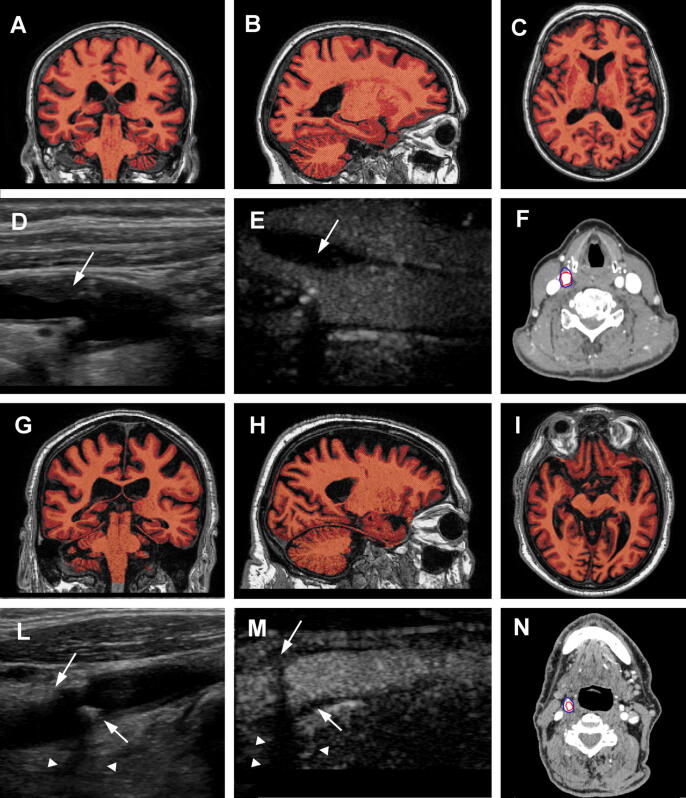
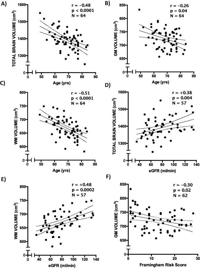
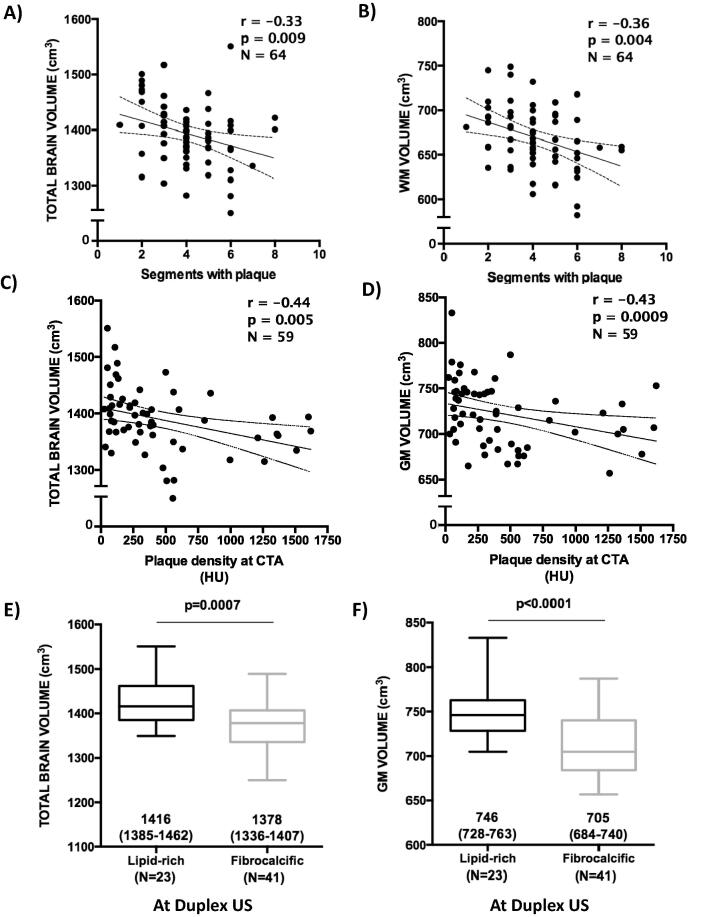
Methods and results: Sixty-four patients (aged 69 ± 8 years, 45% females) with carotid stenosis <70% based on Doppler flow velocity were enrolled in the study. Potential causes of cerebral damage other than atherosclerosis, including history of atrial fibrillation, heart failure, previous cardiac or neurosurgery and neurological disorders were excluded. All subjects underwent carotid computed tomography angiography, contrast enhanced ultrasound for assessment of plaque neovascularization and brain magnetic resonance imaging for measuring brain volumes. On multivariate regression analysis, age and fibrocalcific plaques were independently associated with lower total brain volumes (β = -3.13 and β = -30.7, both p < 0.05). Fibrocalcific plaques were also independently associated with lower gray matter (GM) volumes (β = -28.6, p = 0.003). On the other hand, age and extent of carotid atherosclerosis were independent predictors of lower white matter (WM) volumes.
Conclusions: WM and GM have different susceptibility to processes involved in parenchymal loss. Contrary to common belief, our results show that presence of fibrocalcific plaques is associated with brain atrophy.
Keywords: Brain atrophy; Brain magnetic resonance imaging; Brain volumes; CC-IMT, common carotid intima media thickness; CEUS, contrast enhanced ultrasound; CV, cardiovascular; Cardiovascular risk factors; Carotid atherosclerosis; GFR, glomerular filtration rate; GM, gray matter; TBV, total brain volume; TPA, total plaque area; Vulnerable plaque; WM, white matter.
© 2020 The Authors. Published by Elsevier B.V.
Figures
Similar articles
-
Progression of brain white matter hyperintensities in asymptomatic patients with carotid atherosclerotic plaques and no indication for revascularization.Atherosclerosis. 2019 Aug;287:171-178. doi: 10.1016/j.atherosclerosis.2019.04.230. Epub 2019 May 6. Atherosclerosis. 2019. PMID: 31101367
-
Relation between characteristics of carotid atherosclerotic plaques and brain white matter hyperintensities in asymptomatic patients.Sci Rep. 2017 Sep 5;7(1):10559. doi: 10.1038/s41598-017-11216-x. Sci Rep. 2017. PMID: 28874779 Free PMC article.
-
Contrast-enhanced ultrasonography vs B-mode ultrasound for visualization of intima-media thickness and detection of plaques in human carotid arteries.Echocardiography. 2017 May;34(5):723-730. doi: 10.1111/echo.13513. Epub 2017 Mar 19. Echocardiography. 2017. PMID: 28317160
-
Carotid intima-media thickness and plaque in cardiovascular risk assessment.JACC Cardiovasc Imaging. 2014 Oct;7(10):1025-38. doi: 10.1016/j.jcmg.2013.11.014. Epub 2014 Jul 16. JACC Cardiovasc Imaging. 2014. PMID: 25051948 Review.
-
Contrast-Enhanced Ultrasound to Assess Carotid Intraplaque Neovascularization.Ultrasound Med Biol. 2020 Mar;46(3):466-478. doi: 10.1016/j.ultrasmedbio.2019.10.020. Epub 2019 Nov 29. Ultrasound Med Biol. 2020. PMID: 31791553
Cited by
-
Associations of carotid atherosclerosis with cognitive function and brain health: Findings from a UK tri-ethnic cohort study (Southall and Brent Revisited).Atheroscler Plus. 2024 Jan 30;55:39-46. doi: 10.1016/j.athplu.2024.01.002. eCollection 2024 Mar. Atheroscler Plus. 2024. PMID: 38371883 Free PMC article.
-
Association between cerebral lesions and the severity of diabetic cardiovascular disease, retinopathy, and nephropathy-new lessons to learn from neuroimaging.J Endocrinol Invest. 2025 May 27. doi: 10.1007/s40618-025-02600-w. Online ahead of print. J Endocrinol Invest. 2025. PMID: 40423899 Review.
-
Non-alcoholic fatty liver disease increases risk of carotid atherosclerosis and ischemic stroke: An updated meta-analysis with 135,602 individuals.Clin Mol Hepatol. 2022 Jul;28(3):483-496. doi: 10.3350/cmh.2021.0406. Epub 2022 Mar 2. Clin Mol Hepatol. 2022. PMID: 35232007 Free PMC article.
-
Linking Cerebrovascular Dysfunction to Age-Related Hearing Loss and Alzheimer's Disease-Are Systemic Approaches for Diagnosis and Therapy Required?Biomolecules. 2022 Nov 19;12(11):1717. doi: 10.3390/biom12111717. Biomolecules. 2022. PMID: 36421731 Free PMC article. Review.
References
-
- den Hartog A.G., Achterberg S., Moll F.L., Kappelle L.J., Visseren F.L.J., van der Graaf Y. Asymptomatic carotid artery stenosis and the risk of ischemic stroke according to subtype in patients with clinical manifest arterial disease. Stroke. 2013;44(4):1002–1007. - PubMed
LinkOut - more resources
Full Text Sources
