. 2020 Aug 21:12:7569-7578.
doi: 10.2147/CMAR.S264474. eCollection 2020.

Clinicopathological Characteristics of Pseudomyxoma Peritonei Originated from Ovaries

Affiliations

Clinicopathological Characteristics of Pseudomyxoma Peritonei Originated from Ovaries

Fengcai Yan et al. Cancer Manag Res. .

Abstract

Objective: This study aims to demonstrate clinicopathological characteristics and immunohistopathological phenotypes of pseudomyxoma peritonei (PMP) originated from ovaries.

Methods: The primary origin of PMP was explored by reviewing H&E sections retrospectively and performing a series of immunohistochemical staining on CK7, CK20, CDX2, CEA, Villin, SATB2, CA125, ER, PR, and MUC.

Results: Among 310 PMP patients, a few originated from extra-appendix, whereas eight cases were of ovarian origin (2.6%), including three teratoma-associated ovarian mucinous tumors and five primary ovarian mucinous tumors with spontaneous or iatrogenic rupture, respectively. Most peritoneal metastases were acellular mucin or low-grade mucinous carcinoma peritonei (6/8, 75%), while the rest were high-grade mucinous carcinoma peritonei (2/8, 25%). Tumors were positive for CK20, CDX2, CEA, and Villin. SATB2 was specifically diffuse positive in teratoma-associated ovarian mucinous tumors, and negative in primary ovarian mucinous tumors. Differential expression of MUC was observed in these tumors.

Conclusion: PMP of ovarian origin is extremely rare. The precise diagnosis requires serial sections of the appendix or suspicious tissue to exclude appendiceal mucinous neoplasms, as well as comprehensive analysis of clinical features, surgical findings, histopathological characteristics, and immunohistochemistry on specific biomarkers.

Keywords: PMP; mucinous tumor; ovary; pseudomyxoma peritonei.


Conflict of interest statement

The authors report no conflicts of interest in this work.

Figures

Figure 1
Figure 1
Clinical features of (A) ovarian PMPs vs (B) purely mucinous cystadenoma. Ovarian origin had much thinner mucus than appendiceal PMP that metastasizes to ovaries (which presented with significant gelatinous mucus).
Figure 2
Figure 2
Histopathological features of ovarian PMPs. (A1) Ovarian mucinous borderline tumor with multiple-grade papillary structures (H&E, magnification ×100). (B1) Skin appendages with gastrointestinal-type mucinous gland (black arrow) (H&E, magnification ×200). (A2) Mucus in serous layer of appendix (H&E, magnification ×50). (B2) Low-grade neoplastic epithelium in serous layer of appendix (H&E, magnification ×25). (A3) Teratoma-associated mucinous neoplasm. Note obvious subepithelial cleft similar to that in low-grade appendiceal mucinous neoplasm involving ovary. (B3). Scanning magnification of primary ovarian gastrointestinal-type mucinous tumor, containing scattered goblet cells. (A3, B3) H&E, magnification ×200.
Figure 3
Figure 3
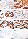
Immunohistochemical staining in PMPs originated from ovaries. Teratoma-associated mucinous tumors were (A1) Negative for CK7, (A2) Strongly positive for CK20, (A3) Positive for CDX2, (A4) Strongly positive for SATB2. PMPs originated from purely ovarian mucinous tumor were (B1) Strongly positive for CK7, (B2) Scattered positive of goblet cell for CK20, (B3) Weakly positive for CDX2, (B4) Negative for SATB2. (A1-B4) EnVision staining ×200.
Figure 4
Figure 4
Immunohistochemical staining in PMPs originated from ovaries. Teratoma-associated mucinous tumors were (A1) Weakly positive for MUC1, (A2) Diffusely strong positive for MUC2, (A3) Diffusely positive for MUC5AC, (A4) Negative for MUC6. Primary ovarian mucinous tumor was (B1) Weakly positive for MUC1, (B2) Scattered positive of goblet cell for MUC2, (B3) Diffusely positive for MUC5AC, (B4) Focal positive for MUC6. (A1-B4) EnVision staining ×200.

Similar articles

Cited by

References

    1. Carr NJ, Cecil TD, Mohamed F, et al. A consensus for classification and pathologic reporting of pseudomyxoma peritonei and associated appendiceal neoplasia the results of the PSOGI. Am J Surg Pathol. 2016;40:14–26. doi:10.1097/PAS.0000000000000535 - DOI - PubMed
    1. Mittal R, Chandramohan A, Moran B. Pseudomyxoma peritonei: natural history and treatment. Int J Hyperthermia. 2017;33:511–519. doi:10.1080/02656736.2017.1310938 - DOI - PubMed
    1. Ronnett BM, Shmookler BM, Sugarbaker PH, et al. Pseudomyxoma peritonei: new concepts in diagnosis, origin, nomenclature, and relationship to mucinous borderline (low malignant potential) tumors of the ovary. Anat Pathol. 1997;2:197–226. - PubMed
    1. Stewart CJ, Ardakani NM, Doherty DA, et al. An evaluation of the morphologic features of low-grade mucinous neoplasms of the appendix metastatic in the ovary, and comparison with primary ovarian mucinous tumors. Int J Gynecol Pathol. 2014;33:1–10. doi:10.1097/PGP.0b013e318284e070 - DOI - PubMed
    1. van Ruth S, Acherman YI, van de Vijver MJ, et al. Pseudomyxoma peritonei: a review of 62 cases. Eur J Surg Oncol. 2003;29:682–688. doi:10.1016/s0748-7983(03)00149-5 - DOI - PubMed