. 2020 Jul-Aug;53(4):255-261.
doi: 10.1590/0100-3984.2020.0030.

Thoracic tomographic manifestations in symptomatic respiratory patients with COVID-19

Affiliations

Thoracic tomographic manifestations in symptomatic respiratory patients with COVID-19

Lucas de Pádua Gomes de Farias et al. Radiol Bras. 2020 Jul-Aug.

Abstract

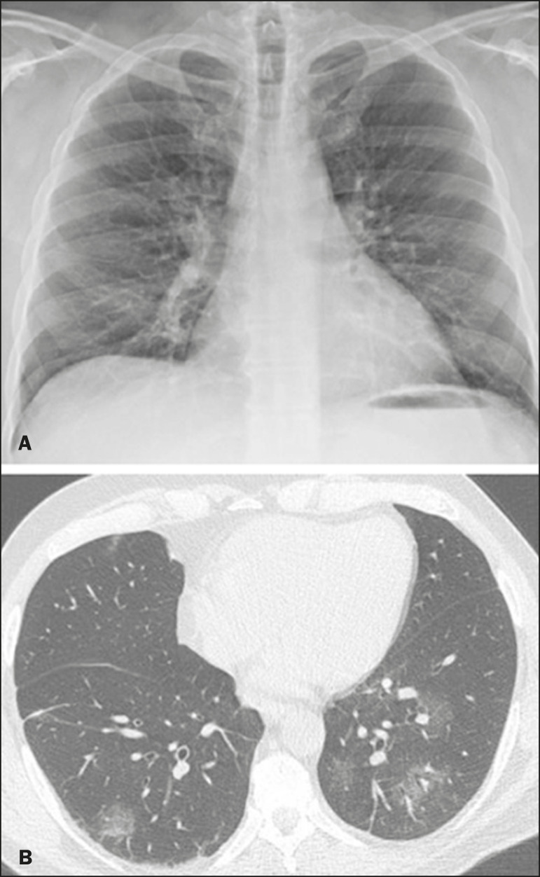
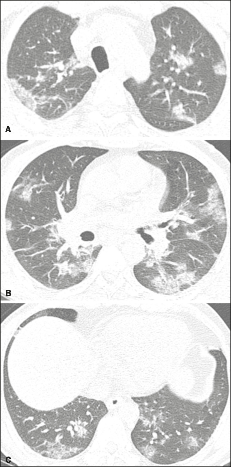
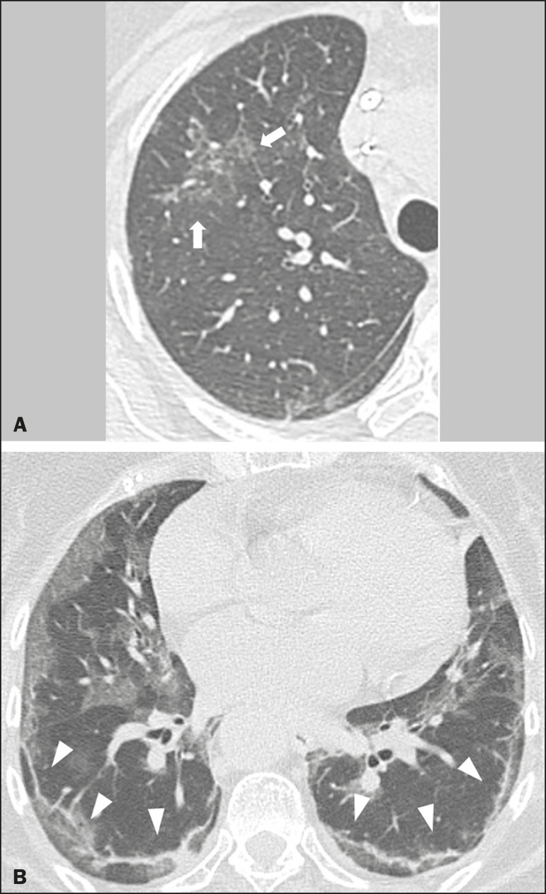
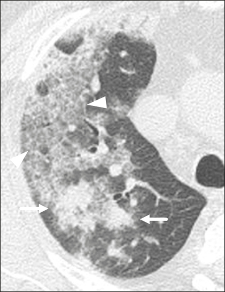
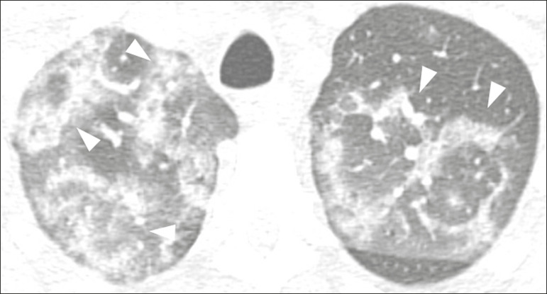
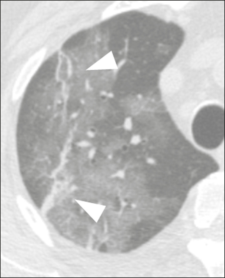
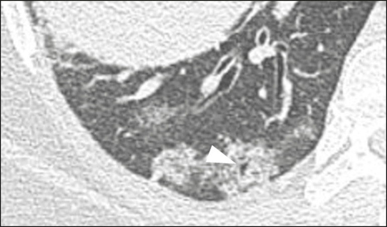
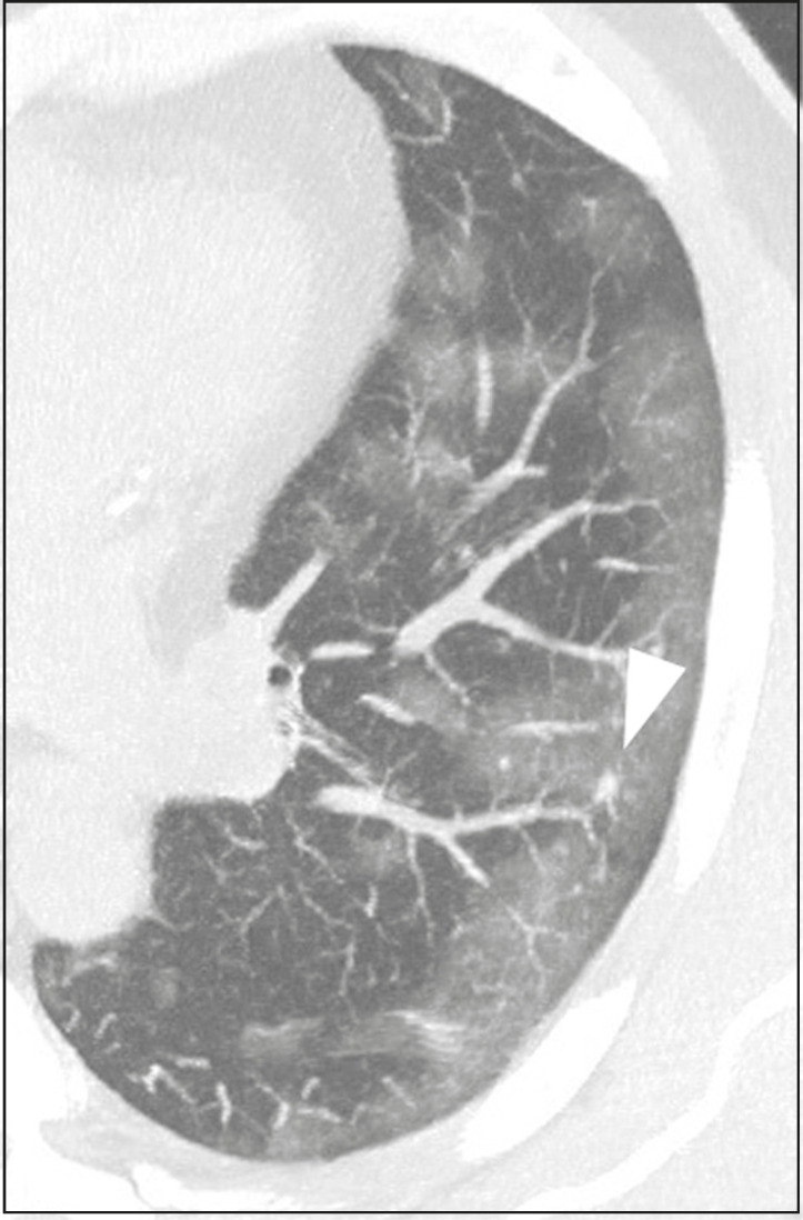
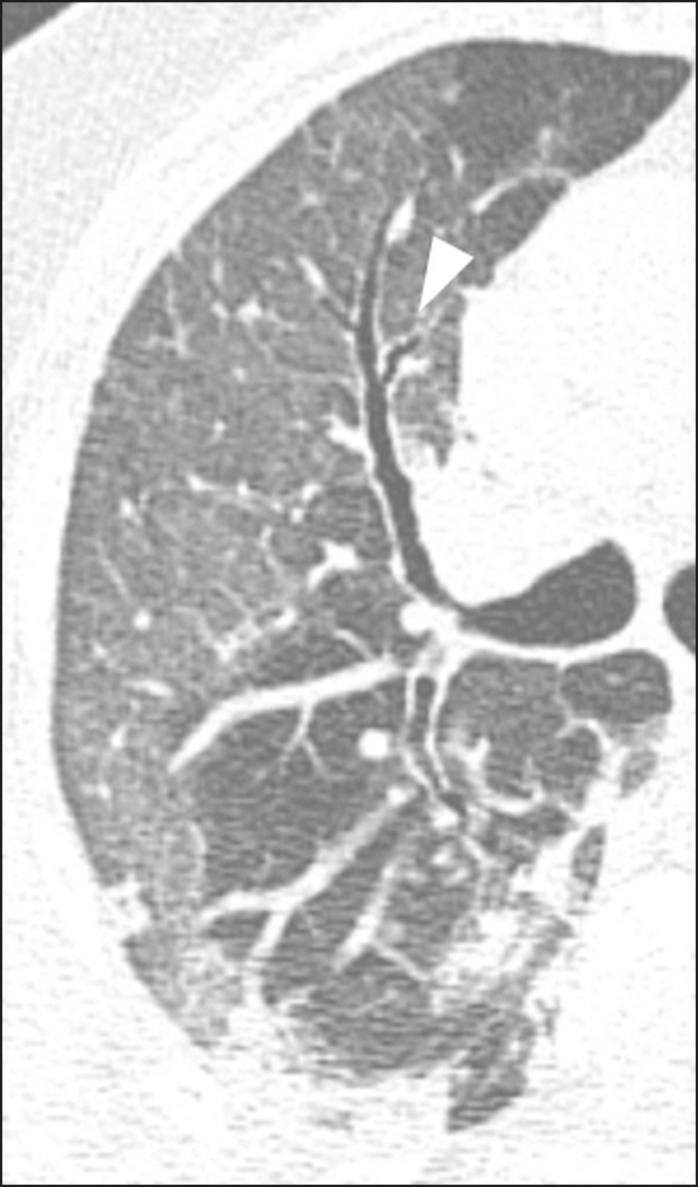
China was the epicenter for the novel coronavirus disease (COVID-19), which quickly spread to other Asian countries and later to Western countries; subsequently, COVID-19 was categorized as a pandemic by the World Health Organization. Diagnosis primarily depends on viral detection in respiratory samples; however, available kits are limited, lack high sensitivity, and have a long turnaround time for providing results. In this scenario, computed tomography has emerged as an efficient and available high-sensitivity method, allowing radiologists to readily recognize findings related to COVID-19. The objective of this article is to demonstrate the main tomographic findings in symptomatic respiratory patients with COVID-19 to assist medical professionals during this critical moment.

A doença pelo novo coronavírus (COVID-19) teve epicentro na China e rapidamente se espalhou pelos demais países asiáticos e, posteriormente, para os países ocidentais, sendo definida como pandemia pela Organização Mundial da Saúde. O diagnóstico da COVID-19 é primariamente dependente da pesquisa do vírus nas vias aéreas superiores, mas os kits para sua confirmação ainda são limitados, não apresentam sensibilidade elevada e os resultados são demorados. Nesse cenário, a tomografia computadorizada surge como método eficiente e disponível e com alta sensibilidade, cabendo a nós radiologistas reconhecer prontamente os achados relacionados a essa doença. O objetivo deste artigo é demonstrar os principais achados tomográficos de tórax em pacientes sintomáticos respiratórios infectados pela COVID-19, de modo a auxiliar os colegas nesse momento crítico.

Keywords: COVID-19; Coronavirus; Multislice computed tomography.


Figures

Figure 1
Figure 1
Male patient, 35 years old. Posteroanterior chest radiography (A) showing no pulmonary changes. MDCT (B) on the same day shows opacities with ground-glass attenuation in the lower lobes. Positive RT-PCR for SARS-CoV-2.
Figure 2
Figure 2
Male patient, 48 years old. MDCT shows ground-glass opacities and diffuse consolidation foci in the upper (A), middle (B), and lower (C) lung fields with predominantly peripheral distribution in the posterior regions. Positive RTPCR for SARS-CoV-2.
Figure 3
Figure 3
Female patients, 51 (A) and 68 years old (B). MDCT shows reticular opacities (arrows) in the anterior segment of the right upper lobe and bilateral subpleural curvilinear lines (arrowheads) in the lower lobes. Additionally, other multifocal opacities with ground-glass attenuation are present. Positive RT-PCR for SARS-CoV-2 in both patients.
Figure 4
Figure 4
Male patient, 59 years old. MDCT shows ground-glass opacity in the right lung associated with interlobular and intralobular septal thickening, characterized by the crazy-paving pattern (arrowheads) and some consolidation foci (arrows). Positive RT-PCR for SARS-CoV-2.
Figure 5
Figure 5
Male patient, 56 years old. MDCT shows ground-glass attenuation opacities, partially or surrounded by a consolidation ring, characterized by the reversed halo sign (arrowheads) in both lungs. Positive RT-PCR for SARS-CoV-2.
Figure 6
Figure 6
Male patient, 70 years old. MDCT shows parenchymal bands/fibroatelectatic band opacities on the pulmonary periphery. Additionally, other opacities with ground-glass attenuation are observed. Positive RT-PCR for SARS-CoV-2.
Figure 7
Figure 7
Male patient, 41 years old. MDCT shows pseudo-cavity associated with consolidation and ground-glass opacity on the right lower lobe (arrowhead). Positive RT-PCR for SARS-CoV-2.
Figure 8
Figure 8
Male patient, 44 years old. Axial MDCT image with maximum intensity projection reconstruction shows extensive peripheral opacity with groundglass attenuation and some ectatic vessels inside (arrowhead). Positive RTPCR for SARS-CoV-2.
Figure 9
Figure 9
Male patient, 69 years old. MDCT shows diffuse ground-glass opacity in both lungs and consolidation foci associated with a minor bilateral pleural effusion, indicating restrictive atelectasis of the adjacent lung parenchyma. Positive RT-PCR for SARS-CoV-2.
Figure 10
Figure 10
Female patient, 76 years old. MDCT shows air bronchogram between opacity with ground-glass attenuation and some consolidation foci associated with bronchial wall thickening and distortion (arrowhead). Positive RT-PCR for SARS-CoV-2.
Figure 11
Figure 11
Illustrative scheme to measure the cardiac chambers and estimate the volume of pericardial effusion/thickening in chest tomography not coupled to an electrocardiogram. The left atrium (LA) must be measured in the anteroposterior diameter while the other cardiac chambers must be measured in the transverse diameter(27,28).

References

    1. Zhu N, Zhang D, Wang W, et al. A novel coronavirus from patients with pneumonia in China, 2019. N Engl J Med. 2020;382:727–733. - PMC - PubMed
    1. World Health Organization WHO Director-General's opening remarks at the media briefing on COVID-19 - 11 March 2020. [2020 Mar 11]. [2020 Mar 23]. Available from: https://www.who.int/dg/speeches/detail/who-director-general-s-opening-re....
    1. Simpson S, Kay FU, Abbara S, et al. Radiological Society of North America Expert Consensus Statement on Reporting Chest CT Findings Related to COVID-19. Endorsed by the Society of Thoracic Radiology, the American College of Radiology, and RSNA. Radiology: Cardiothoracic Imaging. 2020;2(2) - PMC - PubMed
    1. American College of Radiology ACR Recommendations for the use of chest radiography and computed tomography (CT) for suspected COVID-19 infection. [2020 Mar 22]. [2020 Mar 23]. Available from: https://www.acr.org/Advocacy-and-Economics/ACR-Position-Statements/Recom....
    1. Colégio Brasileiro de Radiologia e Diagnóstico por Imagem Recomendações de uso de métodos de imagem para pacientes suspeitos de infecção pelo COVID-19. [2020 Mar 23]. Available from: https://cbr.org.br/wp-content/uploads/2020/03/CBR_Recomendações-de-uso-d....

LinkOut - more resources