. 2020 Jul 16;2(1):vdaa088.
doi: 10.1093/noajnl/vdaa088. eCollection 2020 Jan-Dec.

Patient-derived cells from recurrent tumors that model the evolution of IDH-mutant glioma

Affiliations

Patient-derived cells from recurrent tumors that model the evolution of IDH-mutant glioma

Lindsey E Jones et al. Neurooncol Adv. .

Abstract

Background: IDH-mutant lower-grade gliomas (LGGs) evolve under the selective pressure of therapy, but well-characterized patient-derived cells (PDCs) modeling evolutionary stages are lacking. IDH-mutant LGGs may develop therapeutic resistance associated with chemotherapy-driven hypermutation and malignant progression. The aim of this study was to establish and characterize PDCs, single-cell-derived PDCs (scPDCs), and xenografts (PDX) of IDH1-mutant recurrences representing distinct stages of tumor evolution.

Methods: We derived and validated cell cultures from IDH1-mutant recurrences of astrocytoma and oligodendroglioma. We used exome sequencing and phylogenetic reconstruction to examine the evolutionary stage represented by PDCs, scPDCs, and PDX relative to corresponding spatiotemporal tumor tissue and germline DNA. PDCs were also characterized for growth and tumor immortality phenotypes, and PDX were examined histologically.

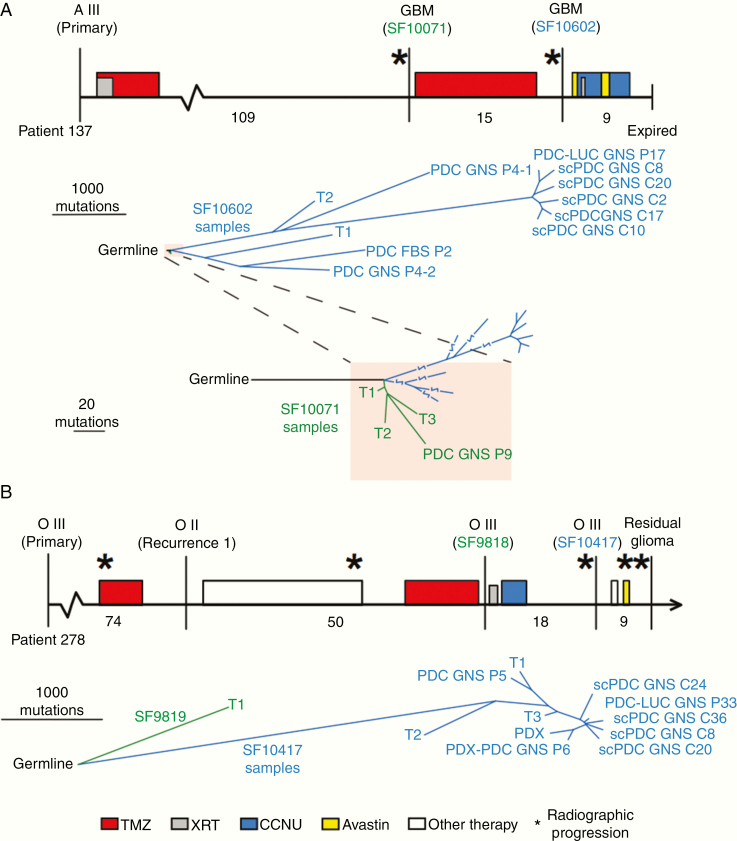
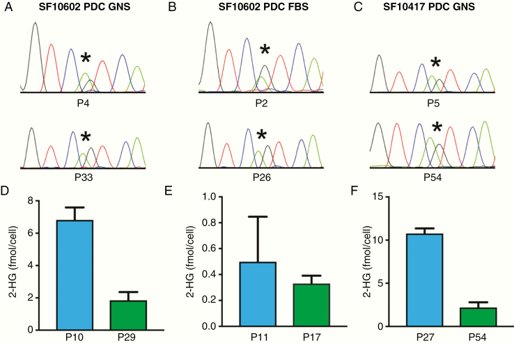
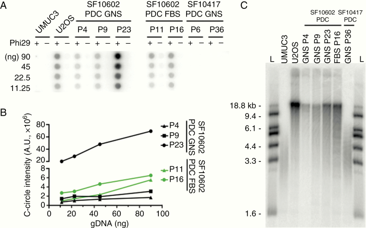
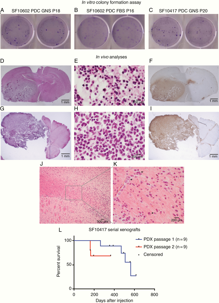
Results: The integrated astrocytoma phylogeny revealed 2 independent founder clonal expansions of hypermutated (HM) cells in tumor tissue that are faithfully represented by independent PDCs. The oligodendroglioma phylogeny showed more than 4000 temozolomide-associated mutations shared among tumor samples, PDCs, scPDCs, and PDX, suggesting a shared monoclonal origin. The PDCs from both subtypes exhibited hallmarks of tumorigenesis, retention of subtype-defining genomic features, production of 2-hydroxyglutarate, and subtype-specific telomere maintenance mechanisms that confer tumor cell immortality. The oligodendroglioma PDCs formed infiltrative intracranial tumors with characteristic histology.

Conclusions: These PDCs, scPDCs, and PDX are unique and versatile community resources that model the heterogeneous clonal origins and functions of recurrent IDH1-mutant LGGs. The integrated phylogenies advance our knowledge of the complex evolution and immense mutational load of IDH1-mutant HM glioma.

Keywords: IDH1-mutant glioma; hypermutation; intracranial xenograft; intratumoral heterogeneity and evolution; patient-derived cells.


Figures

Figure 1.
Figure 1.
Phylogenies of IDH-mutant astrocytoma and oligodendroglioma PDCs, scPDCs, and PDX integrated with spatiotemporal tumor tissue samples. (A, upper panel): The clinical timeline of astrocytoma Patient 137, drawn to scale. Vertical lines indicate surgical resection; stars represent radiographic progression; filled rectangles indicate treatment period and fill color indicates treatment type (see key at bottom). Months between surgical resections are provided below the timeline. The tumor classification is listed above each surgical time point, and surgeries from which samples were sequenced are designated with an SF number. Lower panel: A tumor phylogeny was constructed from single-nucleotide somatic mutations. Line length is proportional to the number of mutations. Lowest panel: A zoomed-in view of the shaded portion of the tree showing the relationship between samples of the non-HM first tumor recurrence (SF10071) and the HM samples of the second recurrence (SF10602). (B, upper panel): The clinical timeline of oligodendroglioma Patient 278. Lower panel: A tumor phylogeny of tumor tissue and PDCs of Patient 278. FBS, cells cultured in media containing fetal bovine serum; GNS, cells cultured in glioma neural stem cell media; PDCs, patient-derived cells; scPDCs, single-cell-derived patient-derived cells; PDC-LUC, luciferase-modified PDC; T1, T2, T3, spatially distinct pieces of tumor tissue; PDX, patient-derived intracranial xenograft; PDX-PDC, PDC derived from PDX; A, astrocytoma; O, oligodendroglioma; TMZ, temozolomide; XRT, radiation therapy; CCNU, lomustine; P#, passage number.
Figure 2.
Figure 2.
PDCs retain heterozygous IDH1 R132H through serial passage and continue to produce 2-HG. (A–C) Each of the 3 PDCs retains IDH1 R132H through multiple passages as determined by Sanger sequencing and (D–F) produces 2-HG through multiple passages, as measured by NMR. *The location of the heterozygous G>A mutation in IDH1.
Figure 3.
Figure 3.
ATRX-mutant PDCs are positive for the ALT phenotype. (A) The ATRX-mutant SF10602 PDC GNS and PDC FBS cultures are positive for C-circle amplification across multiple passages. In contrast, the ATRX wildtype SF10417 GNS culture is negative for C-circles. A human bladder transitional cell carcinoma cell line (UMUC3) and a human osteosarcoma cell line (U2OS) serve as negative and positive controls for C-circle amplification, respectively. (B) Signal intensity was quantified in arbitrary units and plotted. (C) Telomere restriction fragment length analysis shows an accumulation of long chromosomes and a heterogeneous telomere length distribution characteristic of ALT. The ALT cells show a tighter and shorter telomere length distribution. L, DNA ladder.
Figure 4.
Figure 4.
PDCs demonstrate tumorigenic properties in vitro and in vivo. To measure in vitro tumorigenesis, 1000 cells each of (A) astrocytoma SF10602 PDC GNS, (B) SF10602 PDC FBS, and (C) oligodendroglioma SF10417 PDC GNS were seeded into soft agar. After 4 weeks, they were stained with crystal violet and yielded an average of 185, 117, and 218 colonies, respectively. (D–L) To measure in vivo tumorigenesis, 300 000 cells were implanted into the right cerebrum of 5-week-old female athymic mice. (D) H&E staining of a coronal section of the mouse brain demonstrating the infiltrative nature of the tumor. (E) The tumor recapitulates oligodendroglioma histologic features and (F) is positive for IDH1 R132H by IHC. A cell line was established from this tumor and re-implanted in 5-week-old female athymic mice, where (G) it again formed infiltrative tumors that (H) recapitulate oligodendroglioma histology and (I) are positive for IDH1 R132H by IHC. (J) Tumor cells infiltrated from the site of injection in the striatum into and within the corpus callosum, (K) as single cells and as clusters of tumor cells (arrows) along blood vessels. (L) Upon the first xenotransplantation, SF10417 PDC GNS formed tumors in 4 of 9 mice over a protracted time period, consistent with the generally slower growth of this tumor subtype. Upon serial xenotransplantation, SF10417 PDX-PDC GNS formed tumors in 3 of 9 mice in a reduced time period. Tick marks indicate censored animals.

Similar articles

Cited by

References

    1. Louis DN, Perry A, Reifenberger G, et al. . The 2016 World Health Organization classification of tumors of the central nervous system: a summary. Acta Neuropathol. 2016;131(6):803–820. - PubMed
    1. Yip S, Butterfield YS, Morozova O, et al. . Concurrent CIC mutations, IDH mutations, and 1p/19q loss distinguish oligodendrogliomas from other cancers. J Pathol. 2012;226(1):7–16. - PMC - PubMed
    1. Soffietti R, Baumert BG, Bello L, et al. ; European Federation of Neurological Societies Guidelines on management of low-grade gliomas: report of an EFNS-EANO Task Force. Eur J Neurol. 2010;17(9):1124–1133. - PubMed
    1. Shaw EG, Wang M, Coons SW, et al. . Randomized trial of radiation therapy plus procarbazine, lomustine, and vincristine chemotherapy for supratentorial adult low-grade glioma: initial results of RTOG 9802. J Clin Oncol. 2012;30(25):3065–3070. - PMC - PubMed
    1. Fisher BJ, Hu C, Macdonald DR, et al. . Phase 2 study of temozolomide-based chemoradiation therapy for high-risk low-grade gliomas: preliminary results of Radiation Therapy Oncology Group 0424. Int J Radiat Oncol Biol Phys. 2015;91(3):497–504. - PMC - PubMed