. 2020 Sep 22;1(6):100099.
doi: 10.1016/j.xcrm.2020.100099. Epub 2020 Sep 1.

Combined Point-of-Care Nucleic Acid and Antibody Testing for SARS-CoV-2 following Emergence of D614G Spike Variant

Collaborators, Affiliations

Combined Point-of-Care Nucleic Acid and Antibody Testing for SARS-CoV-2 following Emergence of D614G Spike Variant

Petra Mlcochova et al. Cell Rep Med. .

Abstract

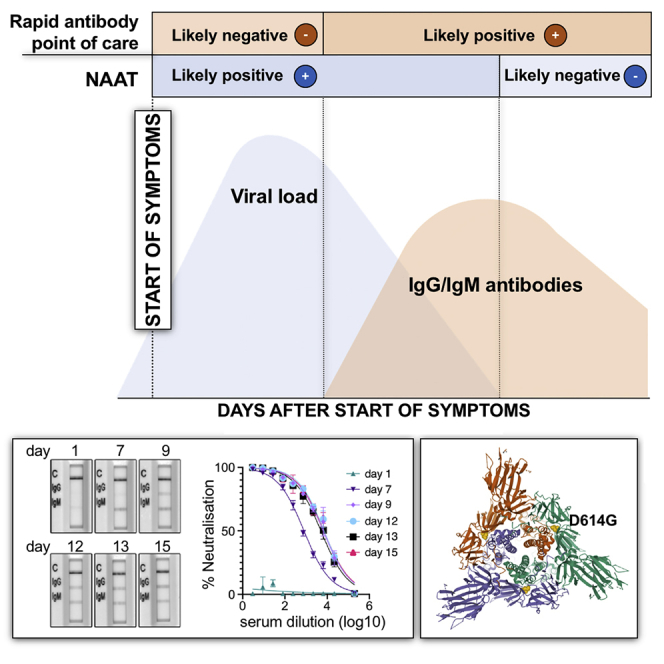
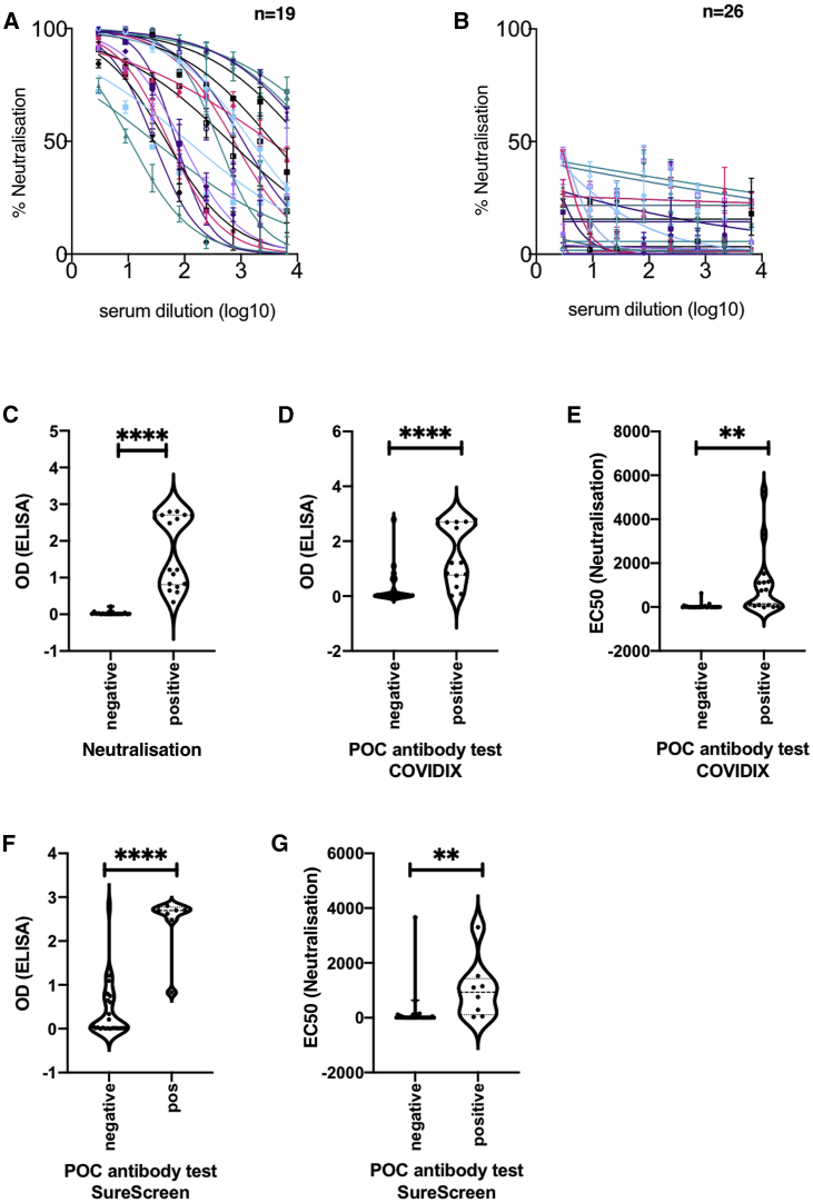
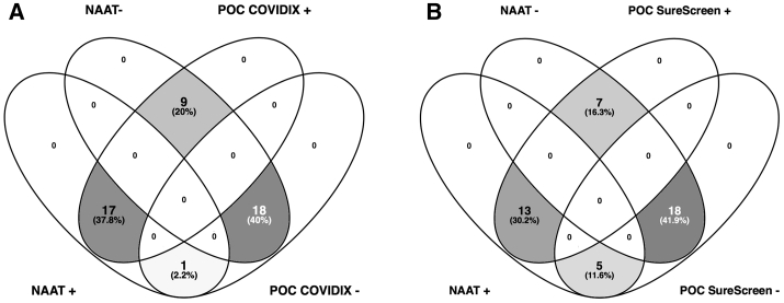
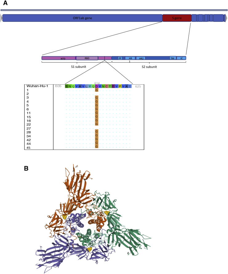
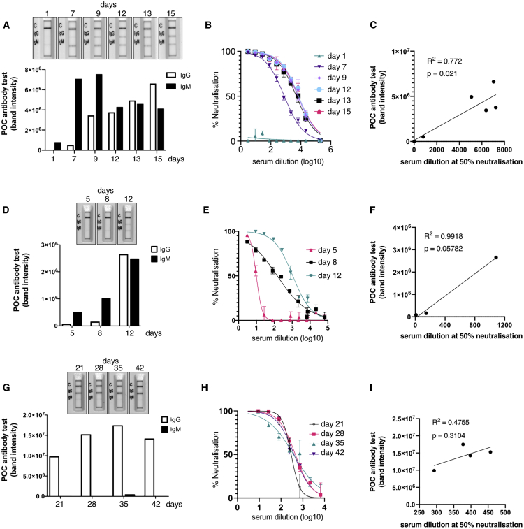
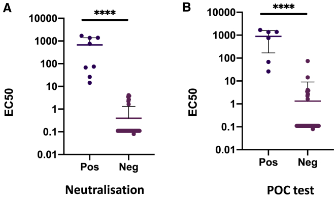
Rapid COVID-19 diagnosis in the hospital is essential, although this is complicated by 30%-50% of nose/throat swabs being negative by SARS-CoV-2 nucleic acid amplification testing (NAAT). Furthermore, the D614G spike mutant dominates the pandemic and it is unclear how serological tests designed to detect anti-spike antibodies perform against this variant. We assess the diagnostic accuracy of combined rapid antibody point of care (POC) and nucleic acid assays for suspected COVID-19 disease due to either wild-type or the D614G spike mutant SARS-CoV-2. The overall detection rate for COVID-19 is 79.2% (95% CI 57.8-92.9) by rapid NAAT alone. The combined point of care antibody test and rapid NAAT is not affected by D614G and results in very high sensitivity for COVID-19 diagnosis with very high specificity.

Keywords: COVID-19; D614G; SARS-CoV-2; point of care testing; rapid diagnoses.


Conflict of interest statement

The authors declare no competing interests.

Figures

None
Graphical abstract
Figure 1
Figure 1
Antibody Detection for SARS-CoV-2: Cross-Validation of Lateral Flow Diagnostic Tests (POC Antibody Tests) with ELISA and SARS-CoV-2 Pseudotype Virus Neutralization Assays (A and B) Serum from COVID-19 suspected participants inhibited (n = 19) (A) or did not inhibit (n = 26) (B) SARS-CoV-2 pseudotype virus infection in a neutralization assay. Serum from a healthy donor was used and a negative control. The error bars represent SEMs. (C) Comparison between ELISA and positive/negative results from neutralization assay; n = 37, p < 0.0001. (D) Comparison between ELISA Spike protein reactivity and positive/negative POC antibody test results (COVIDIX SARS-CoV-2 IgM/IgG test); n = 38, p < 0.0001. (E) Comparison between half-maximal effective concentration (EC50) dilution titer from neutralizing assay and positive/negative POC antibody test results (COVIDIX SARS-CoV-2 IgM/IgG test); n = 44, p = 0.0025. (F) Comparison between ELISA IgG and positive/negative POC IgG band results for SureScreen SARS-CoV-2 IgM/IgG test; n = 38, p < 0.0001. (G) Comparison between EC50 dilution titer from neutralization assay and positive/negative SureScreen SARS-CoV-2 IgM/IgG antibody band test results; n = 43, p = 0.005. The assays were performed in duplicate.
Figure 2
Figure 2
Venn Diagrams Comparing Positive and Negative Diagnostic Test Results in Hospitalized Patients Testing by NAAT and POC antibody testing by (A) COVIDIX Healthcare IgM/IgG kit (n = 45) and (B) SureScreen IgM/IgG kit (n = 43).
Figure 3
Figure 3
Spike D614G Characterization in the Phase 1 Clinical Cohort (A) Genome map of SARS-CoV-2, with overall topography of Spike expanded. FP, fusion peptide; HR1, heptad repeat 1; HR2, heptad repeat 2; IC, intracellular domain; NTD, N-terminal domain; RBD, receptor-binding domain; TM, transmembrane region. The aligned sequence of 10 amino acids on either side of D614 is shown for 16 participants for whom sequence data were available. A dot represents where the amino acid is unchanged from wild type, the mutant glycine is represented by G. (B) Top view of SARS-CoV-2 Spike glycoprotein trimeric structure in a closed state, with position 614 in yellow in each protomer. Structure determined by cryoelectron microscopy. RCSB PDB: 6VXX.
Figure 4
Figure 4
Longitudinal Antibody Responses in Patients Infected with D614G Mutant SARS-CoV-2 Detected by Rapid Lateral Flow and Neutralization Assays (A, D, and G) An immunochromatographic lateral flow rapid diagnostic test (POC antibody test-COVIDIX SARS-CoV-2 IgM IgG test) on longitudinal samples in individual patients detecting SARS-CoV-2 IgM and IgG bands. Band intensities were acquired using the ChemiDoc MP Imaging System and quantified using Image Lab software. (B, E, and H) SARS-CoV-2 pseudotyped virus neutralization assay from longitudinal serum samples in individual patient examples. The assays were performed in duplicate. The error bars represent SEMs. (C, F, and I) Comparison of IgG band intensities from lateral flow rapid diagnostic test with EC50 neutralization titers from SARS-CoV-2 pseudotyped virus neutralization assay in individual patients. The correlations were estimated by linear regression analysis.
Figure 5
Figure 5
Distribution of Serum Neutralization Activity against SARS-CoV-2 in Hospitalized Patients during the Implementation Phase (A) Neutralization EC50 dilution titer interpreted as positive or negative using a cutoff for positive neutralization of 1:4 dilution. (B) Neutralization data for individual patients stratified by POC antibody test result (both tests were fully concordant in phase 2). The data points represent the reciprocal dilution of serum required to inhibit 50% of infection by lentivirus pseudotyped with the SARS-CoV-2 Spike glycoprotein. The assays were performed in duplicate. The line represents the mean and the bar represents the standard deviation (n = 101 sera tested).

Similar articles

Cited by

References

    1. Dong E., Du H., Gardner L. An interactive web-based dashboard to track COVID-19 in real time. Lancet Infect. Dis. 2020;20:533–534. - PMC - PubMed
    1. Collier D.A., Assennato S.M., Warne B., Sithole N., Sharrocks K., Ritchie A., Ravji P., Routledge M., Sparkes D., Skittrall J. Point of Care Nucleic Acid Testing for SARS-CoV-2 in Hospitalized Patients: A Clinical Validation Trial and Implementation Study. Cell Rep. Med. 2020;1:100062. - PMC - PubMed
    1. Arevalo-Rodriguez I., Buitrago-Garcia D., Simancas-Racines D., Zambrano-Achig P., del Campo R., Ciapponi A., Sued O., Martinez-Garcia L., Rutjes L., Low N. False-negative results of initial RT-PCR assays for COVID-19: a systematic review. medRxiv. 2020 doi: 10.1101/2020.04.16.20066787. - DOI - PMC - PubMed
    1. Wang W., Xu Y., Gao R., Lu R., Han K., Wu G., Tan W. Detection of SARS-CoV-2 in Different Types of Clinical Specimens. JAMA. 2020;323:1843–1844. - PMC - PubMed
    1. Fang Y., Zhang H., Xie J., Lin M., Ying L., Pang P., Ji W. Sensitivity of Chest CT for COVID-19: Comparison to RT-PCR. Radiology. 2020;296:E115–E117. - PMC - PubMed

Publication types

MeSH terms

Substances

Supplementary concepts