

Diagnosis of thyroid nodules for ultrasonographic characteristics indicative of malignancy using random forest
- PMID: 32905307
- PMCID: PMC7469308
- DOI: 10.1186/s13040-020-00223-w
Diagnosis of thyroid nodules for ultrasonographic characteristics indicative of malignancy using random forest
Abstract
Background: Various combinations of ultrasonographic (US) characteristics are increasingly utilized to classify thyroid nodules. But they lack theories, and heavily depend on radiologists' experience, and cannot correctly classify thyroid nodules. Hence, our main purpose of this manuscript is to select the US characteristics significantly associated with malignancy and to develop an efficient scoring system for facilitating ultrasonic clinicians to correctly identify thyroid malignancy.
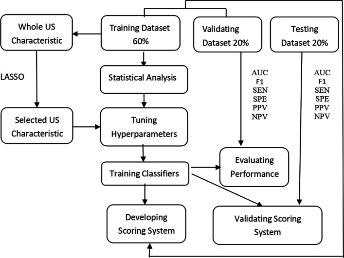
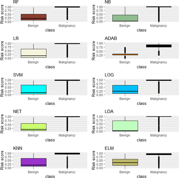
Methods: A logistic regression (LR) model is utilized to identify the potential thyroid malignancy, and the least absolute shrinkage and selection operator (LASSO) method is adopted to simultaneously select US characteristics significantly associated with malignancy and estimate parameters in LR model. Based on the selected US characteristics, we calculate the probability for each of thyroid nodules via random forest (RF) and extreme learning machine (ELM), and develop a scoring system to classify thyroid nodules. For comparison, we also consider eight state-of-the-art methods such as support vector machine (SVM), neural network (NET), etc. The area under the receiver operating characteristic curve (AUC) is employed to measure the accuracy of various classifiers.
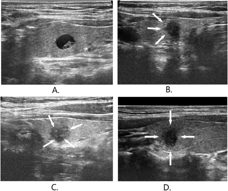
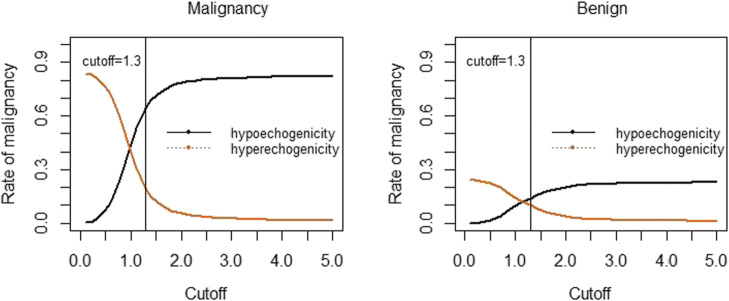
Results: The US characteristics: nodule size, AP/T≥1, solid component, micro-calcifications, hackly border, hypoechogenicity, presence of halo, unclear border, irregular margin, and central vascularity are selected as the significant predictors associated with thyroid malignancy via the LASSO LR (LLR). Using the developed scoring system, thyroid nodules are classified into the following four categories: benign, low suspicion, intermediate suspicion, and high suspicion, whose rates of malignancy correctly identified for RF (ELM) method on the testing dataset are 0.0% (4.3%), 14.3% (50.0%), 58.1% (59.1%) and 96.1% (97.7%), respectively.
Conclusion: LLR together with RF performs better than other methods in identifying malignancy, especially for abnormal nodules, in terms of risk scores. The developed scoring system can well predict the risk of malignancy and guide medical doctors to make management decisions for reducing the number of unnecessary biopsies for benign nodules.
Keywords: Random forest; Risk score; Thyroid nodule; Ultrasonographic characteristic.
© The Author(s) 2020.
Conflict of interest statement
Competing interestsThe authors declare that they have no competing interests.
Figures


References
-
- Kwak JY, Han KH, Yoon JH, Moon HJ, Son EJ, Park SH, Jung HK, Choi JS, Kim BM, Kim E-K. Thyroid imaging reporting and data system for US features of nodules: a step in establishing better stratification of cancer risk. Radiology. 2011;260(3):892–9. - PubMed
-
- Morris LF, Ragavendra N, Yeh MW. Evidence-based assessment of the role of ultrasonography in the management of benign thyroid nodules. World J Surg. 2008;32(7):1253–63. - PubMed
-
- Horvath E, Majlis S, Rossi R, Franco C, Niedmann JP, Castro A, Dominguez M. An ultrasonogram reporting system for thyroid nodules stratifying cancer risk for clinical management. J Clin Endocrinol Metab. 2009;94(5):1748–51. - PubMed
LinkOut - more resources
Full Text Sources
Research Materials
Miscellaneous
