. 2020 Aug 15:417:213343.
doi: 10.1016/j.ccr.2020.213343. Epub 2020 May 7.

Sulfhydryl groups as targets of mercury toxicity

Affiliations

Sulfhydryl groups as targets of mercury toxicity

Olga P Ajsuvakova et al. Coord Chem Rev. .

Abstract

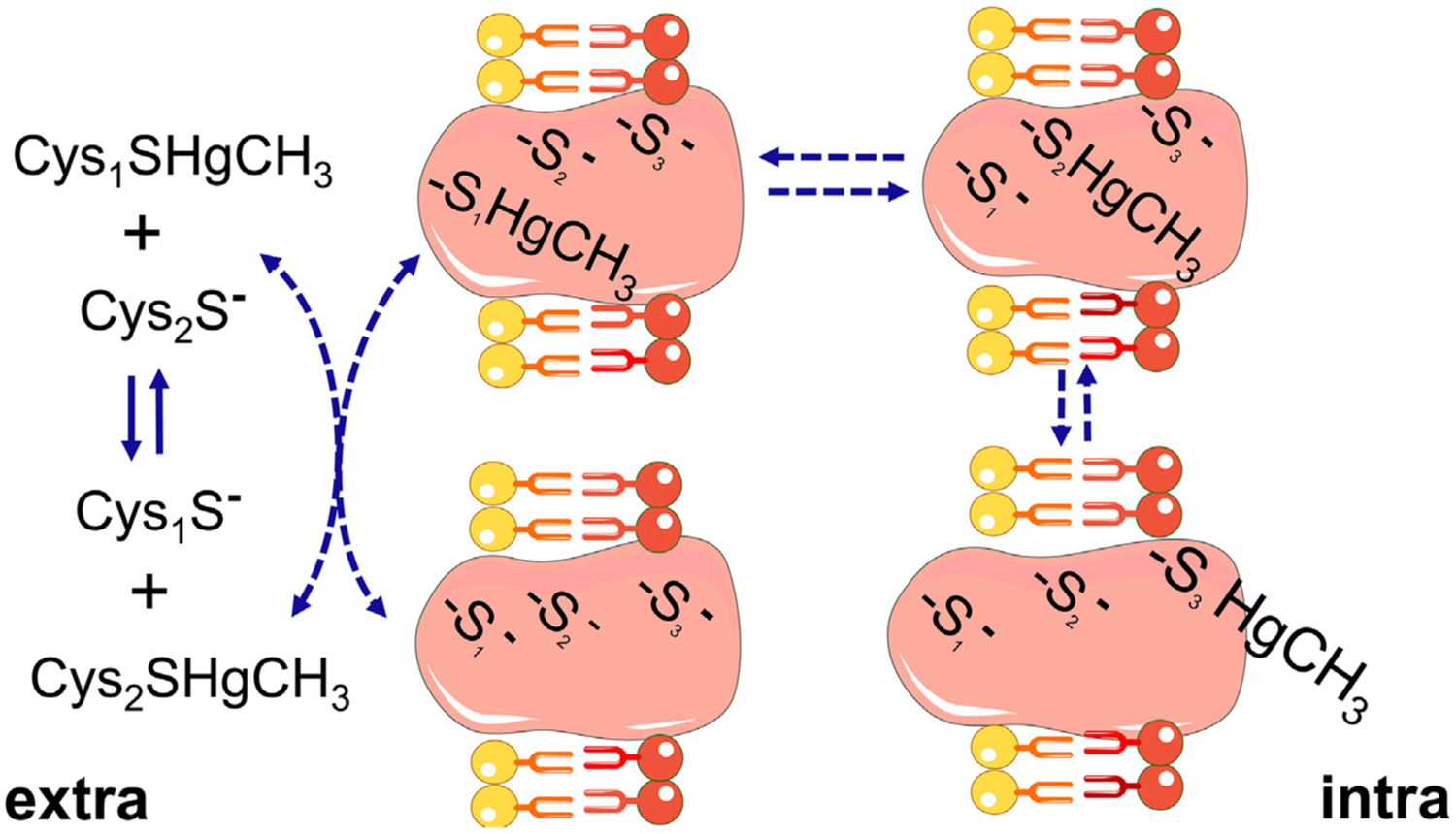
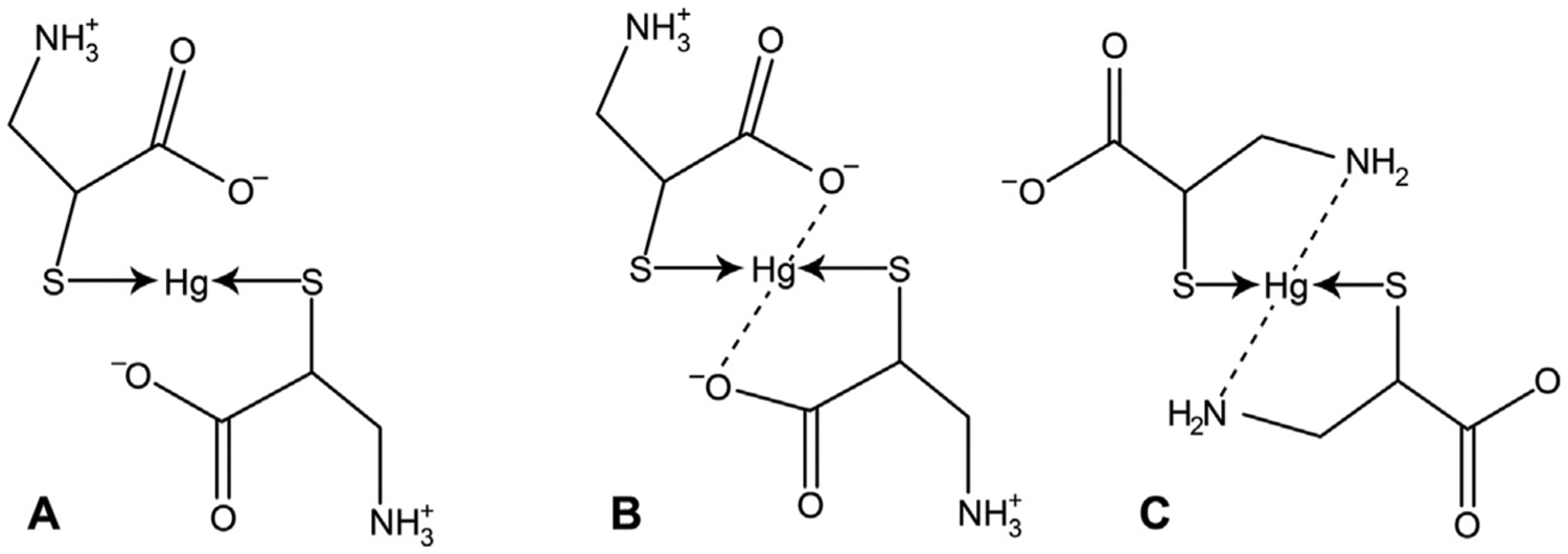
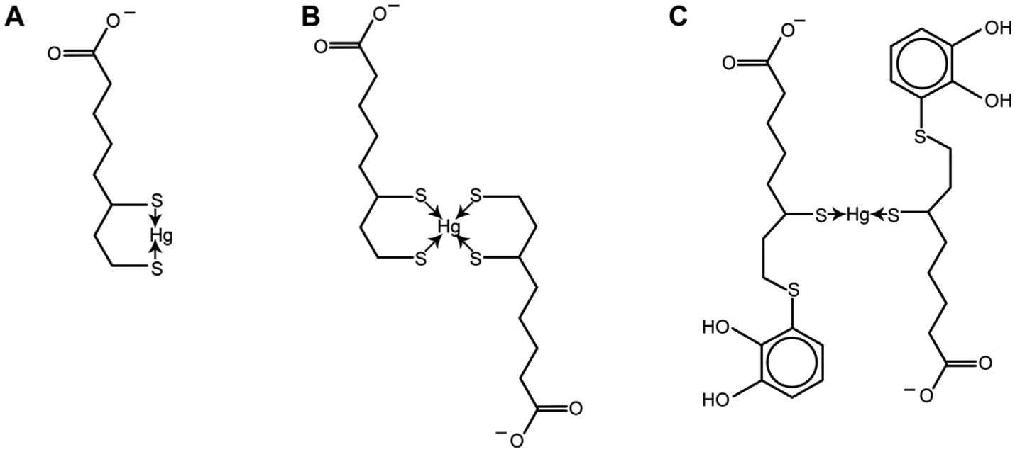
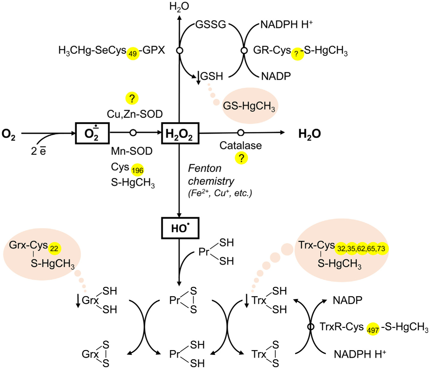
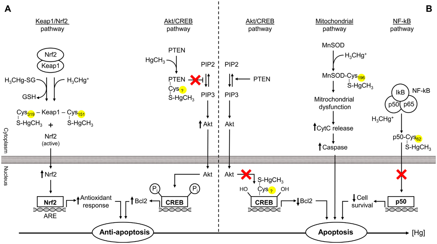
The present study addresses existing data on the affinity and conjugation of sulfhydryl (thiol; -SH) groups of low- and high-molecular-weight biological ligands with mercury (Hg). The consequences of these interactions with special emphasis on pathways of Hg toxicity are highlighted. Cysteine (Cys) is considered the primary target of Hg, and link its sensitivity with thiol groups and cellular damage. In vivo, Hg complexes play a key role in Hg metabolism. Due to the increased affinity of Hg to SH groups in Cys residues, glutathione (GSH) is reactive. The geometry of Hg(II) glutathionates is less understood than that with Cys. Both Cys and GSH Hg-conjugates are important in Hg transport. The binding of Hg to Cys mediates multiple toxic effects of Hg, especially inhibitory effects on enzymes and other proteins that contain free Cys residues. In blood plasma, albumin is the main Hg-binding (Hg2+, CH3Hg+, C2H5Hg+, C6H5Hg+) protein. At the Cys34 residue, Hg2+ binds to albumin, whereas other metals likely are bound at the N-terminal site and multi-metal binding sites. In addition to albumin, Hg binds to multiple Cys-containing enzymes (including manganese-superoxide dismutase (Mn-SOD), arginase I, sorbitol dehydrogenase, and δ-aminolevulinate dehydratase, etc.) involved in multiple processes. The affinity of Hg for thiol groups may also underlie the pathways of Hg toxicity. In particular, Hg-SH may contribute to apoptosis modulation by interfering with Akt/CREB, Keap1/Nrf2, NF-κB, and mitochondrial pathways. Mercury-induced oxidative stress may ensue from Cys-Hg binding and inhibition of Mn-SOD (Cys196), thioredoxin reductase (TrxR) (Cys497) activity, as well as limiting GSH (GS-HgCH3) and Trx (Cys32, 35, 62, 65, 73) availability. Moreover, Hg-thiol interaction also is crucial in the neurotoxicity of Hg by modulating the cytoskeleton and neuronal receptors, to name a few. However, existing data on the role of Hg-SH binding in the Hg toxicity remains poorly defined. Therefore, more research is needed to understand better the role of Hg-thiol binding in the molecular pathways of Hg toxicology and the critical role of thiols to counteract negative effects of Hg overload.

Keywords: Apoptosis; Conjugates; Cysteine; Mercury; S-mercuration.


Conflict of interest statement

Declaration of Competing Interest The authors declare that they have no known competing financial interests or personal relationships that could have appeared to influence the work reported in this paper.

Figures

Fig. 1.
Fig. 1.
Movement of methylmercury (MeHg) from one thiol to other thiol groups. The exchange of MeHg from one thiol to other of similar reactivity can occur between low molecular mass thiols (LMM-SH, represented by to free cysteine (Cys) molecules, Cys1-SH or Cys2-SH) or between LMM-SH and thiol-containing proteins (HMM-SH; in the figure the integral transmembrane protein has three thiol groups located in different topological places of the membrane. The first thiol or thiolate (–S1) is accessible to the extracellular space, which allows this group to exchange with the complex Cys2-S-HgMe. The complex protein-S1-HgMe can then exchange with a more buried free thiol group (–S2) to form the protein-S2-HgMe complex. The second complex can react with a third thiol/thiolate group (–S3) facing the intracellular surface of the membrane. The complex protein-S3-HgMe can exchange either with thiol-containing proteins or with cysteine or reduced glutathione molecules found inside the cell. The scheme is based on the exchange reactions of MeHg and thiol groups first described by Rabenstein et al. The migration of MeHg from the extracellular to the intracellular side of the membrane is based on indirect experimental data [74,79], but can explain additional mechanisms of MeHg transport other than those carried out by the LAT-1 transporter [132,152,234].
Fig. 2.
Fig. 2.
The proposed coordination of mercury(II) ion with cysteine molecules. A – Non-chelate complex formed using only sulfur atoms; B – The role of sulfur and oxygen atoms in metal–ligand coordination; C – The potential involvement of sulfur and nitrogen atoms in coordination between Hg2+ and cysteine molecules.
Fig. 3.
Fig. 3.
The potential mechanisms of Hg(II) binding to alpha-lipoic acid (ALA). A – Monoligand complex of Hg(II) with dihydrolipoic acid (DHLA); B – Biligand complex of mercuric Hg with dihydrolipoic acid (DHLA); C – Biligand complex of Hg2+ with 5-S-Lipoylhydroxytyrosol.
Fig. 4.
Fig. 4.
The role of Hg-SH binding in Hg-induced decrease in antioxidant system activity. Mercury was shown to inhibit antioxidant enzymes, including Cu,Zn-SOD, Mn-SOD, catalase, GPX, GR, and TrxR, although the particular role of Hg-SH interaction in this process is unclear. Inhibition of Mn-SOD was shown to be associated with Hg-SH binding at Cys196, whereas the particular location of Hg-binding cysteine residue in GR is not estimated. Inactivation of GPX may occur due to Hg binding to SeCys49 residue. In turn, recent data demonstrate that direct Hg-SH interaction is unlikely to be involved in inhibition of Cu,Zn-SOD, and catalase. In addition to Hg-induced antioxidant enzyme inactivation, Hg may also bind thiol groups of proton-donor cofactors (GSH, Trx, Grx), thus limiting their availability for reduction.
Fig. 5.
Fig. 5.
Mercury-sulfhydryl-dependent effects on apoptosis regulation. Mercury exposure may induce both anti- (low dose) and pro-apoptotic (high-dose) signaling. Covalent modification of Keap1 through Hg binding to Cys151 (MeHg) or Cys319 (MeHg-SG) results in Nrf2 activation and subsequent downstream signaling leading to up-regulation of antioxidant enzymes and anti-apoptotic signals. Akt/CREB pathway may be involved both in the anti- and pro-apoptotic effects of Hg. PTEN S-mercuration results in Akt activation and subsequent Bcl2 up-regulation. In contrast, increasing Hg levels bind Cys286 residue in CREB, thus disrupting Akt/CREB/Bcl-2 signaling and induction of proapoptotic signaling. NF-κB may also partially mediate the effects of Hg on apoptosis through S-mercuration of p50 (p50-Cys62-S-HgCH3) and alteration of its antiapoptotic effects. Finally, inhibition of mitochondrial Mn-SOD through Cys196-S-HgCH3 binding may result in mitochondrial oxidative stress and dysfunction, causing increased cytochrome c (CytC) leakage and caspase-mediated apoptosis.

References

    1. International Programme on Chemical Safety, in, World Health Organization, Geneva, 2010.
    1. Evers DC, Keane SE, Basu N, Buck D, Sci. Total Environ 569 (2016) 888–903. - PubMed
    1. Streets DG, Horowitz HM, Lu Z, Levin L, Thackray CP, Sunderland EM, Atmos. Environ 201 (2019) 417–427.
    1. Pacyna EG, Pacyna J, Sundseth K, Munthe J, Kindbom K, Wilson S, Steenhuisen F, Maxson P, Atmos. Environ 44 (2010) 2487–2499.
    1. Sundseth K, Pacyna J, Pacyna E, Pirrone N, Thorne R, Int. J. Environ. Res. Public Health 14 (2017) 105. - PMC - PubMed

LinkOut - more resources