. 2020 Aug 20;5(34):21815-21823.
doi: 10.1021/acsomega.0c02886. eCollection 2020 Sep 1.

Effects of Mutations in the Phenamacril-Binding Site of Fusarium Myosin-1 on Its Motor Function and Phenamacril Sensitivity

Affiliations

Effects of Mutations in the Phenamacril-Binding Site of Fusarium Myosin-1 on Its Motor Function and Phenamacril Sensitivity

Tong Ni et al. ACS Omega. .

Abstract

Phenamacril is a Fusarium-specific fungicide used for Fusarium head blight management. The target of phenamacril is FgMyo1, the sole class I myosin in Fusarium graminearum. The point mutation S217L in FgMyo1 is responsible for the high resistance of F. graminearum to phenamacril. Recent structural studies have shown that phenamacril binds to the 50 kDa cleft of the FgMyo1 motor domain, forming extensive interactions, including a hydrogen bond between the cyano group of phenamacril and the hydroxyl group of S217. Here, we produced FgMyo1IQ2, a truncated FgMyo1 composed of the motor domain and two IQ motifs complexed with the F. graminearum calmodulin in insect Sf9 cells. Phenamacril potently inhibited both the basal and the actin-activated ATPase activities of FgMyo1IQ2, with an IC50 in a micromolar range. S217 mutations of FgMyo1IQ2 substantially increased the IC50 of phenamacril. S217T or S217L each increased the IC50 of phenamacril for ∼60-fold, while S217A only increased the IC50 for ∼4-fold. These results indicate that the hydroxyl group of S217 plays an important, but nonessential role in phenamacril binding and that the bulky side chain at the position 217 sterically hinders phenamacril binding. On the other hand, S217P, which might alter the local conformation of the phenamacril-binding site, completely abolished the phenamacril inhibition. Because the cyano group of phenamacril does not form discernible interactions with FgMyo1 other than the nonessential hydrogen bond with the S217 hydroxyl group, we propose the cyano group of phenamacril as a key modification site for the development of novel fungicides.


Conflict of interest statement

The authors declare no competing financial interest.

Figures
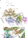
Figure 1
Figure 1
Structure of phenamacril-bound FgMyo1. (A) Chemical structure of fungicide phenamacril. (B) Three-dimensional structure of the phenamacril-bound FgMyo1 (PDB code 6UI4). This structure contains the motor domain and the first IQ motif in complex with FgCaM. The upper panel shows the whole structure, and the lower panel shows an enlarged view of the boxed region in the upper panel. The four subdomains of the motor domain are shown in different colors as follows: the 25 kDa subdomain (light green), the upper 50 kDa subdomain (light orange), the lower 50 kDa subdomain (light blue), and the 20 kDa subdomain (gray). FgCaM is colored in magenta. Phenamacril is shown as spheres. ATPγS is shown as sticks, and Mg2+ is shown as a red sphere. S217 and E420 are shown as green sticks, and other residues studied in this work are shown as cyan sticks.
Figure 2
Figure 2
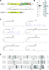
Identification of FgCaM as the light chain of FgMyo1. (A) Diagram of the FgMyo1 domains. TH1 and TH2, class I myosin tail homology domain 1 and 2; SH3, Src homology 3 domain; and CA, the C-terminal acid-rich region. (B) SDS-PAGE of purified FgMyo1IQ2. Lane 1, FgMyo1IQ2 purified from Sf9 cells infected with a recombinant baculovirus encoding FgMyo1IQ2. Lane 2, re-purified FgMyo1IQ2 after incubating with the crude extract of myosin light chain from F. graminearum. The arrowhead indicates FgMyo1IQ2, and the arrow indicates the co-purified light chain, which was subjected to MS/MS analysis. The numbers on the left show the molecular mass in kDa. (C) MS/MS analysis of the ∼20 kDa band in lane 2 in panel (B) reveal eight peptides (P-1 to P-8) matching the FgCaM sequence. (D) Sequence alignment of human calmodulin (HsCaM) and FgCaM. The residues different between HsCaM and FgCaM are gray-shaded. The peptide sequences (P-1 to P-8) identified in MS/MS are underlined.
Figure 3
Figure 3
Phenamacril potently inhibits the ATPase activity of FgMyo1IQ2. (A) Actin dependence of FgMyo1IQ2 ATPase activity. Values are the mean ± std of three independent preparations of FgMyo1IQ2. (B, C) Effects of phenamacril on (B) the basal ATPase activity and (C) the actin-activated ATPase activity of FgMyo1IQ2. The basal ATPase activity was measured in the absence of actin and in the presence of 0–4 μM phenamacril. The actin-activated ATPase activity was measured in the presence of 40 μM actin and 0–4 μM phenamacril. The IC50 was obtained by a hyperbolic fit. Curve fitting shows that both the basal and the actin-activated ATPase activities of FgMyo1IQ2 are almost completely inhibited by the saturated concentration of phenamacril. Values are the mean ± std from four independent assays of two independent preparations of FgMyo1IQ2. (D, E) Effect of phenamacril on the ATP-dependent interaction between FgMyo1 and actin. (D) About 1 μM FgMyo1 was incubated with 10 μM actin in the absence or presence of 1 mM phenamacril in 20 mM Tris-HCl (pH 7.5), 2 mM MgCl2, 1 mM DTT, and 50 mM KCl on ice for ∼10 min. Just prior to centrifugation at 85,000 rpm for 15 min, 1 mM (final concentration) ATP or equal volume of H2O was added to the above solutions. Equal portions of the supernatants and the pellets were subjected to SDS-PAGE separation and Coomassie Brilliant Blue staining. (E) In the presence of ATP, 45.4 ± 3.9% and 28.5 ± 8.2% of FgMyo1IQ2 were sedimented with actin with the absence and the presence of phenamacril, respectively. Values are the mean ± std from three independent assays.
Figure 4
Figure 4
Purification of FgMyo1IQ2 S217 mutants and their ATPase activities. (A) SDS-PAGE showing the purified FgMyo1IQ2 wild-type (WT) and five S217 mutants (S217L, S217A, S217T, S217G, and S217P). The arrowhead indicates the FgMyo1IQ2 heavy chain, and the arrow indicates the co-purified FgCaM. (B) Basal and the actin-activated ATPase activities of FgMyo1IQ2 WT and S217 mutants. The basal ATPase activities were measured in the absence of actin, and the actin-activated ATPase activities were measured in the presence of 40 μM actin. Values are the mean ± std from three independent assays of two independent preparations.
Figure 5
Figure 5
Effects of phenamacril on the actin-activated ATPase activity of FgMyo1IQ2 S217 mutants. The actin-activated ATPase activities of FgMyo1IQ2 S217 mutants were measured in the presence of 40 μM actin and various concentrations of phenamacril. Values are the mean ± std from four independent assays of two independent preparations of FgMyo1IQ2. For comparison, the amino acid structures at position 217 of FgMyo1IQ2 variants are shown in each panel. Note that the differences between the actin-activated ATPase activities of FgMyo1IQ2 in the absence of phenamacril in Figure 4B and those in this figure are due to the presence of 5% DMSO in the latter.
Figure 6
Figure 6
Effects of phenamacril on the actin-activated ATPase activities of two double-mutants of FgMyo1IQ2. (A) SDS-PAGE showing the purified FgMyo1IQ2-K216Q/N380K and -V151A/S418T mutants. The arrowhead indicates the FgMyo1IQ2 heavy chain, and the arrow indicates the co-purified FgCaM. (B) Basal and the actin-activated ATPase activities of FgMyo1IQ2 K216Q/N380K and V151A/S418T. The ATPase assays were performed as described in Figure 4B. Values are the mean ± std from three independent assays of two independent preparations of FgMyo1IQ2. (C) Effects of phenamacril on the actin-activated ATPase activities of FgMyo1IQ2 K216Q/N380K and V151A/S418T. The ATPase assays were performed, as described in Figure 5. Values are the mean ± std from four independent assays of two independent preparations of FgMyo1IQ2.

Similar articles

Cited by

References

    1. Xu X.; Nicholson P. Community ecology of fungal pathogens causing wheat head blight. Annu. Rev. Phytopathol. 2009, 47, 83–103. 10.1146/annurev-phyto-080508-081737. - DOI - PubMed
    1. Dean R.; Van Kan J. A. L.; Pretorius Z. A.; Hammond-Kosack K. E.; Di Pietro A.; Spanu P. D.; Rudd J. J.; Dickman M.; Kahmann R.; Ellis J.; Foster G. D. The Top 10 fungal pathogens in molecular plant pathology. Mol. Plant Pathol. 2012, 13, 414–430. 10.1111/j.1364-3703.2011.00783.x. - DOI - PMC - PubMed
    1. Li H.; Diao Y.; Wang J.; Chen C.; Ni J.; Zhou M. JS399-19, a new fungicide against wheat scab. Crop Protection 2008, 27, 90–95. 10.1016/j.cropro.2007.04.010. - DOI
    1. Wollenberg R. D.; Donau S. S.; Nielsen T. T.; Sørensen J. L.; Giese H.; Wimmer R.; Søndergaard T. E. Real-time imaging of the growth-inhibitory effect of JS399-19 on Fusarium. Pestic. Biochem. Physiol. 2016, 134, 24–30. 10.1016/j.pestbp.2016.05.002. - DOI - PubMed
    1. Zhang C.; Chen Y.; Yin Y.; Ji H. H.; Shim W. B.; Hou Y.; Zhou M.; Li X. D.; Ma Z. A small molecule species specifically inhibits Fusarium myosin I. Environ. Microbiol. 2015, 17, 2735–2746. 10.1111/1462-2920.12711. - DOI - PubMed