. 2020 Aug 1;10(8):2464-2479.
eCollection 2020.

NAT1 promotes osteolytic metastasis in luminal breast cancer by regulating the bone metastatic niche via NF-κB/IL-1B signaling pathway

Affiliations

NAT1 promotes osteolytic metastasis in luminal breast cancer by regulating the bone metastatic niche via NF-κB/IL-1B signaling pathway

Chenglong Zhao et al. Am J Cancer Res. .

Abstract

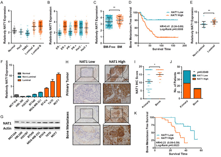
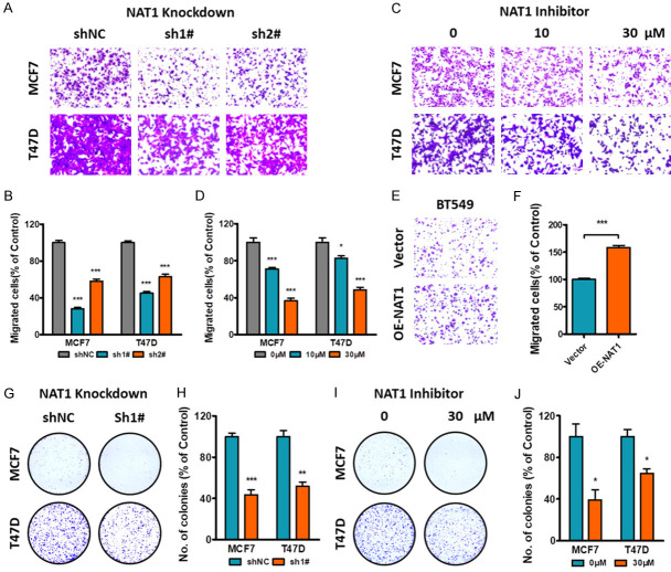
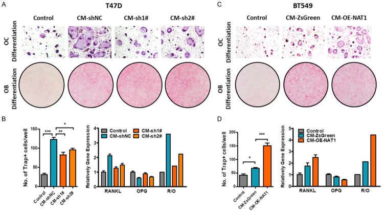
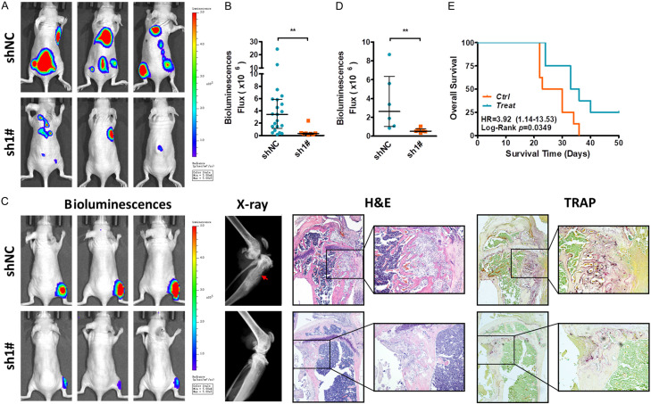
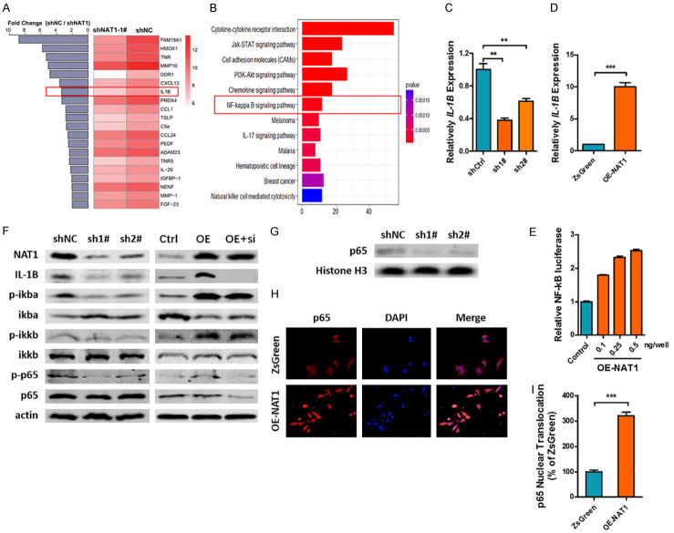
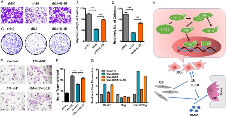
Breast cancer is a molecularly heterogeneous disease that can be subdivided into different subtypes. Compared with the other subtypes, luminal breast cancer (LBC) is considered more susceptible to bone metastasis. However, the intrinsic mechanisms remain elusive. Bioinformatics analysis of the preset study showed that N-acetyltransferase 1 (NAT1) was specifically expressed in LBC and closely correlated with bone metastasis. In addition, NAT1 could promote LBC cell migration and clonal formation, induce osteoclast differentiation and raise the Rankl/Opg ratio in osteoblasts. Our in vivo experiment demonstrated that NAT1 promoted LBC bone metastasis and bone destruction, which could be reversed by NAT1 inhibitor treatment. The result of cytokine array showed that NAT1 could significantly over activate the NF-κB signaling pathway and up-regulate the expression of IL-1B, which further worked as downstream factors in these processes. All these results demonstrated NAT1 was up-regulated in LBC and promoted the formation of bone metastatic niche and osteolytic bone metastasis through the NAT1/NF-κB/IL-1B axis. This finding may provide a new pathway to help understand the mechanisms of LBC bone metastasis and suggest a novel therapeutic and diagnostic target for its treatment.

Keywords: IL-1B; Luminal breast cancer; N-acetyltransferase 1 (NAT1); bone metastasis; bone microenvironment.


Conflict of interest statement

None.

Figures

Figure 1
Figure 1
NAT1 was up-regulated in LBC and correlated with bone metastasis. (A, B) The TCGA database indicate that NAT1 was upregulated in LBC. Meanwhile, the expression of NAT1 was correlated closely with ER and PR. but interrelated little with the expression of Her2. (C) Compared with bone metastasis -free patients. Bone metastatic patients had a higher NAT1 expression in their primary tumors. **P=0.0038 by Mann-Whitney U test. (D) Kaplan-Meier plot of BMFS for patients with primary BCs. nHigh=203. nLow=83. Log-Rank p=0.0008. (E) Expression of NAT1 between Luminal and non-Luminal BC cell lines. **P=0.0018 by Mann-Whitney U test. (F, G) NAT1 expression in normal and BC cell lines. (H-K) IHC staining of primary BC (n=44) and BC bone metastasis (n=16). The expression of NAT1 was significantly up-regulated in bone metastasis (I). *P=0.0193 by unpaired t test. Expression of NAT1 in primary and metastatic BC samples (J). P=0.0049 by one-way ANOVA. Kaplan-Meier plot of primary BC tumors in bone metastasis-free patients (K). nHigh=24. nLow=20. Log-Rank P=0.0023.
Figure 2
Figure 2
Effect of NAT1 on BC migration and clone formation. A, B. Knockdown of NAT1 in LBC cell lines reduced cell migration by transwell assay. ***P<0.001 by unpaired t test. C, D. Cell migration was depressed by NAT1 inhibitor at indicated concentrations. *P<0.05. ***P<0.001 by unpaired t test. E, F. Upregulation of NAT1 expression in TNBC cell line BT-549 promoted tumor migration. ***P<0.001 by unpaired t test. G-J. NAT1 knockdown or NAT1 inhibitor significantly depressed tumor clonal formation in LBC cell lines. *P<0.05. ***P<0.001 by unpaired t test.
Figure 3
Figure 3
Effect of NAT1 expression on tumor induced bone metastatic niche. (A, B) Knockdown of NAT1 attenuated LBC-promoted bone metastatic niche. Tumor conditioned media were collected and added to the culture systems of OC and OB differentiation to simulate the BC-induced bone metastatic niche. Knockdown of NAT1 significantly decreased tumor induced OC differentiation (A up and B left. One-way ANOVA. *P<0.05. **P<0.01. ***P<0.001) but had little effect on OB differentiation (A down). However. knockdown of NAT1 significantly decreased tumor induced Rankl expression in OBs and upregulated Opg expression. One-way ANOVA. pRankl=0.0003. pOpg=0.0027. (C, D) Upregulation of NAT1 in BT-549 cells promoted tumor induced OC differentiation (C up and D left. One-way ANOVA. *P<0.05. ***P<0.001), and increased Rankl expression but decreased Opg expression in OBs. One-way ANOVA. pRankl=0.0111. pOpg=0.0050.
Figure 4
Figure 4
Inhibition of NAT1 attenuates LBC osteolytic bone metastasis. A, B. Luciferase labeled T47D-shNC and sh1# cells were inoculated into the left heart ventricle of the nude mice and cultured (n=6 for each group). Mice were sacrificed and metastasis was evaluated after 3 weeks. **P=0.0016 by Mann-Whitney U test. C, D. Luciferase labeled T47D-shNC and sh1# cells were directly injected in to the right tibia bone marrow cavity of the nude mice (n=6 per group). After two-week culture, the mice were sacrificed and local bone destruction was detected by X-ray and Trap staining. **P=0.0043 by Mann-Whitney U test. E. After successful inoculation of T47D cells into the left heart ventricles. 16 nude mice were equally randomized into, and treated either with the NAT1 inhibitor (10 uM. 0.1 ml) or PBS every two days by i.p. injection. Mice were cultured till death or to 50 days after tumor inoculation. Survival time was recorded and Kaplan-Meier plot was drawn accordingly. Log-Rank P=0.0349.
Figure 5
Figure 5
NAT1 promotes IL-1B expression through NF-κB signaling pathway. A, B. After 12-h culture, the conditioned media of T47D-shNAT and sh1# cells were subjected to cytokine array analysis. IL-1B was found to be significantly downregulated after NAT1 knockdown while the KEGG analysis indicated the inhibition of NF-κB signaling pathway. C, D. qRT-PCR confirmed the regulatory effect of NAT1 on IL-1B expression. **P<0.01. ***P<0.001 by unpaired t test. E. Relative NF-κB luciferase activity of BT-549 cells after transfection with OE-NAT1 vectors. One-way ANOVA. P<0.001. F, G. Western blot array indicated that NAT1 significantly affected the phosphorylation of NF-κB signaling pathway and p65 nuclear transportation. H, I. Immunofluorescence indicated that NF-κB p65 nuclear transportation was significantly upregulated after OE-NAT1 transfection in BT-549 cells. ***P<0.001 by unpaired t test.
Figure 6
Figure 6
IL-1B promotes LBC osteolytic bone metastasis. A-G. IL-1B could rescue NAT1 depletion induced downregulation of BC migration. clone formation. OC differentiation and OB Rankl/Opg expression. One-way ANOVA. **P<0.01. ***P<0.001. H. A schematic illustration of the NAT1/NF-κB/IL-1B axis in regulation of the LBC bone metastatic niche and osteolytic bone metastasis.

Similar articles

Cited by

References

    1. Siegel RL, Miller KD, Jemal A. Cancer statistics. CA Cancer J Clin. 2018;68:7–30. - PubMed
    1. Chen W, Zheng R, Baade PD, Zhang S, Zeng H, Bray F, Jemal A, Yu XQ, He J. Cancer statistics in China. 2015. CA Cancer J Clin. 2016;66:115–132. - PubMed
    1. Warner E. Clinical practice. Breast-cancer screening. N Engl J Med. 2011;365:1025–1032. - PubMed
    1. Li Z, Kang Y. Emerging therapeutic targets in metastatic progression: a focus on breast cancer. Pharmacol Ther. 2016;161:79–96. - PMC - PubMed
    1. Cai WL, Huang WD, Li B, Chen TR, Li ZX, Zhao CL, Li HY, Wu YM, Yan WJ, Xiao JR. microRNA-124 inhibits bone metastasis of breast cancer by repressing Interleukin-11. Mol Cancer. 2018;17:9. - PMC - PubMed

LinkOut - more resources