

Circulating CD14+ HLA-DRlo/- monocytic cells as a biomarker for epithelial ovarian cancer progression
- PMID: 32905653
- PMCID: PMC7897210
- DOI: 10.1111/aji.13343
Circulating CD14+ HLA-DRlo/- monocytic cells as a biomarker for epithelial ovarian cancer progression
Abstract
Problem: Previous studies identified circulating CD14+ HLA-DRlo/- monocytic cells as an immune suppressive subset in solid malignancies, such as prostate, renal cell carcinoma, and pancreatic cancer. Such monocytic cells have been implicated not only in tumour progression but also as a potential barrier for immunotherapy. This study examined the relationship between the frequency of circulating monocytic cells and epithelial ovarian cancer (EOC) progression pre- and post-frontline chemotherapy, defined by disease stage, which is a leading prognostic factor for this malignancy.
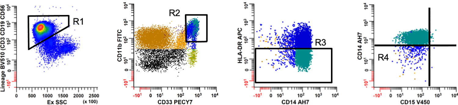
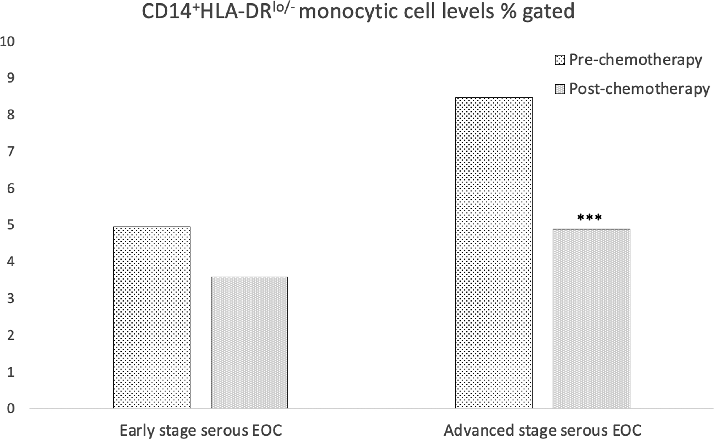
Method of study: Incident cases of 236 women with EOC were recruited and comprehensive flow cytometry was utilized to assess the frequency of peripheral blood CD33+ CD11b+ HLA-DR-/low CD14+ CD15- monocytic cells, henceforth termed CD14+ HLA-DRlo/- monocytic cells, prior to and after completion of frontline chemotherapy. Multivariable odds ratios (OR) were used to estimate the association between CD14+ HLA-DRlo/- monocytic cell percentages and disease stage. Wilcoxon signed-rank tests evaluated changes in these monocytic cell levels pre- and post-chemotherapy in a patient subset (n = 70).
Results: Patients with elevated frequencies of circulating CD14+ HLA-DRlo/- monocytic cells at diagnosis were at 3.33-fold greater odds of having advanced stage (III/IV) EOC (CI: 1.04-10.64), with a significant trend in increasing CD14+ HLA-DRlo/- monocytic cell levels (P = .04). There was a 2.02% median decrease of these monocytic cells post-chemotherapy among a subset of patients with advanced stage disease (P < .0001).
Conclusion: These findings support the potential clinical relevance of CD14+ HLA-DRlo/- monocytic cells in EOC for prognosis and may indicate a non-invasive biomarker to measure disease progression.
Keywords: MDSC; biomarker; monocytes; monocytic cells; ovarian cancer.
© 2020 John Wiley & Sons A/S. Published by John Wiley & Sons Ltd.
Conflict of interest statement
Figures
References
-
- Abraham JGJ, Allegra CJ. The Bethesda Handbook of Clinical Oncology. In 4 Edition Wolters Kluwer; 2014.
Publication types
MeSH terms
Substances
Grants and funding
LinkOut - more resources
Full Text Sources
Medical
Research Materials
