. 2020 Oct:37:101699.
doi: 10.1016/j.redox.2020.101699. Epub 2020 Aug 25.

Low density lipoprotein receptor related protein 6 (LRP6) protects heart against oxidative stress by the crosstalk of HSF1 and GSK3β

Affiliations

Low density lipoprotein receptor related protein 6 (LRP6) protects heart against oxidative stress by the crosstalk of HSF1 and GSK3β

Ying Wang et al. Redox Biol. 2020 Oct.

Erratum in

Abstract

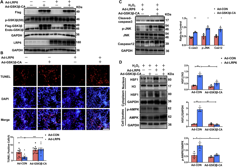
Low density lipoprotein receptor-related protein 6 (LRP6), a Wnt co-receptor, induces multiple functions in various organs. We recently reported cardiac specific LRP6 deficiency caused cardiac dysfunction in mice. Whether cardiomyocyte-expressed LRP6 protects hearts against ischemic stress is largely unknown. Here, we investigated the effects of cardiac LRP6 in response to ischemic reperfusion (I/R) injury. Tamoxifen inducible cardiac specific LRP6 overexpression mice were generated to build I/R model by occlusion of the left anterior descending (LAD) coronary artery for 40 min and subsequent release of specific time. Cardiac specific LRP6 overexpression significantly ameliorated myocardial I/R injury as characterized by the improved cardiac function, strain pattern and infarct area at 24 h after reperfusion. I/R induced-apoptosis and endoplasmic reticulum (ER) stress were greatly inhibited by LRP6 overexpression in cardiomyocytes. LRP6 overexpression enhanced the expression of heat shock transcription factor-1(HSF1) and heat shock proteins (HSPs), the level of p-glycogen synthase kinase 3β(GSK3β)(S9) and p-AMPK under I/R. HSF1 inhibitor deteriorated the apoptosis and decreased p-GSK3β(S9) level in LRP6 overexpressed -cardiomyocytes treated with H2O2. Si-HSF1 or overexpression of active GSK3β significantly attenuated the increased expression of HSF1 and p-AMPK, and the inhibition of apoptosis and ER stress induced by LRP6 overexpression in H2O2-treated cardiomyocytes. AMPK inhibitor suppressed the increase in p-GSK3β (S9) level but didn't alter HSF1 nucleus expression in LRP6 overexpressed-cardiomyocytes treated with H2O2. Active GSK3β, but not AMPK inhibitor, attenuated the inhibition of ubiquitination of HSF1 induced by LRP6-overexpressed-cardiomyocytes treated with H2O2. LRP6 overexpression increased interaction of HSF1 and GSK3β which may be involved in the reciprocal regulation under oxidative stress. In conclusion, cardiac LRP6 overexpression significantly inhibits cardiomyocyte apoptosis and ameliorates myocardial I/R injury by the crosstalk of HSF1 and GSK3β signaling.

Keywords: Apoptosis; GSK3β; HSF1; Ischemic reperfusion; LRP6.


Conflict of interest statement

We declare that there are no competing interests.

Figures

Fig. 1
Fig. 1
LRP6 is involved in myocardial ischemia reperfusion. Western blot analysis of LRP6, p-LRP6, p-AKT(T308), p-AKT(S473), p-GSK3α and p-GSK3β(S9) expression in ischemia area of heart tissue from mice subjected to I/R surgery at different reperfusion time points (15min, 30min, 1 h) after ischemia for 45min. A. Representative images. B. Quantitative analysis of p-LRP6 and LRP6 level. C. Quantitative analysis of p-AKT(T308) and p-AKT(S473) level. D. Quantitative analysis of p-GSK3α and p-GSK3β(S9) level. Values are the means ± S.E.M. N = 3/group. *p < 0.05, **p < 0.005.
Fig. 2
Fig. 2
Cardiac specific LRP6 overexpression protects heart from I/R injury. A. The overall experimental protocol of in vivo to explore the effects of cardiac LRP6 overexpression in mice subjected to I/R. 6–8 weeks age male LRP6-CTG or MCM mice were intraperitoneally injected with tamoxifen (30 mg/kg) for 3 continuous days respectively, 2 weeks later, these mice (LRP6-CTG mice and MCM mice) were subjected to I/R surgery or sham operation. The MCM mice were used as control group. B. The expression of LRP6 and p-LRP6 in MCM mice and LRP6-CTG mice were analyzed by western blot. N = 3/group. C. Echocardiographic analysis at 24 h after I/R surgery or sham operation in MCM and LRP6-CTG mice. Enjection fraction (EF) and fraction shortening (FS) were quantified at 24 h after I/R injury. N = 4–8/group. D. Radial strain analysis in MCM mice and LRP6-CTG mice at 24 h after I/R were obatined from VevoStrain analysis software. Upper lane: Representative images of radial strain curves. Colored lines represent 6 standard myocardial regions; 7th black line calculates average (global) strain at each time point. Down lane: Quantitative analysis of radial strain both globally and in infarct area (anterior apex), and maximal opposite wall delay in time. N = 6/group. E. Representative TTC staining of the heart sections from Evans blue perfused hearts was shown for each group. The bar = 500 μm. The ratio of infarct area (IA) to area at risk (AAR), and area at risk/left ventricle (AAR/LV) were quantified at 24 h after I/R injury, as detailed in the Methods. N = 12–15/group. Values are means ± S.E.M. *p < 0.05, **p < 0.005,***p < 0,001 and ****p < 0.0001 vs. MCM group. (For interpretation of the references to colour in this figure legend, the reader is referred to the Web version of this article.)
Fig. 3
Fig. 3
Cardiac specific LRP6 overexpression inhibits cardiomyocytes apoptosis following I/R. A. Terminal deoxynucleotidyl transferase-mediated dUTP (deoxyuridine triphosphate)-biotin nick-end labeling (TUNEL) in heart tissue from MCM and LRP6-CTG mice at 24 h reperfusion following 45min ischemia. Scale bar, 50 μm. The percentage of TUNEL positive cells was calculated. N = 4/group. B–C. Western blot analysis of apoptosis-related proteins (cleaved-caspase3; p-JNK; JNK) expression and an ER stress marker protein (caspase12) in the ischemia zone of hearts from MCM and LRP6-CTG mice at 24 h reperfusion after 45min ischemia. N = 3/group. In vitro, neonatal rat cardiomyocytes were cultured and transfected Ad-LRP6 (LRP6 OVER) or control adenovirus (CON), 72 h later, 200 μM H2O2 was treated to induce oxidative stress, and PBS treatment was as control. D. TUNEL staining analysis of control or LRP6 overexpressed-cardiomyocytes treated with H2O2 for 24 h, scale bar, 200 μm. The percentage of TUNEL positive cells (apoptosis) was calculated. N = 11/group. E. Western blot analysis for apoptosis-related proteins (cleaved-caspase3; p-JNK; JNK) expression and ER stress marker protein (caspase12) expression in control or LRP6 overexpressed-cardiomyocytes treated with H2O2 or PBS for 24 h. N = 3/group. Values are means ± S.E.M. *p < 0.05, **p < 0.005; ***p < 0.001. and ****p < 0.0001.
Fig. 4
Fig. 4
Cardiac specific LRP6overexpression promotes HSF1 nucleus translocation and phosphorylation of GSK3β and AMPK after I/R. A. Western blot analysis of LRP6, p-LRP6, p-AKT(T308), p-AKT(S473), AKT, p-GSK3β(S9), GSK3β, p-ERK, ERK in the ischemia zone of hearts from MCM and LRP6-CTG mice at 3 h reperfusion following 45min ischemia. N = 3/group. B–C. Western blot analysis of the active β-catenin and p-AMPK level in the ischemia zone of hearts from MCM and LRP6-CTG mice at 3 h reperfusion following 45min ischemia. N = 3/group. In vitro, human cardiomyocytes (AC16 cell line) were cultured and transfected LRP6 adenovirus (Ad-LRP6) or control adenovirus (Ad-CON), 72 h later, 200 μM H2O2 was treated to induce oxidative stress, and PBS treatment was as control. D. Western blot for p-AMPK, AMPK, p-GSK3β and GSK3β in control or LRP6 overexpressed-cardiomyocytes treated with 200 μM H2O2 or PBS for 3 h. N = 3/group. E. Western blot analysis of HSF1 in nucleus and cytoplasm lysates from the ischemia zone of hearts of MCM and LRP6-CTG mice at 3 h reperfusion following 45min ischemia. N = 3/group. F. Western blot analysis of HSF1 in nucleus and cytoplasm lysates from control or LRP6 overexpressed-cardiomyocytes treated with 200 μM H2O2 or PBS for 3 h. N = 3/group. Values are means ± S.E.M. *p < 0.05, **p < 0.005,***p < 0.001 and ****p < 0.0001.
Fig. 4
Fig. 4
Cardiac specific LRP6overexpression promotes HSF1 nucleus translocation and phosphorylation of GSK3β and AMPK after I/R. A. Western blot analysis of LRP6, p-LRP6, p-AKT(T308), p-AKT(S473), AKT, p-GSK3β(S9), GSK3β, p-ERK, ERK in the ischemia zone of hearts from MCM and LRP6-CTG mice at 3 h reperfusion following 45min ischemia. N = 3/group. B–C. Western blot analysis of the active β-catenin and p-AMPK level in the ischemia zone of hearts from MCM and LRP6-CTG mice at 3 h reperfusion following 45min ischemia. N = 3/group. In vitro, human cardiomyocytes (AC16 cell line) were cultured and transfected LRP6 adenovirus (Ad-LRP6) or control adenovirus (Ad-CON), 72 h later, 200 μM H2O2 was treated to induce oxidative stress, and PBS treatment was as control. D. Western blot for p-AMPK, AMPK, p-GSK3β and GSK3β in control or LRP6 overexpressed-cardiomyocytes treated with 200 μM H2O2 or PBS for 3 h. N = 3/group. E. Western blot analysis of HSF1 in nucleus and cytoplasm lysates from the ischemia zone of hearts of MCM and LRP6-CTG mice at 3 h reperfusion following 45min ischemia. N = 3/group. F. Western blot analysis of HSF1 in nucleus and cytoplasm lysates from control or LRP6 overexpressed-cardiomyocytes treated with 200 μM H2O2 or PBS for 3 h. N = 3/group. Values are means ± S.E.M. *p < 0.05, **p < 0.005,***p < 0.001 and ****p < 0.0001.
Fig. 5
Fig. 5
Active GSK3β overexpression restrains the increased level of HSF1 and p-AMPK, and the inhibition of cardiomyocytes apoptosis mediated by LRP6 overexpression in cardiomyocytes under oxidative stress. In vitro, neonatal rat cardiomyocytes were cultured and transfected LRP6 adenovirus (Ad-LRP6) and/or GSK3β-CA adenovirus (Ad-GSK3β-CA tagged with flag) or control adenovirus (Ad-CON), 72 h later, 200 μM H2O2 was treated to induce oxidative stress. A. Western blot analysis for the expression of flag, p-GSK3β(S9), GSK3β and LRP6 in cultured cardiomyocytes (grouped as above) treated with H2O2 for 24 h. N = 3/group. B. TUNEL staining of cultured cardiomyocytes (grouped as above) treated with H2O2 for 24 h. scale bar, 200 μm. The percentage of TUNEL positive cells was calculated. N = 10/group. C. Western blot analysis for p-JNK, JNK, cleaved-caspase3, and caspase12 in cultured LRP6 overexpressed-cardiomyocytes transfected with GSK3β-CA adenovirus (Ad-GSK3β-CA) or control adenovirus (Ad-CON), and these cardiomyocytes were treated with 200 μM H2O2 for 24 h. N = 3/group. D. Western blot for HSF1 in nucleus and cytoplasm, p-AMPK and AMPK in whole cell lysates from GSK3β-CA adenovirus (Ad-GSK3β-CA) and/or LRP6 adenovirus (Ad-LRP6) transfected cardiomycoytes treated with 200 μM H2O2 for 3 h. N = 3/group. Values are means ± S.E.M. *p < 0.05, **p < 0.01, ***p < 0.001, ****p < 0.0001.
Fig. 6
Fig. 6
Si-HSF1 blunts the increase in p-GSK3β(S9) and p-AMPK level and attenuates the inhibition of apoptosis induced by LRP6 overexpression in cardiomyocytes during oxidative stress. A. Western blot analysis for HSF1 expression level in cultured cardiomyocytes with transfection of si-Scramble (si-Scram) or si-HSF1. N = 3/group. B. The apoptosis was assessed by flow cytometry analysis for annexin-V and propidium iodide (PI) staining in cultured control or LRP6 overexpressed-cardiomyocytes treated with 200 μM H2O2 for 24 h, and these cardiomyocytes were pre-transfected into si-scramble (si-Scram) or si-HSF1. N = 3–6/group. C. Western blot analysis for p-AMPK, AMPK, p-GSK3β (S9) and GSK3β in cultured control or LRP6 overexpressed-cardiomyocytes treated with 200 μM H2O2 for 24 h, and these cardiomyocytes were pre-transfected into si-Scramble or si-HSF1. N = 3/group. A-C, Human cardiomyocytes (AC16 cells) were transfected control adenovirus (Ad-CON) or LRP6 adenovirus (Ad-LRP6) as control or LRP6 overexpressed-cardiomyocytes respectively. D. Western blot analysis for HSP70, p-GSK3β (S9) and GSK3β in cultured control or LRP6 overexpressed-cardiomyocytes (NRCMs) treated with 200 μM H2O2 for 3 h, and these cardiomyocytes were pre-treated with DMSO or KRIBB11(20 μM) for 48 h. N = 3/group. E. TUNEL staining of cultured cardiomyocytes (grouped as above) treated with H2O2 for 24 h. scale bar, 200 μm. The percentage of TUNEL positive cells was calculated. N = 4/group.Values are means ± S.E.M. *p < 0.05, **p < 0.005,***p < 0.001 and ****p < 0.0001.
Fig. 7
Fig. 7
AMPK inhibitor suppresses the increased level in p-GSK3β(S9) but not HSF1 induced by LRP6 overexpression in cardiomyocytes during oxidative stress. Human cardiomyocytes (AC16 cells) were transfected control adenovirus (Ad-CON) or LRP6 adenovirus (Ad-LRP6) as control or LRP6 overexpressed-cardiomyocytes respectively. A. Western blot analysis for LRP6, p-AMPK, AMPK, p-GSK3β (S9), GSK3β in cultured control or LRP6 overexpressed-cardiomyocytes treated with 200 μM H2O2 for 3 h, and these cardiomyocytes were pre-treated with PBS or Compound C (20 μM) for 24 h. N = 3/Group. B. Western blot analysis of HSF1 in nucleus and cytoplasm in cultured control or LRP6 overexpressed-cardiomyocytes treated with 200 μM H2O2 for 3 h, and these cardiomyocytes were pre-treated with PBS or Compound C (20 μM) for 24 h. N = 3/group. Values are means ± S.E.M. *p < 0.05, **p < 0.005, ***p < 0.001.
Fig. 8
Fig. 8
LRP6 overexpression inhibits the ubiquitination of HSF1 by regulation of GSK3β signaling in cardiomyocytes under oxidative stress. A. Real-time PCR analysis of HSF1 mRNA level in cultured cardiomyocytes treated with 200 μM H2O2 for 3 h. N = 3/Group. Values are means ± S.E.M. ***p < 0.001. B. Analysis of the ubiquitination of HSF1 in cardiomyocytes. Human cardiomyocytes (AC16 cells) were pre-transfected with control adenovirus (Ad-CON) or LRP6 adenovirus (Ad-LRP6), and then treated with 200 μM H2O2 or PBS for 3 h. C. The ubiquitination of HSF1 was analyzed in neonatal rat cardiomyocytes. These cardiomyocytes were pre-transfected with GSK3β-CA adnovirus (GSK3β-CA) and/or control adenovirus (Ad-CON) or LRP6 adenovirus (Ad-LRP6), and treated with 200 μM H2O2 for 3 h. D. The ubiquitination of HSF1 was analyzed in cardiomyocytes (AC16 cells). These cardiomyocytes were pre-transfected with control adenovirus (Ad-CON) or LRP6 adenovirus (Ad-LRP6). After incubation of Compund C (20 μM) or PBS for 24 h, these cardiomyocytes were treated with 200 μM H2O2 for 3 h. B–D: Whole-cell lysates (WCL) were immunoprecipitated using an anti-HSF1 antibody, and the ubiquitin levels were determined by western blotting using an anti-ubiquitin antibody. Ubi, ubiquitin. The experiments were repeated at least 3 times. E. The interaction between HSF1 and GSK3β was investigated by co-immunoprecipitation. HSF1 and GSK3β was precipitated from AC16 cell lysate with anti-HSF1 antibody and blotted with anti-GSK3β and p-GSK3β antibody, and vice versa. The experiments were repeated at least 3 times.
Fig. 9
Fig. 9
The summary of the study. The present study indicated that cardiomyocyte-expressed-LRP6 inhibited ER stress and apoptosis by the crosstalk of HSF1 and GSK3β pathway which protects heart against myocardial I/R injury, and AMPK may be involved the process.

References

    1. Yellon D.M., Hausenloy D.J. Myocardial reperfusion injury. N. Engl. J. Med. 2007;357:1121–1135. - PubMed
    1. Abbate A., Narula J. Role of apoptosis in adverse ventricular remodeling. Heart Fail. Clin. 2012;8:79–86. - PubMed
    1. Chen Z., Li Y., Wang Y., Qian J., Ma H., Wang X. Cardiomyocyte-restricted low density lipoprotein receptor-related protein 6 (LRP6) deletion leads to lethal dilated cardiomyopathy partly through Drp1 signaling. Theranostics. 2018;8:627–643. - PMC - PubMed
    1. Wang Y., Yin C., Chen Z., Li Y., Zou Y., Wang X. Cardiac-specific LRP6 knockout induces lipid accumulation through Drp1/CPT1b pathway in adult mice. Cell Tissue Res. 2020;380:143–153. - PubMed
    1. Niehrs C., Shen J. Regulation of Lrp6 phosphorylation. Cell. Mol. Life Sci. : CMLS. 2010;67:2551–2562. - PMC - PubMed

Publication types

MeSH terms

Substances