. 2020 Nov;41(11):1979-1998.
doi: 10.1002/humu.24106. Epub 2020 Oct 1.

Cytogenetically visible inversions are formed by multiple molecular mechanisms

Affiliations

Cytogenetically visible inversions are formed by multiple molecular mechanisms

Maria Pettersson et al. Hum Mutat. 2020 Nov.

Abstract

Cytogenetically detected inversions are generally assumed to be copy number and phenotypically neutral events. While nonallelic homologous recombination is thought to play a major role, recent data suggest the involvement of other molecular mechanisms in inversion formation. Using a combination of short-read whole-genome sequencing (WGS), 10X Genomics Chromium WGS, droplet digital polymerase chain reaction and array comparative genomic hybridization we investigated the genomic structure of 18 large unique cytogenetically detected chromosomal inversions and achieved nucleotide resolution of at least one chromosomal inversion junction for 13/18 (72%). Surprisingly, we observed that seemingly copy number neutral inversions can be accompanied by a copy-number gain of up to 350 kb and local genomic complexities (3/18, 17%). In the resolved inversions, the mutational signatures are consistent with nonhomologous end-joining (8/13, 62%) or microhomology-mediated break-induced replication (5/13, 38%). Our study indicates that short-read 30x coverage WGS can detect a substantial fraction of chromosomal inversions. Moreover, replication-based mechanisms are responsible for approximately 38% of those events leading to a significant proportion of inversions that are actually accompanied by additional copy-number variation potentially contributing to the overall phenotypic presentation of those patients.

Keywords: chromosomal inversions; nonallelic homologous recombination; nonhomologous end-joining; recombinant chromosomes; replication-based repair mechanisms; whole-genome sequencing.


Conflict of interest statement

James R. Lupski has stock ownership in 23andMe, is a paid consultant for Regeneron Pharmaceuticals and is a coinventor on multiple United States and European patents related to molecular diagnostics for inherited neuropathies, eye diseases, and bacterial genomic fingerprinting. The Department of Molecular and Human Genetics at Baylor College of Medicine derives revenue from the chromosomal microarray analysis and clinical exome sequencing offered in the Baylor Genetics Laboratory (https://www.baylorgenetics.com).

Figures

Figure 1
Figure 1
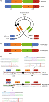
Examples of resolved classic and complex inversions using distinct methodologies. (a) Fluorescence in situ hybridization (FISH) data (left) showing both p and q arm probe signals in a classic heterozygous inversion case (inv(3)(p25.3q28)) initially detected by karyotyping. Two different probe colors are placed on either side of the pericentric inversion junctions allowing for confirmation of the event. In a complex inversion case (inv(X)(p22.31q28)) FISH data (right) shows the p and q probe signals switching arms. Complexities are only detected with additional experiments. (b) Array comparative genomic hybridization confirms copy number neutral state in the classic inversion case (left) but reveals the p and q arm duplications flanking the inversion in the complex case (right). (c) Proposed chromosomal architecture of the classic and complex inversion. (d) Integrative Genomics Viewer (IGV) screenshot of the classic inversion showing the discordant mapped reads as well as split‐reads clustering together. In contrast, the complex inversion does not show clustering of the discordant mapped reads as it is disrupted by a copy number event. Of note, both IGV screenshots are representative figures for such junctions in whole‐genome sequencing data. (e) Final nucleotide‐level resolution for each inversion breakpoint junction alignment based on Sanger‐sequencing for both inversion carriers.
Figure 2
Figure 2
Recombinant chromosomes allow for the characterization of breakpoints in inversion carriers. (a) Reference structure as well as the inverted structure of chromosome 3 highlighting the two junctions (jct1 and jct2) with genomic segments aligned during recombination event. (b) The two possible results, rec(3)dup(3p) or rec(3)dup(3q) of a recombination event. Each result can only carry one of the junctions (either jct1 or jct2). (c) For classic inversions, where the array shows no apparent genomic alteration, we can infer the presence of both inversion junctions through mapping the location of the DEL/DUP recombinant structure. Color matching arrows representing the primer locations for each predicted junction are displayed. Using these predicted locations we were able to Sanger validate the breakpoints of jct1 and jct2 in the inversion carrier (BAB12196) as well jct2 in the recombinant chromosome (BAB12195)
Figure 3
Figure 3
Nuclotide‐level resolution for jct2 was obtained in two individuals with a recombinant chromosome X. (a) Custom aCGH showing DEL/DUP structure of recombinant chromosome X in patients BAB3037 and BAB3038. (b) Sanger sequencing of jct2 was obtained from individual‐specific PCR products based on aCGH CNV positions. Sequencing revealed microhomology (bold black) and templated insertions (see text for details) suggesting replicative mechanism such as MMBIR underlies the formation of the origional inversions. aCGH, array comparative genomic hybridization; MMBIR, microhomology‐mediated break‐induced replication
Figure 4
Figure 4
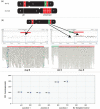
Unexpected complexity in P5371_206 revealed by whole‐genome sequencing (WGS) and array comparative genomic hybridization. (a) WGS revealed a complex rearrangement in individual P5371_206 with a pericentric inversion on chromosome 12 (inv(12)(p11.2q24.1)), which appeared to be balanced on karyotyping. The rearrangement consisted of six genomic segments, of which two were duplicated (red segments B and E) and one was lost (green segment D). (b) A 1 M microarray confirmed the duplications and the deletion that had first been identified by WGS. Screenshots from Agilent Technologies Genomic Workbench microarray software (top, B) and Integrative Genomics Viewer (below, B). (c) Droplet digital PCR confirmed the structure of the chromosome with junction 2 present twice.
Figure 5
Figure 5
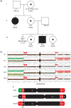
Complex pericentric inversion on chromosome X, segregates in three generations and produces two independent recombinant chromosomes. (a)The family was referred for clinical investigation due to an intrauterine fetal death in gestational week 40 (III:3), which revealed an apparently balanced inv(X)(p22.31q28) in four individuals, and an unbalanced recombinant chromosome in the fetus as well as the sister of the proband. (b) The targeted array comparative genomic hybridization (aCGH) analysis provided with detailed information on the structure of the rearranged chromosomes in both inversion and recombinant chromosome carriers in the family. The duplications were found to originate from the same allele as the inversion and had hence been formed concomitantly with the inversion. (c) The proposed genomic architecture for both the inversion and recombinant chromosome using aCGH and whole‐genome sequencing revealed additional complexity with two duplications on each side of the inversion (red segments B and D).
Figure 6
Figure 6
Proposed mechanism of formation of inv(X) with additional complexities and formation of unbalanced recombinants. (a) The karyotypically balanced inv(X) was found to have two duplications flanking the inversion (DUP–INV–DUP). Phasing of the duplications B and D supported the hypothesis that the duplications had formed concomitantly to the inversion. (b) The family history revealed that two individuals in the family had the same unbalanced recombinant chromosome formed through recombination between the normal allele and the allele with inversion, with duplication of segments D and E and deletion of segment A. The recombinant chromosome in this family is highlighted by the dashed red line
Figure 7
Figure 7
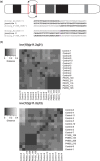
Two founder inversions detected in multiple unrelated individuals. (a) The pericentric inversion on chromosome 12, inv(12)(p11.2q13), was identified in three unrelated Swedish families with identical breakpoint junctions in all individuals. (b) In addition to the inv(12) founder inversion, a previously published and known founder inversion (Gilling et al., 2006) was identified in the cohort (inv(10)(p11.2q21)) (breakpoint junctions: Figure S7). Heatmaps were generated through analysis and comparison of haplotypes performed on all founder inversion carriers, and 11 unrelated individuals of Swedish descent. Both analyses showed that the founder inversion carriers shared a significant amount of common haplotypes and clustered tightly. Distance; the fraction of dissimilar single nucleotide variants (SNVs) between individuals. The darker color indicates a higher amount of shared SNVs

References

    1. Abyzov, A. , Urban, A. E. , Snyder, M. , & Gerstein, M. (2011). CNVnator: An approach to discover, genotype, and characterize typical and atypical CNVs from family and population genome sequencing. Genome Research, 21(6), 974–984. - PMC - PubMed
    1. Ameur, A. , Dahlberg, J. , Olason, P. , Vezzi, F. , Karlsson, R. , Martin, M. , … Gyllensten, U. (2017). SweGen: A whole‐genome data resource of genetic variability in a cross‐section of the Swedish population. European Journal of Human Genetics, 25(11), 1253–1260. - PMC - PubMed
    1. Bahrambeigi, V. , Song, X. , Sperle, K. , Beck, C. R. , Hijazi, H. , Grochowski, C. M. , … Lupski, J. R. (2019). Distinct patterns of complex rearrangements and a mutational signature of microhomeology are frequently observed in PLP1 copy number gain structural variants. Genome Medicine, 11(1), 80. - PMC - PubMed
    1. Beck, C. R. , Carvalho, C. M. B. , Akdemir, Z. C. , Sedlazeck, F. J. , Song, X. , Meng, Q. , … Lupski, J. R. (2019). Megabase length hypermutation accompanies human structural variation at 17p11.2. Cell, 176(6), 1310–1324e10. - PMC - PubMed
    1. Beck, C. R. , Carvalho, C. M. , Banser, L. , Gambin, T. , Stubbolo, D. , Yuan, B. , … Lupski, J. R. (2015). Complex genomic rearrangements at the PLP1 locus include triplication and quadruplication. PLoS Genetics, 11(3), e1005050. - PMC - PubMed

Publication types