Review
. 2020 Sep 7;12(9):2534.
doi: 10.3390/cancers12092534.

Therapeutically Significant MicroRNAs in Primary and Metastatic Brain Malignancies

Affiliations
Review

Therapeutically Significant MicroRNAs in Primary and Metastatic Brain Malignancies

Akilandeswari A Balachandran et al. Cancers (Basel). .

Abstract

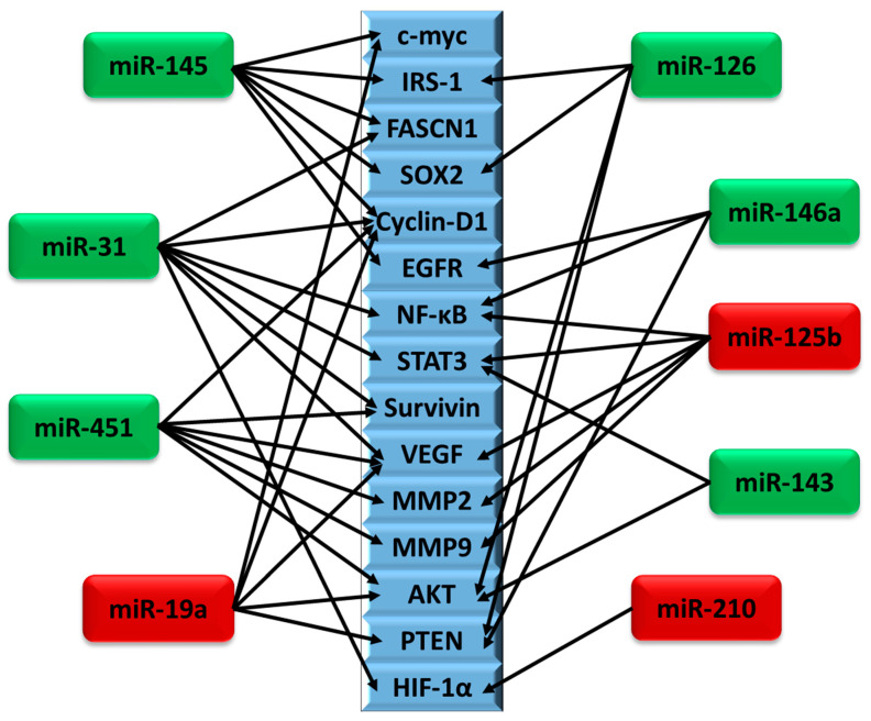
Brain cancer is one among the rare cancers with high mortality rate that affects both children and adults. The most aggressive form of primary brain tumor is glioblastoma. Secondary brain tumors most commonly metastasize from primary cancers of lung, breast, or melanoma. The five-year survival of primary and secondary brain tumors is 34% and 2.4%, respectively. Owing to poor prognosis, tumor heterogeneity, increased tumor relapse, and resistance to therapies, brain cancers have high mortality and poor survival rates compared to other cancers. Early diagnosis, effective targeted treatments, and improved prognosis have the potential to increase the survival rate of patients with primary and secondary brain malignancies. MicroRNAs (miRNAs) are short noncoding RNAs of approximately 18-22 nucleotides that play a significant role in the regulation of multiple genes. With growing interest in the development of miRNA-based therapeutics, it is crucial to understand the differential role of these miRNAs in the given cancer scenario. This review focuses on the differential expression of ten miRNAs (miR-145, miR-31, miR-451, miR-19a, miR-143, miR-125b, miR-328, miR-210, miR-146a, and miR-126) in glioblastoma and brain metastasis. These miRNAs are highly dysregulated in both primary and metastatic brain tumors, which necessitates a better understanding of their role in these cancers. In the context of the tumor microenvironment and the expression of different genes, these miRNAs possess both oncogenic and/or tumor-suppressive roles within the same cancer.

Keywords: brain cancer; cancer metastasis; glioblastoma; glioma; miRNA.


Conflict of interest statement

The authors declare no conflict of interest.

Figures

Figure 1
Figure 1
miRNA biogenesis and mechanism of action. miRNAs are transcribed by RNA polymerase II or III, producing primary transcripts (pri-miRNA). The pri-miRNA is then cleaved by the Drosha and DGCR8 complex, producing precursor miRNA (pre-miRNA). Pre-miRNA assembles into a complex with Exportin-5 (XPO5) and Ran/GTP. Once in the cytoplasm, pre-miRNA is further processed by Dicer complex into mature miRNA. The passenger strand of mature miRNA is degraded, while the other strand is loaded into argonaute protein (Ago1–4) and incorporated into the RNA-induced silencing complex (RISC). Binding of RISC to its target mRNA results in degradation and/or translational repression of the target gene, by either slicer-independent or slicer-dependent silencing. When the miRNA is extensively base-paired, slicer-dependent silencing mechanisms proceed. GW182 family of proteins is recruited by AGO. GW182 interacts with PABPC, promoting efficient mRNA deadenylation through the recruitment of PAN2–PAN3 and CCR4–NOT complexes. Cleavage begins with the deadenylation of the mRNA to remove the poly (A) tail. Deadenylation promotes subsequent mRNA decapping and degradation by Xrn1p. Alternatively, degradation can occur via the exosome (vesicles with a size of 30 to 100nm, with 3′5′ exonuclease activity). In slicer-independent silencing, multiple complementary sites with imperfect base-pairing create bulges in the RNA duplex propelling slicer-independent gene silencing mechanisms. miRNAs can repress translation determined by the target mRNA promoter. Alternatively, miRNA can repress translation indirectly by segregating mRNA into P-Bodies. Ultimately, mRNA can be isolated for storage or be targeted for decay via Xrn1p or exosome degradation.
Figure 2
Figure 2
Venn diagram showing the number of miRNAs that are in common between upregulated and downregulated miRNAs in glioblastoma (GBM) and brain metastasis (BrM). Upregulation and downregulation are abbreviated by ‘Up’ and ‘Down’, respectively.
Figure 3
Figure 3
Examples of nucleotide analogues used in oligonucleotide synthesis. 2′-OMe, 2′-O-methyl [250,251]; 2′-MOE, 2′-O-methoxyethyl [252]; 2′-NH2, 2′-amino [253]; 2′-F, 2′-fluoro [254]; 2′-FANA, 2′-fluroarabino nucleic acid [255]; TNA, threose nucleic acid [256]; 4′-S, 4′-thio [257]; LNA, locked nucleic acid [258,259,260]; ENA, 2′-O, 4′-C-ethylene-bridged nucleic acid [261]; PNA, peptide nucleic acid [262]; PMO, phosphorodiamidate morpholino oligomer [263]; MNA, morpholino nucleic acid [264]; Phosphorothioate, PS [265]; HNA, 1,5-anhydro hexitol nucleic acid [266]; CeNA, cyclohexenyl nucleic acid [266]; ANA, altritol nucleic acid [266].
Figure 4
Figure 4
Schematic representation of genes regulated by multiple miRNAs in GBM and BrM. Among the genes that are reported to be regulated by the miRNAs reviewed above, the most common genes that are regulated by at least two miRNAs are represented (blue rectangles). The arrows indicate interactions between miRNAs and their targets. miRNAs indicated in green and red are predominantly tumor suppressors and oncomiRs, respectively. Expression of Cyclin-D1, VEGF, and AKT, which plays a major role in cell proliferation, angiogenesis, and metastasis, was shown to be modulated by four different miRNAs. Although miR-328 is reviewed above, it did not share any common targets with other miRNAs.

Similar articles

Cited by

References

    1. McNeill K.A. Epidemiology of brain tumors. Neurol. Clin. 2016;34:981–998. doi: 10.1016/j.ncl.2016.06.014. - DOI - PubMed
    1. Shah V., Kochar P. Brain cancer: Implication to disease, therapeutic strategies and tumor targeted drug delivery approaches. Recent Pat. Anticancer Drug Discov. 2018;13:70–85. doi: 10.2174/1574892812666171129142023. - DOI - PubMed
    1. Franchino F., Ruda R., Soffietti R. Mechanisms and therapy for cancer metastasis to the brain. Front. Oncol. 2018;8:161. doi: 10.3389/fonc.2018.00161. - DOI - PMC - PubMed
    1. Lapointe S., Perry A., Butowski N.A. Primary brain tumours in adults. Lancet. 2018;392:432–446. doi: 10.1016/S0140-6736(18)30990-5. - DOI - PubMed
    1. Lukas R.V., Wainwright D.A., Ladomersky E., Sachdev S., Sonabend A.M., Stupp R. Newly diagnosed glioblastoma: A review on clinical management. Oncology. 2019;33:91–100. - PMC - PubMed

LinkOut - more resources