. 2020 Sep 7;9(9):834.
doi: 10.3390/antiox9090834.

Cardamonin Inhibits Oxazolone-Induced Atopic Dermatitis by the Induction of NRF2 and the Inhibition of Th2 Cytokine Production

Affiliations

Cardamonin Inhibits Oxazolone-Induced Atopic Dermatitis by the Induction of NRF2 and the Inhibition of Th2 Cytokine Production

Ok-Kyung Yoo et al. Antioxidants (Basel). .

Abstract

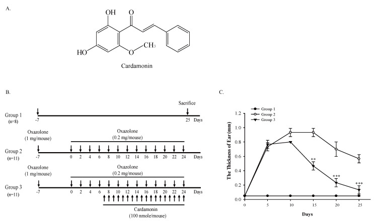
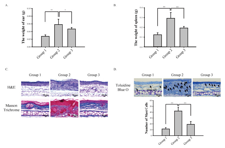
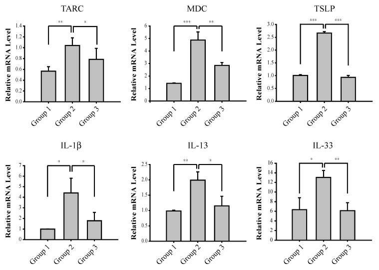
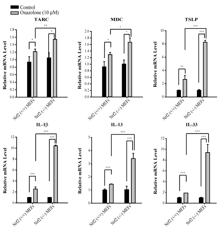
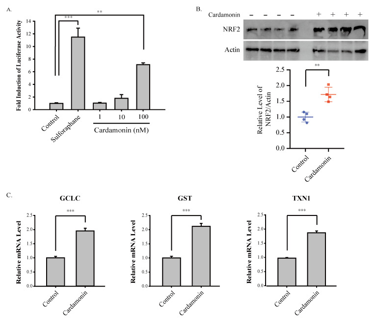
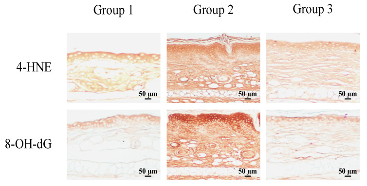
The skin is constantly exposed to various types of chemical stresses that challenge the immune cells, leading to the activation of T cell-mediated hypersensitivity reactions including atopic dermatitis. Previous studies have demonstrated that a variety of natural compounds are effective against development of atopic dermatitis by modulating immune responses. Cardamonin is a natural compound abundantly found in cardamom spices and many other medicinal plant species. In the present study, we attempted to examine whether cardamonin could inhibit oxazolone-induced atopic dermatitis in vivo. Our results show that topical application of cardamonin onto the ear of mice suppressed oxazolone-induced inflammation in the ear and hyperplasia in the spleen. Cardamonin also inhibited oxazolone-induced destruction of connective tissues and subsequent infiltration of mast cells into the skin. In addition, we found that the production of Th2 cytokines is negatively regulated by NRF2, and the induction of NRF2 by cardamonin contributed to suppressing oxazolone-induced Th2 cytokine production and oxidative damages in vivo. Together, our results demonstrate that cardamonin is a promising natural compound, which might be effective for treatment of atopic dermatitis.

Keywords: NF-E2-related factor 2 (NRF2); T helper 2 (Th2) cytokines; cardamonin; oxazolone.


Conflict of interest statement

The authors declare no conflict of interest.

Figures

Figure 1
Figure 1
Cardamonin suppresses oxazolone-induced inflammation in mouse skin. (A) Chemical structure of cardamonin. (B) Experimental scheme evaluating the effect of cardamonin on oxazolone-induced atopic dermatitis. (C) Cardamonin inhibits oxazolone-induced inflammation in mouse skin. The thickness of ear in mice was measured using a caliper every five days. Asterisks indicate a statistical significance of ** p < 0.01 and *** p < 0.001.
Figure 2
Figure 2
Cardamonin suppresses oxazolone-induced atopic dermatitis by maintaining the integrity of the skin and inhibiting the infiltration of mast cells. (A) Cardamonin inhibits an increase in the weight of ear induced by oxazolone. At sacrifice, mouse ears were excised and their weight was measured. Asterisks indicate a statistical significance of * p < 0.05 and ** p < 0.01. (B) Topical application of oxazolone onto the ear of mouse induces splenomegaly in mice, which is inhibited by cardamonin. At sacrifice, the spleen of mouse was excised and the weight was measured. Asterisks indicate a statistical significance of ** p < 0.01. (C) Cardamonin inhibits oxazolone-induced destruction of connective tissues in the ear of mouse. Gross morphology of mouse skin tissues in the ear are stained by H&E staining (upper panel) and the collagens in connective tissues are stained by Masson trichrome staining (lower panel). (D) Cardamonin inhibits oxazolone-induced infiltration of mast cells into the skin. Mast cells are stained by Toluidine Blue O staining (Upper Panel) and the number of mast cells under the bright field was counted (Lower Panel). Asterisks indicate a statistical significance of ** p < 0.01.
Figure 3
Figure 3
Cardamonin mediates transcriptional suppression of oxazolone-induced production of Th2 cytokines in vivo. The mRNA levels of Th2 cytokines in mouse skin were measured by real-time RT-PCR analysis. Asterisks indicate a statistical significance of * p < 0.05, ** p < 0.01 and *** p < 0.001.
Figure 4
Figure 4
Transcriptional activation of Th2 cytokines by oxazolone is significantly higher in Nrf2 (−/−) MEFs compared with Nrf2 (+/+) MEFs. Primary Nrf2 (+/+) and Nrf2 (−/−) MEFs were exposed to oxazolone (10 μM) for 24 h and real-time PCR was performed using specific primers against Th2 cytokines. Asterisks indicate a statistical significance of * p < 0.05, ** p < 0.01 and *** p < 0.001.
Figure 5
Figure 5
Cardamonin activates ARE-luciferase activity in HaCaT cells and induces the expression NRF2 and its target genes in mice. (A) Cardamonin induces ARE activation in HaCaT-ARE-GFP-luciferase cells. HaCaT-ARE-GFP-luciferase cells were exposed to cardamonin at various concentrations for 24 h and the luciferase activity was measured. The experiment was performed in triplicate and sulforaphane (10 μM) was included as a positive control. Asterisks indicate a statistical significance of ** p < 0.01 and *** p < 0.001. (B) Cardamonin induces NRF2 in vivo. Cardamonin (100 nmole/mouse) was topically applied to the ear of mouse for 24 h and Western blotting was performed against NRF2 and actin (Upper Panel). The relative level of NRF2 was normalized against actin (Lower Panel). Asterisks indicate a statistical significance of ** p < 0.01. (C) Cardamonin induces NRF2 target genes in vivo. Cardamonin (100 nmole/mouse) was topically applied to the ear of mouse for 24 h and real-time RT-PCR was performed against NRF2 target genes (GCLC, GST, TXN1). The experiment was performed in quadruplicate. Asterisks indicate a statistical significance of *** p < 0.001.
Figure 6
Figure 6
Cardamonin protects against oxazolone-induced oxidative damage in vivo. The formation of oxidative damage markers in mouse skin was visualized by immunohistochemistry using 4-HNE and 8-OH-dG antibodies. A representative slide is provided.

Similar articles

Cited by

References

    1. Yang E.J., Sekhon S., Sanchez I.M., Beck K.M., Bhutani T. Recent Developments in Atopic Dermatitis. Pediatrics. 2018;142 doi: 10.1542/peds.2018-1102. - DOI - PubMed
    1. Weidinger S., Novak N. Atopic dermatitis. Lancet. 2016;387:1109–1122. doi: 10.1016/S0140-6736(15)00149-X. - DOI - PubMed
    1. Walker J.A., McKenzie A.N.J. TH2 cell development and function. Nat. Rev. Immunol. 2018;18:121–133. doi: 10.1038/nri.2017.118. - DOI - PubMed
    1. Mack M.R., Kim B.S. The Itch-Scratch Cycle: A Neuroimmune Perspective. Trends Immunol. 2018;39:980–991. doi: 10.1016/j.it.2018.10.001. - DOI - PMC - PubMed
    1. Lin Y.T., Wang C.T., Chiang B.L. Role of bacterial pathogens in atopic dermatitis. Clin. Rev. Allergy Immunol. 2007;33:167–177. doi: 10.1007/s12016-007-0044-5. - DOI - PubMed

LinkOut - more resources