Review
. 2020 Sep 7;8(3):17.
doi: 10.3390/jdb8030017.

A Series of Tubes: The C. elegans Excretory Canal Cell as a Model for Tubule Development

Affiliations
Review

A Series of Tubes: The C. elegans Excretory Canal Cell as a Model for Tubule Development

Matthew Buechner et al. J Dev Biol. .

Abstract

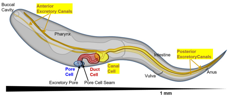
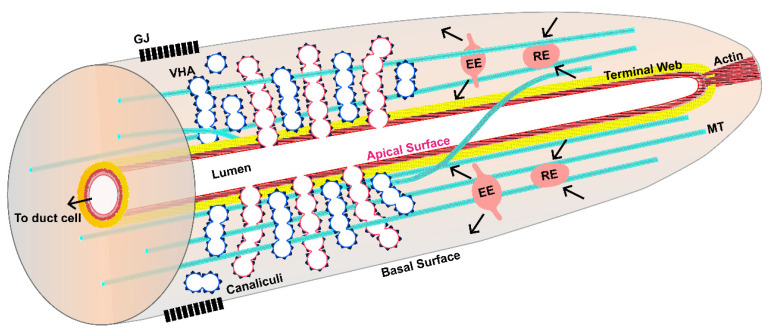
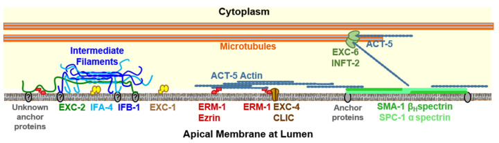
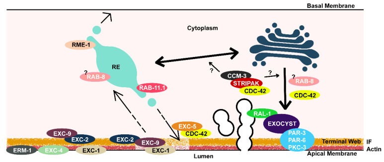
Formation and regulation of properly sized epithelial tubes is essential for multicellular life. The excretory canal cell of C. elegans provides a powerful model for investigating the integration of the cytoskeleton, intracellular transport, and organismal physiology to regulate the developmental processes of tube extension, lumen formation, and lumen diameter regulation in a narrow single cell. Multiple studies have provided new understanding of actin and intermediate filament cytoskeletal elements, vesicle transport, and the role of vacuolar ATPase in determining tube size. Most of the genes discovered have clear homologues in humans, with implications for understanding these processes in mammalian tissues such as Schwann cells, renal tubules, and brain vasculature. The results of several new genetic screens are described that provide a host of new targets for future studies in this informative structure.

Keywords: IRG protein; apical surface; epithelial tube; exocytosis; intermediate filaments; terminal web; vacuolar ATPase; vesicle transport.


Conflict of interest statement

The authors declare no conflict of interest.

Figures

Figure 1
Figure 1
Perspective diagram (anterior close, posterior far) showing position of the cells of the excretory system, and the long tubular canals stretching the length of the nematode (1 mm total length, about 50 μm in diameter). The excretory pore cell (seamed) is in blue, excretory duct cell (seam present at birth, then removed to become seamless) in red, and excretory canal cell in yellow. The canals can collect excess liquid from the entire length of the animal to transport to the duct and pore cells for removal.
Figure 2
Figure 2
Diagram of canal section, showing subcellular morphology of lumen and canal tip. The apical membrane is shown in red surrounding the central lumen (in white), while the basal membrane is shown in grey. The apical surface is coated by actin filaments (thick red) and intermediate filaments (thick yellow) that together form the terminal web. Filaments extend to the distal terminus of the canal, presumably to help in canal extension. Small canalicular vesicles appear as separate vesicles (in blue) or connected to the lumen (in red) to form canaliculi. These vesicles are coated with vacuolar ATPase (VHA, black spikes on vesicles). Microtubules (cyan) extend along the length of the canal, interspersed with and outside the canaliculi. Some microtubules appear helically wound around the lumen. Early endosomes (EE) and recycling endosomes (RE) move along the canal length (See Supplementary Video S1). Gap junctions (black) connect the canal cytoplasm to neighboring hypoderm.
Figure 3
Figure 3
Diagram (not to scale) of cytoskeletal elements at the excretory canal apical membrane. Microtubules are located both near and far from the apical membrane. Note that not all locations and interactions have been proven to occur within the excretory canals of C. elegans. Tips of microtubules are located at organizing centers, where formin EXC-6 (and possibly formin INFT-2) nucleate actin filaments both along the length of the canal and towards its apical surface [55]. Fibers of ACT-5 actin are held close to the apical surface and organized there through interactions with the β-heavy spectrin SMA-1 [30,31,56] (presumably anchored by the Band 4.1 homologue FRM-1 [57]) and with the ezrin/radixin/moesin homologue ERM-1 [22,29,58]. In mammals, intracellular chloride channel (CLIC) channels homologous to EXC-4 are also associated with ezrin [59]. Intermediate filaments are associated with the luminal membrane by unknown anchor proteins, though spacing of these filaments depends on ERM-1 and SMA-1 [22,29]. The intermediate filaments associate by their central filament domain to form heterologous filaments that extend farther from the membrane than do the bulk of the apical actin filaments [60]. EXC-1 is another protein affecting canal structure located exclusively at the apical membrane [22].
Figure 4
Figure 4
Speculative model of formation and maintenance of canal luminal shape. Lumen on bottom, basal membrane and neighboring hypodermal cell on top. Ezrin/Radixin/Moesin (ERM)-1 and EXC-1(IRG) proteins are seen at an apical membrane. EXC-2/IF and two other intermediate filaments, along with actin, make up the terminal web. The EXC-4/CLIC channel is apical. EXC-9 is retained to the apical surface by EXC-2. If terminal web is damaged or thinned during growth (light area shade of terminal web), EXC-9 can make contact with and presumably activate EXC-1. Ras domains of EXC-1 presumably trigger trafficking machinery at the recycling endosome (RE). Trafficking may be directed from the RE to apical surface, or possibly via Golgi via the STRiatin-Interacting Phosphatase And Kinase (STRIPAK) complex. EXC-5/FGD Guanine Exchange Factor activates CDC-42 to polymerize actin to bring vesicles to apical surface where the Exocyst complex and PAR complex complete fusion.

Similar articles

Cited by

References

    1. Lubarsky B., Krasnow M.A. Tube morphogenesis: Making and shaping biological tubes. Cell. 2003;112:19–28. doi: 10.1016/S0092-8674(02)01283-7. - DOI - PubMed
    1. Plattner H. The contractile vacuole complex of protists—new cues to function and biogenesis. Crit. Rev. Microbiol. 2015;41:218–227. doi: 10.3109/1040841X.2013.821650. - DOI - PubMed
    1. Maruyama R., Andrew D.J. Drosophila as a model for epithelial tube formation. Dev. Dyn. 2012;241:119–135. doi: 10.1002/dvdy.22775. - DOI - PMC - PubMed
    1. Rosa J.B., Metzstein M.M., Ghabrial A.S. An Ichor-dependent apical extracellular matrix regulates seamless tube shape and integrity. PLoS Genet. 2018;14:e1007146. doi: 10.1371/journal.pgen.1007146. - DOI - PMC - PubMed
    1. Samakovlis C., Hacohen N., Manning G., Sutherland D.C., Guillemin K., Krasnow M.A. Development of the Drosophila tracheal system occurs by a series of morphologically distinct but genetically coupled branching events. Development. 1996;122:1395–1407. - PubMed

LinkOut - more resources