. 2020 Sep 7;12(9):995.
doi: 10.3390/v12090995.

The Husavirus Posa-Like Viruses in China, and a New Group of Picornavirales

Affiliations

The Husavirus Posa-Like Viruses in China, and a New Group of Picornavirales

Zhenzhi Han et al. Viruses. .

Abstract

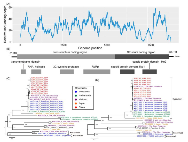
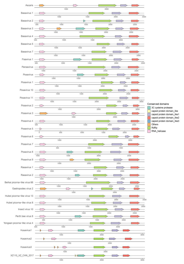
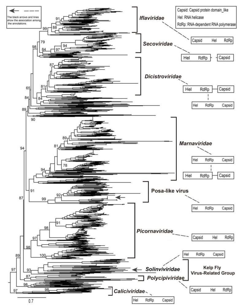
Novel posa-like viral genomes were first identified in swine fecal samples using metagenomics and were designated as unclassified viruses in the order Picornavirales. In the present study, nine husavirus strains were identified in China. Their genomes share 94.1-99.9% similarity, and alignment of these nine husavirus strains identified 697 nucleotide polymorphism sites across their full-length genomes. These nine strains were directly clustered with the Husavirus 1 lineage, and their genomic arrangement showed similar characteristics. These posa-like viruses have undergone a complex evolutionary process, and have a wide geographic distribution, complex host spectrum, deep phylogenetic divergence, and diverse genomic organizations. The clade of posa-like viruses forms a single group, which is evolutionarily distinct from other known families and could represent a distinct family within the Picornavirales. The genomic arrangement of Picornavirales and the new posa-like viruses are different, whereas the posa-like viruses have genomic modules similar to the families Dicistroviridae and Marnaviridae. The present study provides valuable genetic evidence of husaviruses in China, and clarifies the phylogenetic dynamics and the evolutionary characteristics of Picornavirales.

Keywords: Picornavirales; evolution; husavirus; phylogeny; posa-like virus.


Conflict of interest statement

The authors declare no conflict of interest.

Figures

Figure 1
Figure 1
Maximum-likelihood phylogenetic tree of different conserved domain amino acid sequences. (AD) show the phylogenetic trees based on the picorna-like capsid protein domain 1, picorna-like capsid protein domain 2, RNA-dependent RNA polymerase, and helicase, respectively. The black arrows represent the major husaviruses reported, and the box shows the husavirus strains besides the previously reported Husavirus 1. The genomes of six known families of Picornavirales were used as outgroups. The names are colored according to the types of hosts in the inset. Scale bars indicate the substitutions per site per year. The numbers at each node indicate the bootstrap support value, with 1000 bootstrap replicates.
Figure 2
Figure 2
(A) Relative sequencing depth across the reference genome. (B) The classical genomic organization of posa-like viruses. (C) The maximum-likelihood phylogenetic tree of husaviruses and the neighboring posa-like viruses, constructed using nucleotide sequences. (D) The maximum-likelihood phylogenetic tree of husaviruses and the neighboring posa-like viruses was constructed using amino acid sequences. Scale bars indicate the substitutions per site per year, and taxa are colored according to the countries of origin. The box show the previously reported Husavirus 1 strain. The numbers at each node indicate the bootstrap support value, with 1000 bootstrap replicates.
Figure 3
Figure 3
The genomic organization of the representative posa-like viruses and a representative husavirus identified in the present study. The arrow blocks show the conserved protein domains and their positions along the genome. Different colors indicate different protein domains. All conserved domains are drawn to scale with the genome size. For clear display, the label “others” refers to the non-conserved domains, including the Picornavirus core protein 2A, Peptidase_C3G superfamily, Poliovirus 3A protein like domain, and CRPV capsid protein-like domains.
Figure 4
Figure 4
The maximum-likelihood phylogenetic tree of posa-like viruses, other known families, and unclassified viruses of Picornavirales. The conserved amino acid sequences of RdRp were used to generate phylogenetic trees. Scale bars indicate the substitutions per site per year. The numbers at each node indicate the SH-like approximate likelihood ratio test (SH-aLRT) support value, with 1000 iterations. The black arrows represent the husavirus strains identified in the present study. The genomic organization on the right shows the major non-structural and structural domains, with the arrangement of open reading frames (ORFs) based on previous reports [2,3,5].
Figure 5
Figure 5
The first two principal components of the genomic sequences of Picornavirales, with the hosts and available sample locations used as the prior groups. Eigenvalues of the analysis (PCA and DA) are displayed in the inset, along which the black modules represent the dimensions retained and the gray modules show the dimensions eliminated in the datasets. Groups are shown in different colors, with dots representing individual strains. (A) The scatterplot using the host information as a prior cluster. (B) The scatterplot using the location information as a prior cluster. PCA, principal component analysis; DA, discriminant analysis.

Similar articles

Cited by

References

    1. Adams M.J., Lefkowitz E.J., King A.M.Q., Harrach B., Harrison R.L., Knowles N.J., Kropinski A.M., Krupovic M., Kuhn J., Mushegian A., et al. Changes to taxonomy and the International Code of Virus Classification and Nomenclature ratified by the International Committee on Taxonomy of Viruses (2017) Arch. Virol. 2017;162:2505–2538. doi: 10.1007/s00705-017-3358-5. - DOI - PubMed
    1. Le Gall O., Christian P., Fauquet C.M., King A.M.Q., Knowles N.J., Nakashima N., Stanway G., Gorbalenya A.E. Picornavirales, a proposed order of positive-sense single-stranded RNA viruses with a pseudo-T = 3 virion architecture. Arch. Virol. 2008;153:715–727. doi: 10.1007/s00705-008-0041-x. - DOI - PubMed
    1. Zell R., Delwart E., Gorbalenya A.E., Hovi T., King A.M.Q., Knowles N.J., Lindberg A.M., Pallansch M.A., Palmenberg A.C., Reuter G., et al. ICTV Virus Taxonomy Profile: Picornaviridae. J. Gen. Virol. 2017;98:2421–2422. doi: 10.1099/jgv.0.000911. - DOI - PMC - PubMed
    1. Shi M., Lin X.-D., Chen X., Tian J.-H., Chen L.-J., Li K., Wang W., Eden J.-S., Shen J.-J., Liu L., et al. The evolutionary history of vertebrate RNA viruses. Nature. 2018;556:197–202. doi: 10.1038/s41586-018-0012-7. - DOI - PubMed
    1. Shi M., Lin X.-D., Tian J.-H., Chen L.-J., Chen X., Li C.-X., Qin X.-C., Li J., Cao J., Eden J.-S., et al. Redefining the invertebrate RNA virosphere. Nature. 2016;540:539–543. doi: 10.1038/nature20167. - DOI - PubMed

Publication types

LinkOut - more resources