Review
. 2020 Sep 7;9(9):2042.
doi: 10.3390/cells9092042.

"Repair Me if You Can": Membrane Damage, Response, and Control from the Viral Perspective

Affiliations
Review

"Repair Me if You Can": Membrane Damage, Response, and Control from the Viral Perspective

Coralie F Daussy et al. Cells. .

Abstract

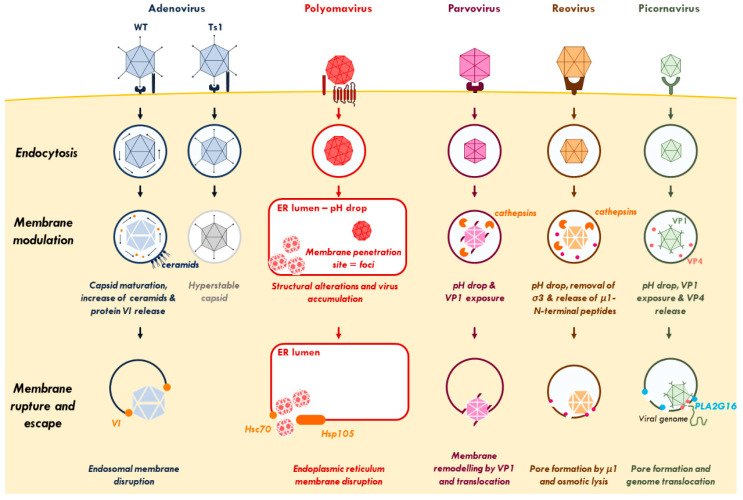
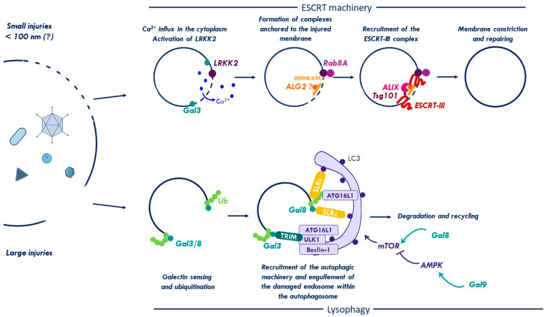
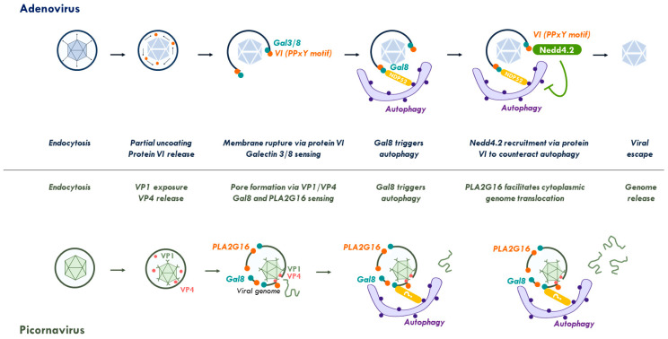
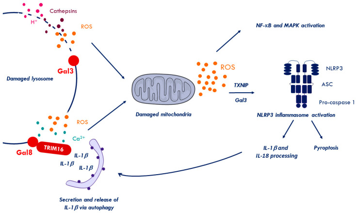
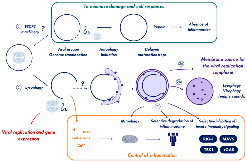
Cells are constantly challenged by pathogens (bacteria, virus, and fungi), and protein aggregates or chemicals, which can provoke membrane damage at the plasma membrane or within the endo-lysosomal compartments. Detection of endo-lysosomal rupture depends on a family of sugar-binding lectins, known as galectins, which sense the abnormal exposure of glycans to the cytoplasm upon membrane damage. Galectins in conjunction with other factors orchestrate specific membrane damage responses such as the recruitment of the endosomal sorting complex required for transport (ESCRT) machinery to either repair damaged membranes or the activation of autophagy to remove membrane remnants. If not controlled, membrane damage causes the release of harmful components including protons, reactive oxygen species, or cathepsins that will elicit inflammation. In this review, we provide an overview of current knowledge on membrane damage and cellular responses. In particular, we focus on the endo-lysosomal damage triggered by non-enveloped viruses (such as adenovirus) and discuss viral strategies to control the cellular membrane damage response. Finally, we debate the link between autophagy and inflammation in this context and discuss the possibility that virus induced autophagy upon entry limits inflammation.

Keywords: ESCRT machinery; adenovirus; antiviral autophagy; bacterial invasion; galectin; inflammation; interferon; lysophagy; membrane damage; virus entry.


Conflict of interest statement

The authors declare no conflict of interest. The funders had no role in the design of the study; in the collection, analyses, or interpretation of data; in the writing of the manuscript, or in the decision to publish the results.

Figures

Figure 1
Figure 1
Virus-inflicted membrane damage. After binding to cell-surface receptors, viruses are internalized through endocytosis. Once in the endosome, adenovirus capsid undergoes partial disassembly and releases protein VI. The increase of ceramide concentration enhances the binding of protein VI to the endosomal membrane and its subsequent rupture. Polyomavirus-containing endosomes are targeted to the endoplasmic reticulum (ER) where the virus undergoes conformational changes to penetrate the ER-membrane and escape to the cytosol. Parvovirus and reovirus require a pH drop and the action of endosomal cathepsins to induce conformational rearrangements, disrupt the endosome, and reach the cytosol. After endocytosis and conformational changes, picornaviruses rely on a cellular lipid-modifying enzyme (PLA2G16) to facilitate the translocation of its genome via selective pores across the endosomal membrane. See Section 1 for further details. Abbreviations: ER, endoplasmic reticulum; Hsc70, Heat shock cognate 71 kDa protein; Hsp105, Heat shock protein 105 kDa; PLA2G16, phospholipase A2 group XVI.
Figure 2
Figure 2
Membrane damage repair and removal. The endosome is constantly challenged by pathogens (bacteria, virus, and fungi), protein aggregates, or chemicals that can disrupt its membrane and provoke injuries of different sizes. Small disruptions (<100 nm) trigger a leakage of Ca2+ into the cytoplasm and activate LRKK2. LRKK2 and Ca2+ effectors mediate the recruitment of the ESCRT machinery to promote repair of the injured organelle. Galectin-3 (Gal3) is recruited to damage sites and may promote ESCRT assembly. If the injury is too large, the cell will trigger a process of degradation called lysophagy. During lysophagy, damaged vacuoles are sensed and tagged by galectins (Gal) and ubiquitin (Ub). Both signals mediate the recruitment of the autophagic machinery (either directly via autophagic receptors, such as the sequestosome like receptors (SLRs) or indirectly via TRIMs). The membrane remnant is engulfed in a double-membrane vesicle called autophagosome, which fuses with lysosomes for content degradation and recycling. Autophagy is also controlled by metabolic kinase mechanistic target of rapamycin (mTOR) through Gal8. Moreover, Gal9 can also control autophagy induction by directly activating AMP-activated protein kinase (AMPK) in response to endosomal damage to inhibit mTOR. See text for details. Abbreviations: ESCRT, endosomal sorting complexes required for transport; Gal8, galectin8; LRRK2, Leucine-rich repeat kinase 2; ROS, reactive oxygen species; Ub, ubiquitin.
Figure 3
Figure 3
Viral control of the membrane damage response. Top: endocytosed adenoviruses partially uncoat and release the membrane lytic capsid protein VI for endosomal membrane lysis. Membrane damage is sensed by galectins 3 and 8. Galectin 8 recruits autophagic receptors and triggers autophagy. Adenoviruses stall autophagy through a short PPxY peptide motif in protein VI that recruits the ubiquitin ligase Nedd4.2. As a consequence, they avoid degradation and escape into the cytoplasm. Bottom: After endocytosis and acidification of the endosome, picornaviruses undergo conformational changes to expose capsid protein VP1 and release of VP4. Both proteins attach to the endosomal membrane creating membrane-penetrating pores. Membrane damage is then independently sensed by galectin 8 activating autophagy and PLA2G16. PLA2G16 facilitates genome translocation into the cytoplasm preventing autophagic clearance. See Section 2 and Section 3 for further details. Abbreviations: Gal, galectin; NDP52, Nuclear dot protein 52; Nedd4.2, neural precursor cell expressed, developmentally down-regulated 4.2; PLA2G16, phospholipase A2 group XVI.
Figure 4
Figure 4
Membrane damage signaling. Endolysosomal rupture triggers the leakage of harmful components (e.g., ROS, H+, cathepsins or Ca2+), resulting in mitochondrial damage and increased ROS production. Increasing levels of ROS activate nuclear factor-kappa B (NF-κB) and mitogen-activated protein kinase (MAPK) signaling and triggers the release of the thioredoxin-interacting protein (TXNIP) from thioredoxin. Soluble TXNIP mediates Nod-like receptor family, pyrin domain-containing 3 (NLRP3)-inflammasomes activation and association with their adapters (ASC), thereby triggering pro-caspase conversion [149,150]. Activated inflammasomes process pro-inflammatory cytokines (IL-1β and IL-18) and may trigger cell death through pyroptosis. Processed IL-1β in turn can be recruited by Gal8 and TRIM16, that coordinate the autophagic machinery to secrete IL-1β via an unconventional secretory pathway [105,151]. Abbreviations: Gal3, galectin3; Gal8, galectin8; ROS, reactive oxygen species.
Figure 5
Figure 5
Model for non-enveloped virus membrane penetration. Non-enveloped viruses breach endo-lysosomal membranes for particle escape into the cytosol or cytosolic genome translocation. (1) If the membrane damage is small enough, Ca2+-dependent recruitment of the ESCRT machinery will repair the endosomes limiting time and extend of the damage. This strategy could provide enough time for the virus/genome to reach the cytosol without activating the inflammatory pathways. (2) If this repair fails or the inflicted damage is too large, “eat-me signals” (galectins and ubiquitin) will accumulate at the damage site and initiate autophagy. Viruses use viral factors (e.g., Ad) or cellular factors (e.g., PV) to delay the autophagy response until capsids/viral genomes reach the safety of the cytosol. The ensuing autophagy response than may continue and work in favor of the virus. Activating autophagy via membrane damage could (2a) provide a membranes source for viral replication, and (2b) remove and recycle membrane remnants, damaged lysosomes, and empty viral capsids to avoid excessive cells responses. Additionally, autophagy could (2c) limit excessive inflammation through autophagic degradation of damaged mitochondria and by removing activated inflammasomes and effectors of the innate immunity signaling pathways (see text for details).

References

    1. Lombard J. Once upon a time the cell membranes: 175 years of cell boundary research. Biol. Direct. 2014;9:1–35. doi: 10.1186/s13062-014-0032-7. - DOI - PMC - PubMed
    1. Thurston T.L.M.M., Wandel M.P., Von Muhlinen N., Foeglein Á., Randow F., Foeglein A., Randow F., Foeglein Á., Randow F. Galectin 8 targets damaged vesicles for autophagy to defend cells against bacterial invasion. Nature. 2012;482:414–418. doi: 10.1038/nature10744. - DOI - PMC - PubMed
    1. Montespan C., Marvin S.A., Austin S., Burrage A.M., Roger B., Rayne F., Faure M., Campell E.M., Schneider C., Reimer R., et al. Multi-layered control of Galectin-8 mediated autophagy during adenovirus cell entry through a conserved PPxY motif in the viral capsid. PLoS Pathog. 2017;13 doi: 10.1371/journal.ppat.1006217. - DOI - PMC - PubMed
    1. Jia J., Abudu Y.P., Claude-Taupin A., Gu Y., Kumar S., Choi S.W., Peters R., Mudd M.H., Allers L., Salemi M., et al. Galectins Control mTOR in Response to Endomembrane Damage. Mol. Cell. 2018;70:120–135.e8. doi: 10.1016/j.molcel.2018.03.009. - DOI - PMC - PubMed
    1. Vietri M., Radulovic M., Stenmark H. The many functions of ESCRTs. Nat. Rev. Mol. Cell Biol. 2020;21:25–42. doi: 10.1038/s41580-019-0177-4. - DOI - PubMed

Publication types

LinkOut - more resources