Review
. 2020 Sep 7;21(18):6538.
doi: 10.3390/ijms21186538.

Advanced Bioluminescence System for In Vivo Imaging with Brighter and Red-Shifted Light Emission

Affiliations
Review

Advanced Bioluminescence System for In Vivo Imaging with Brighter and Red-Shifted Light Emission

Mizuki Endo et al. Int J Mol Sci. .

Abstract

In vivo bioluminescence imaging (BLI), which is based on luminescence emitted by the luciferase-luciferin reaction, has enabled continuous monitoring of various biochemical processes in living animals. Bright luminescence with a high signal-to-background ratio, ideally red or near-infrared light as the emission maximum, is necessary for in vivo animal experiments. Various attempts have been undertaken to achieve this goal, including genetic engineering of luciferase, chemical modulation of luciferin, and utilization of bioluminescence resonance energy transfer (BRET). In this review, we overview a recent advance in the development of a bioluminescence system for in vivo BLI. We also specifically examine the improvement in bioluminescence intensity by mutagenic or chemical modulation on several beetle and marine luciferase bioluminescence systems. We further describe that intramolecular BRET enhances luminescence emission, with recent attempts for the development of red-shifted bioluminescence system, showing great potency in in vivo BLI. Perspectives for future improvement of bioluminescence systems are discussed.

Keywords: bioluminescence; bioluminescence resonance energy transfer; luciferase; luciferin.


Conflict of interest statement

The authors declare no conflict of interest associated with this manuscript.

Figures

Figure 1
Figure 1
3D structures of luciferases used in in vivo bioluminescence imaging (BLI). (a) Photinus pyralis firefly luciferase (FLuc); PDB ID: 5DVQ. (b) Renilla reniformis luciferase (RLuc); PDB ID: 2PSD. (c) NanoLuc luciferase (NLuc); PDB ID: 5IBO.
Figure 2
Figure 2
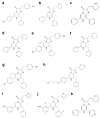
Chemical structures of d-luciferin and its red-shifted derivatives: (a) d-luciferin, (b) NH2-NpLH2, (c) OH-NpLH2, (d) 6′-aminoluciferin, (e) CycLuc1, (f) CycLuc2, (g) CycLuc7, (h) CycLuc10 (i) Infra-luciferin, (j) Akalumine-HCl, (k) PhOH-Luc.
Figure 3
Figure 3
Coelenterazine derivatives for brighter or red-shifted bioluminescence systems: (a) Coelenterazine, (b) Furimazine, (c) Diphenylterazine, (d) Selenoterazine, (e) Coelenterazine-v, (f) DeepBlueC, (g) methoxy-eCoelenterazine, (h) BBlue2.3, (i) Hydrofurimazine, (j) Fluorofurimazine, (k) 8pyDTZ.
Figure 4
Figure 4
In vivo BLI with brighter or red-shifted bioluminescence systems. (a) Video-rate BLI of Nano-lantern-expressing tumor cells in an unshaved mouse; Scale bar, 1 cm. Reprinted with permission from ref. [34]. White arrow indicates the luminescence signal from Nano-lantern-expressing cells. Copyright 2012 Nature Publishing Group. (b) AkaBLI with 4-year-old female marmoset expressing AkaLuc in the right striatum 12 months after injection. Reprinted with permission from ref. [18]. Copyright 2018 The American Association for the Advancement of Science.

Similar articles

Cited by

References

    1. Yan Y., Shi P., Song W., Bi S. Chemiluminescence and Bioluminescence Imaging for Biosensing and Therapy: In Vitro and In Vivo Perspectives. Theranostics. 2019;9:4047–4065. doi: 10.7150/thno.33228. - DOI - PMC - PubMed
    1. Welsh D.K., Noguchi T. Cellular Bioluminescence Imaging. Cold Spring Harb. Protoc. 2012;2012 doi: 10.1101/pdb.err072298. - DOI - PubMed
    1. Pozzo T., Akter F., Nomura Y., Louie A.Y., Yokobayashi Y. Firefly Luciferase Mutant with Enhanced Activity and Thermostability. Acs Omega. 2018;3:2628–2633. doi: 10.1021/acsomega.7b02068. - DOI - PMC - PubMed
    1. Law G.H.E., Gandelman O.A., Tisi L.C., Lowe C.R., Murray J.A.H. Mutagenesis of solvent-exposed amino acids in Photinus pyralis luciferase improves thermostability and pH-tolerance. Biochem. J. 2006;397:305–312. doi: 10.1042/BJ20051847. - DOI - PMC - PubMed
    1. Aswendt M., Vogel S., Schäfer C., Jathoul A., Pule M., Hoehn M. Quantitative in vivo dual-color bioluminescence imaging in the mouse brain. Neurophotonics. 2019;6:1–11. doi: 10.1117/1.NPh.6.2.025006. - DOI - PMC - PubMed