. 2020 Sep 7;21(18):6539.
doi: 10.3390/ijms21186539.

Standardization and Validation of Fluorescence-Based Quantitative Assay to Study Human Platelet Adhesion to Extracellular-Matrix in a 384-Well Plate

Affiliations

Standardization and Validation of Fluorescence-Based Quantitative Assay to Study Human Platelet Adhesion to Extracellular-Matrix in a 384-Well Plate

Augusto Martins Lima et al. Int J Mol Sci. .

Abstract

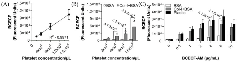
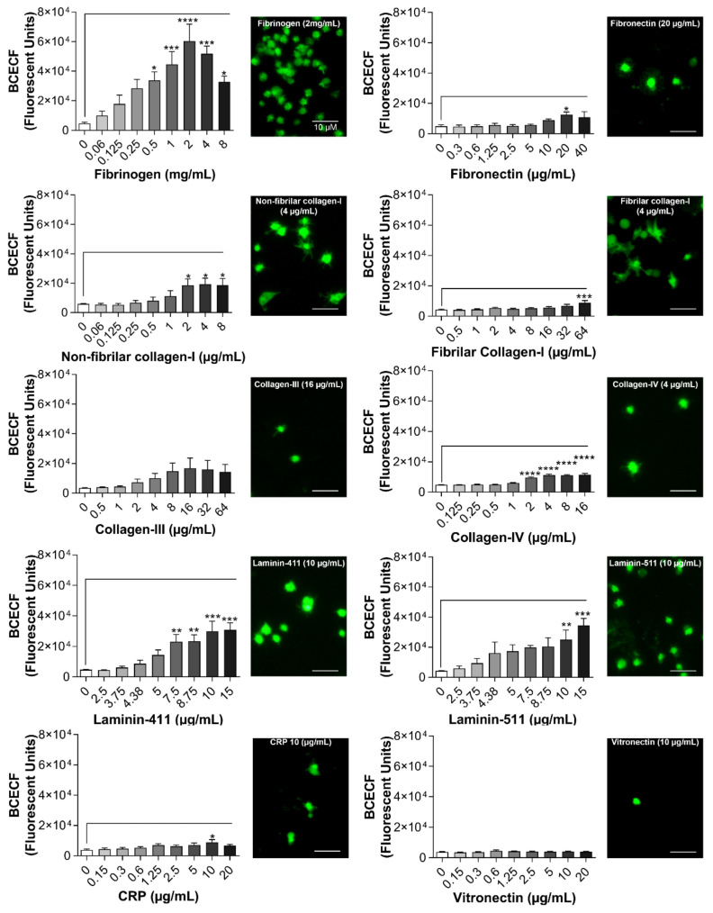
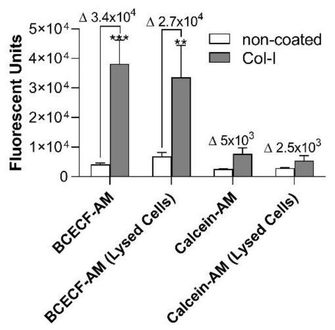
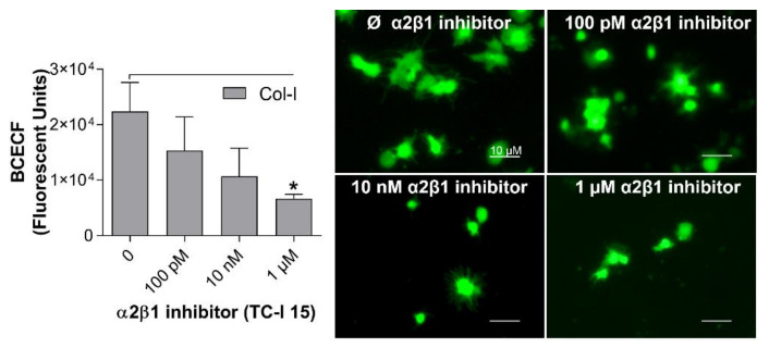
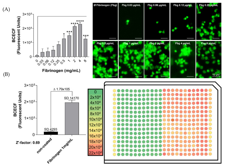
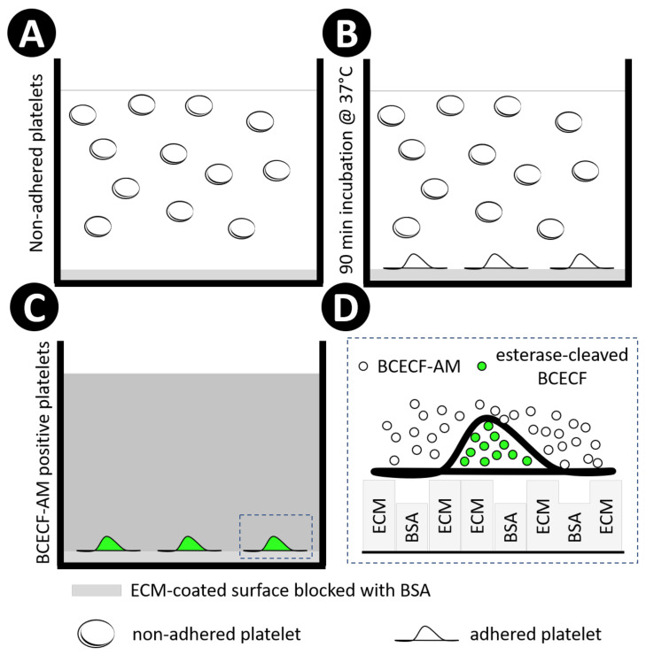
Platelets play a crucial role in the immunological response and are involved in the pathological settings of vascular diseases, and their adhesion to the extracellular matrix is important to bring leukocytes close to the endothelial cells and to form and stabilize the thrombus. Currently there are several methods to study platelet adhesion; however, the optimal parameters to perform the assay vary among studies, which hinders their comparison and reproducibility. Here, a standardization and validation of a fluorescence-based quantitative adhesion assay to study platelet-ECM interaction in a high-throughput screening format is proposed. Our study confirms that fluorescence-based quantitative assays can be effectively used to detect platelet adhesion, in which BCECF-AM presents the highest sensitivity in comparison to other dyes.

Keywords: 384-well plate; BCECF-AM; extracellular matrix; fluorescence-based quantitative assay; high-throughput screening assay; platelet adhesion.


Conflict of interest statement

The authors declare no conflict of interest. The funders had no role in the design of the study; in the collection, analyses, or interpretation of data; in the writing of the manuscript, or in the decision to publish the results.

Figures

Figure 1
Figure 1
Platelet adhesion assay linearity in a 96-well plate. 96-well microplates were coated with collagen-I (8 µg/mL in 50 µL) or incubated with distillated water for 1 h at 37 °C. After blocking the wells with BSA (0.03%), different concentrations of human washed platelets (2 × 10⁴ to 1.6 × 10⁵/µL in 100µL) were added, followed by incubation for 1 h at 37 °C. Next, non-adherent platelets were removed, and adherent platelets were incubated with BCECF-AM (0.25 to 16 µg/mL in 50 µL) for 30 min at 37 °C. Fluorescence intensity was measured using a plate reader (VictorX, PerkinElmer, Waltham, MA, USA). (A) Assay linearity (R2 = 0.9971) was calculated by measuring the fluorescence signal of different platelet concentrations adhered to plastic. (B) The optimum platelet concentration to obtain statistical differences between non-coated (white bars) and collagen-I-coated wells (grey bars) was assessed by using different platelet concentrations. Δ represents delta: (Col-I+BSA) − (BSA). (C) The optimal concentration of BCECF-AM was assessed by comparing the fluorescence signal of adherent platelets (8 × 10⁴/µL in 100µL) on non-coated wells (white bars) and collagen-I-coated wells (grey bars). Plastic was used as positive control (black bars). Δ represents delta (Col-I+BSA) − (BSA). Fluorescence intensity of collagen-coated surfaces blocked with BSA was compared to BSA alone for each experimental group by t-test (* p < 0.05, data are mean ± SEM; n = 4).
Figure 2
Figure 2
BSA blocking on different types of 96-well plates. BSA was used to prevent non-specific adhesion of platelets to the plastic. To find the optimum conditions, 3 types of 96-well plates (Invitrogen™—442404; Greiner Bio-one—655,180 and Falcon—353072) were blocked with different concentrations of BSA (0.00075 to 4% in 50 µL) for 1 hour at 37 °C. Next, human washed platelets (8 × 10⁴/µL in 100 µL) were added, followed by incubation for 1 h at 37 °C. Non-adherent platelets were removed, and adherent platelets were incubated with BCECF-AM (4 µg/mL in 50 µL) for 30 min at 37 °C. Fluorescence intensity was measured using a plate reader (VictorX, PerkinElmer, Waltham, MA, USA). Values were compared to the control condition—without BSA—by one-way ANOVA followed by Dunnett’s post hoc test (**** p < 0.0001, data are mean ± SEM; n = 4).
Figure 3
Figure 3
Platelet adhesion on different ECM proteins measured by BCECF-AM. Coating of 96-well microplates with ECM proteins (50 µL) was performed for 1 h at 37 °C. Different proteins and concentrations of ECM were used: fibrinogen (0.06 to 8 mg/mL); fibronectin (0.3 to 40 µg/mL); non-fibrillar collagen-I (0.06 to 8 µg/mL); fibrillar collagen-I (0.06 to 8 µg/mL); collagen-III (0.5 to 64 µg/mL); collagen-IV (0.125 to 16 µg/mL); laminin-411 (2.5 to 15 µg/mL); laminin-511 (2.5 to 15 µg/mL); collagen-related peptide (CRP) (0.15 to 20 µg/mL); vitronectin (0.15 to 20 µg/mL). In the wells without coating distillated water was added during the coating incubation time. After blocking the wells with BSA (0.03%), platelets (8 × 10⁴/µL in 100µL) were added, followed by incubation for 1 h at 37 °C. Next, non-adherent platelets were removed, and adherent platelets were incubated with BCECF-AM (4 µg/mL in 50 µL) for 30 min at 37 °C. Fluorescence intensity was measured using a plate reader (VictorX, PerkinElmer, Waltham, MA, USA) and images were taken using a fluorescence microscopy (Eclipse Ti2, Nikon, Tokyo, Japan) with a 20× objective (scale bar 10 µM). Values were compared with the non-coated control condition by one-way ANOVA followed by Dunnett’s post hoc test (* p < 0.05, ** p < 0.01, *** p < 0.001, **** p < 0.0001, data are mean ± SEM; n = 4).
Figure 4
Figure 4
Platelet adhesion detection using different techniques: BCECF-AM vs. Calcein-AM. Coating of 96-well microplates with collagen-I (4 µg/mL in 50 µL) or distillated water were performed for 1 h at 37 °C. After blocking the wells with BSA (0.03%), human washed platelets (8 × 10⁴/µL in 100 µL) were added, followed by incubation for 1 h at 37 °C. Next, non-adherent platelets were removed. For the BCECF-AM experimental group, adherent platelets were incubated with BCECF-AM (4 µg/mL in 50 µL) for 30 min at 37 °C. After washing the excess of BCECF-AM, fluorescence intensity was measured using plate reader (VictorX, PerkinElmer, Waltham, MA, USA). In an independent experimental condition, the signal of BCECF-AM was measured after lysing the adhered platelets with lysis buffer. For the experiment using Calcein, AM (2 µg/mL), platelets were pre-stained following the protocol previous described by several authors (see Table 1). Briefly, PRP obtained by centrifugation of the whole blood was incubated with Calcein-AM (2 µg/mL) for 1 h at 37 °C. Platelets were then washed as described in the Section 4.2. Fluorescence intensity was measured using plate reader (VictorX, PerkinElmer, Waltham, MA, USA). Δ represents delta: (Col-I) − (non-coated). Values were compared with the control condition with BSA only (non-coated), by two-way ANOVA followed by Tukey’s post hoc test (** p < 0.01, *** p < 0.001, data are mean ± SEM; n = 4).
Figure 5
Figure 5
Sensitivity of the optimized assay to detect platelet adhesion inhibition. Coating of 96-well microplates with collagen-I (4 µg/mL in 50 µL) or distillated water was performed for 1 h at 37 °C. After blocking the wells with BSA (0.03%), human washed platelets (8 × 10⁴/µL in 100µL) containing different concentrations of TC-I 15, an α2β1 integrin inhibitor, were added to the coated wells, followed by incubation for 1 h at 37 °C. Non-adherent platelets were removed, and adherent platelets were incubated with BCECF-AM (4 µg/mL in 50 µL) for 30 min at 37 °C. Fluorescence intensity was measured using a plate reader (VictorX, PerkinElmer). Values were compared to the control group without TC-I 15 by one-way ANOVA followed by Dunnett’s post hoc test (* p < 0.05, data are mean ± SEM; n = 4). Images were acquired with a fluorescence microscopy (Eclipse Ti2, Nikon, Waltham, MA, USA) with a 20x objective (scale bar 10 µM).
Figure 6
Figure 6
Z′-factor calculation to validate the platelet adhesion assay in a 384-well microplate. (A) Platelet adhesion similarity between 96- and 384-well microplates. Coating of 384-well microplate with different concentrations of fibrinogen (Fbg 0.03 to 8 mg/mL in 20 µL), or incubation with distillated water, were performed for 1 h at 37 °C. After blocking the wells with BSA (0.03%), human washed platelets (8 × 10⁴/µL in 10µL) were added, followed by incubation for 1 h at 37 °C. Next, non-adherent platelets were removed, and adherent platelets were incubated with BCECF-AM (4 µg/mL in 20 µL) for 30 min at 37 °C. Fluorescence intensity was measured using a plate reader (VictorX, PerkinElmer, Waltham, MA, USA). Values were compared with the control condition (non-coated plastic) by one-way ANOVA followed by Dunnett’s post hoc test (* p < 0.05, *** p < 0.001, **** p < 0.000,1 values presented as SEM resulting from duplicate average of four independent experiments). Images were acquired using a fluorescence microscopy (Eclipse Ti2, Nikon) with a 20x objective. (B) Z′-factor calculation to validate the platelet adhesion assay. In a 384-well microplate, 20 µL of water were added to half of the microplate (176 wells—left side). In the other half, fibrinogen (1 mg/mL) diluted in water was added (176 wells—right side). After blocking the wells with BSA (0.03%), human washed platelets (8 × 10⁴/µL in 10 µL) were added, followed by incubation for 1 h at 37 °C. Next, non-adherent platelets were removed, and adherent platelets were incubated with BCECF-AM (4 µg/mL in 20 µL) for 30 min at 37 °C. As a control, platelet adhesion on plastic was measured in the first and last columns. Δ represents delta: (Fibrinogen) − (non-coated). Fluorescence intensity (F.I.) was measured using a plate reader (VictorX, PerkinElmer, Waltham, MA, USA). The data of the graphs showing the Z′-factor were calculated using the equation previously published by Zhang et al. [22].
Figure 7
Figure 7
Schematic representation of the study design using BCECF-AM. Extracellular matrix proteins were employed to coat 96- or 384-well microplates, followed by BSA blocking, both incubated for 1 h at 37 °C. Next, human washed platelets were added into wells (A). and incubated for 90 min at 37 °C (B). Non-adherent platelets were washed away with tyrode buffer, and adherent platelets were incubated with diluted BCECF-AM for 30 min at 37 °C. The excess of BCECF-AM was removed by washing the plate 3 times with tyrode buffer (C). Non-fluorescent BCECF-AM is permeable to the platelet membrane. Once inside the platelet, intracellular esterases cleave the ester bond, releasing BCECF, which is the fluorescent form of the molecule. In addition, the cleavage of lipophilic blocking groups by esterases, leads to a charged form of BCECF, which leaks out of cells more slowly than BCECF-AM [69]. Rectangular boxes represent ECM and BSA coating(D).

Similar articles

Cited by

References

    1. Ruggeri Z.M., Mendolicchio G.L. Adhesion mechanisms in platelet function. Circ. Res. 2007;100:1673–1685. doi: 10.1161/01.RES.0000267878.97021.ab. - DOI - PubMed
    1. Bergmeier W., Hynes R.O. Extracellular matrix proteins in hemostasis and thrombosis. Cold Spring Harb. Perspect. Biol. 2012;4 doi: 10.1101/cshperspect.a005132. - DOI - PMC - PubMed
    1. Frenette P.S., Denis C.V., Weiss L., Jurk K., Subbarao S., Kehrel B., Hartwig J.H., Vestweber D., Wagner D.D. P-Selectin Glycoprotein Ligand 1 (Psgl-1) Is Expressed on Platelets and Can Mediate Platelet–Endothelial Interactions in Vivo. J. Exp. Med. 2000;191:1413–1422. doi: 10.1084/jem.191.8.1413. - DOI - PMC - PubMed
    1. Eriksson A.C., Whiss P.A. Measurement of adhesion of human platelets in plasma to protein surfaces in microplates. J. Pharmacol. Toxicol. Methods. 2005;52:356–365. doi: 10.1016/j.vascn.2005.06.002. - DOI - PubMed
    1. Varga-Szabo D., Pleines I., Nieswandt B. Cell adhesion mechanisms in platelets. Arterioscler. Thromb. Vasc. Biol. 2008;28:403–412. doi: 10.1161/ATVBAHA.107.150474. - DOI - PubMed

LinkOut - more resources